[Preprint]. 2025 Apr 1:2025.03.31.25324695.
doi: 10.1101/2025.03.31.25324695.

De novo variants in KDM2A cause a syndromic neurodevelopmental disorder

Affiliations

De novo variants in KDM2A cause a syndromic neurodevelopmental disorder

Eric N Anderson et al. medRxiv. .

Abstract

Germline variants that disrupt components of the epigenetic machinery cause syndromic neurodevelopmental disorders. Using exome and genome sequencing, we identified de novo variants in KDM2A, a lysine demethylase crucial for embryonic development, in 18 individuals with developmental delays and/or intellectual disabilities. The severity ranged from learning disabilities to severe intellectual disability. Other core symptoms included feeding difficulties, growth issues such as intrauterine growth restriction, short stature and microcephaly as well as recurrent facial features like epicanthic folds, upslanted palpebral fissures, thin lips, and low-set ears. Expression of human disease-causing KDM2A variants in a Drosophila melanogaster model led to neural degeneration, motor defects, and reduced lifespan. Interestingly, pathogenic variants in KDM2A affected physiological attributes including subcellular distribution, expression and stability in human cells. Genetic epistasis experiments indicated that KDM2A variants likely exert their effects through a potential gain-of-function mechanism, as eliminating endogenous KDM2A in Drosophila did not produce noticeable neurodevelopmental phenotypes. Data from Enzymatic-Methylation sequencing supports the suggested gene-disease association by showing an aberrant methylome profiles in affected individuals' peripheral blood. Combining our genetic, phenotypic and functional findings, we establish de novo variants in KDM2A as causative for a syndromic neurodevelopmental disorder.


Conflict of interest statement

Declaration of interests DAC, LMD and SVM are employees of and may own stock in GeneDx, LLC. The other authors declare no competing interests.

Figures

Figure 1.
Figure 1.. Prevalence of clinical findings and variant location on KDM2A protein level.
(A) Radial bar chart illustrating the core symptoms of the KDM2A-related neurodevelopmental disorder sorted by frequency. Numbers denote the frequency if each symptom in the cohort. DD: developmental delay; ID: intellectual disability. (B) Facial appearance of individuals at different ages that harbor missense variants or predicted loss-of-function variants in KDM2A. Epicanthus, upslanted palpebral fissures, thin upper and/or lower lips and low-set ears were noted as recurrent dysmorphic facial features. (C) Linear schematic representation of the KDM2A protein and location of the variants [GenBank: NM_012308.3]). Bold numbers indicate individual within the cohort. Blue variants represent missense, red variants indicate predicted loss-of-function variants.
Figure 2.
Figure 2.. KDM2A variants alter the subcellular distribution of KDM2A protein in mammalian cells.
(a) Representative immunofluorescence images of human embryonic kidney cells 293T (HEK293T) transfected with HA-tagged wild type KDM2A (KDM2A-WT) or variants (P235L, Y141C, or H811N) stained for exogenous (green) and endogenous (red) KDM2A protein. DAPi was used to label nuclei. (b) Exogenous KDM2A nuclear intensity quantification showed that the P235L (****p<0.0001) but not the Y141C or H811N showed significantly decreased nuclear intensity compared to exogenous KDM2A-WT (N = 14–20 cells). (c) Endogenous KDM2A nuclear intensity quantification showed that the P235L (****p<0.0001) but not the Y141C or H811N produced a significant reduction in nuclear intensity of endogenous KDM2A protein as compared to KDM2A-WT (N = 14–20 cells). (d) Western blots (WB) of cytoplasmic (C) and nuclear (N) fractions from HEK cells transfected with KDM2A-WT, P235L, and Y141C variants probed for exogenous KDM2A (anti-HA), endogenous KDM2A (anti-KDM2A), nuclear membrane marker (laminB1), and tubulin. (e) Nuclear-cytoplasmic (N/C) ratio quantification of endogenous KDM2A (n = 3 blots, ***p < 0.001). (f) Nuclear-cytoplasmic (N/C) ratio quantification of exogenous KDM2A (N = 3 blots, ***p < 0.001). (g) WB of HEK cells transfected with KDM2A-WT, P235L, Y141C stained for endogenous KDM2A (anti-KDM2A) and exogenous KDM2A (anti-HA). Tubulin was used as loading control. (h-i) WB quantification of endogenous KDM2A (h) and exogenous KDM2A (i) in HEK cells (*p<0.05, N = 3). One-way ANOVA were preformed to determine significance in panel b, c, e, f, h, and i. All quantifications represent mean ± s.e.m.
Figure 3.
Figure 3.. The P235L mutation alters the stability of the KDM2A protein
(a) Schematic of the cycloheximide (CHX) experiment conducted in HEK293T cells. (b) Representative Western blot showing KDM2A protein levels (anti-HA) in HEK293T cells transfected with either WT or P235L KDM2A plasmid at 0, 6, 12, 24, and 48 hours following CHX addition. Tubulin was used as a normalization control. (c) Quantification of the degradation rate and half-life (t1/2) of KDM2A after CHX treatment revealed an accelerated depletion of KDM2A in P235L-expressing cells compared to exogenous WT expression (nonlinear regression—one-phase decay, n=3).
Figure 4.
Figure 4.. A Drosophila model expressing KDM2A variants display differential toxicity in vivo.
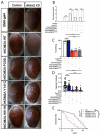
(a) Representative panel of adult Drosophila eyes expressing KDM2A-WT, P235L, Y141C, H811N or luciferase (luc) control. (b) Quantification of eye degeneration severity demonstrated that KDM2A variants significantly enhance toxicity as compared to wild type KDM2A (****p<0.0001, N=15–20). (c) WB of Drosophila expressing KDM2A (WT, P235L, Y141C, and H811N) in the eye (GMR-gal4) stained with anti-KDM2A and anti-tubulin. (d) WB quantification of KDM2A in Drosophila showed that P235L but not the Y141C or H811N variants had a significantly reduced protein level compared to KDM2A-WT (****p<0.0001, N = 3). (e) Quantification of climbing velocity (cm/s) in flies expressing KDM2A-WT, P235L, Y141C, and H811N pan-neuronally (Elav-gal4) compared to Luciferase control or wild type KDM2A (n = 3 trials, 10 animals per trials, **p < 0.01, *p < 0.05). (f) Kaplan-Meier survival curve of flies expressing KDM2A-WT, P235L, Y141C, and H811N in neurons compared with Luciferase control (n = 50–80, ****p < 0.0001). One-way ANOVA was performed in b, d, and c, while Log-rank with Grehan-Breslow-Wilcoxon tests were performed to determine significance for panel f. All quantifications represent mean ± s.e.m.
Figure 5.
Figure 5.. Expression of human KDM2A variants in a Drosophila Kdm2 knockdown or knockout model increases toxicity.
(a) Images of Drosophila eyes expressing human WT or mutant KDM2A in the context of either endogenous Kdm2 or RNAi-mediated Kdm2 knockdown (KD). (b) Quantification shows that human KDM2A variants significantly increase eye degeneration severity in the Kdm2 KD background compared to controls with endogenous Kdm2 (****p<0.0001, n=20). Notably, neither hKDM2A-WT with endogenous Kdm2 KD nor Kdm2 KD alone caused overt eye degeneration. (c-e) Quantification analyses revealed a significant reduction in (c) the percentage of flies capable of climbing (****p<0.0001, n=6 trials/10 flies per trial), (d) climbing velocity (****p<0.0001, n=3 trials/15–20 flies per trial), and (e) lifespan (Kaplan-Meier survival curve, ****p<0.0001, n=80–90 flies) in Kdm2 KO Drosophila expressing human KDM2A variants (P235L, Y141C, and H811N) using the Trojan-Gal4 system, compared to hKDM2A-WT with endogenous Kdm2 KD or KO flies alone. Statistical significance was determined by one-way ANOVA for panels b, c, and d, and by Log-rank with Grehan-Breslow-Wilcoxon tests for panel e. All data are presented as mean ± s.e.m.
Figure 6.
Figure 6.. Episignature of pLoF and missense variants in KDM2A.
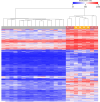
Heatmap displays hierarchical clustering of selected CpG sites of the episignature. Columns represent probes (grey: control probes; yellow: KDM2A pLoF variants; light red: KDM2A missense variants; number represents individual in the cohort). Rows represent CpG sites. Color represents methylation ranging from dark blue (no methylation) to dark red (full methylation). A distinct separation between control samples and those from individuals with variants in KMD2A is observed.

References

    1. Kawakami E., Tokunaga A., Ozawa M., Sakamoto R., and Yoshida N. (2015). The histone demethylase Fbxl11/Kdm2a plays an essential role in embryonic development by repressing cell-cycle regulators. Mechanisms of Development 135, 31–42. 10.1016/j.mod.2014.10.001. - DOI - PubMed
    1. Levy M.A., McConkey H., Kerkhof J., Barat-Houari M., Bargiacchi S., Biamino E., Bralo M.P., Cappuccio G., Ciolfi A., Clarke A., et al. (2022). Novel diagnostic DNA methylation episignatures expand and refine the epigenetic landscapes of Mendelian disorders. Human Genetics and Genomics Advances 3, 100075. 10.1016/j.xhgg.2021.100075. - DOI - PMC - PubMed
    1. Harris J.R., Gao C.W., Britton J.F., Applegate C.D., Bjornsson H.T., and Fahrner J.A. (2023). Five years of experience in the Epigenetics and Chromatin Clinic: what have we learned and where do we go from here? Hum Genet. 10.1007/s00439-023-02537-1. - DOI - PMC - PubMed
    1. Faundes V., Newman W.G., Bernardini L., Canham N., Clayton-Smith J., Dallapiccola B., Davies S.J., Demos M.K., Goldman A., Gill H., et al. (2018). Histone Lysine Methylases and Demethylases in the Landscape of Human Developmental Disorders. The American Journal of Human Genetics 102, 175–187. 10.1016/j.ajhg.2017.11.013. - DOI - PMC - PubMed
    1. van Jaarsveld R.H., Reilly J., Cornips M.-C., Hadders M.A., Agolini E., Ahimaz P., Anyane-Yeboa K., Bellanger S.A., van Binsbergen E., van den Boogaard M.-J., et al. (2022). Delineation of a KDM2B-related neurodevelopmental disorder and its associated DNA methylation signature. Genetics in Medicine, S109836002200942X. 10.1016/j.gim.2022.09.006. - DOI - PMC - PubMed

Publication types

LinkOut - more resources