. 2025 Apr 1:16:1548061.
doi: 10.3389/fmicb.2025.1548061. eCollection 2025.

RNF5 inhibits HBV replication by mediating caspase-3-dependent degradation of core protein

Affiliations

RNF5 inhibits HBV replication by mediating caspase-3-dependent degradation of core protein

Jing Xu et al. Front Microbiol. .

Abstract

The RING finger protein 5 (RNF5), an E3 ubiquitin ligase, has demonstrated significant antiviral activity against various viruses, including severe acute respiratory syndrome coronavirus 2 (SARS-CoV-2) and Kaposi's sarcoma-associated herpesvirus (KSHV). However, its role in hepatitis B virus (HBV) replication has not been previously studied. In this study, we demonstrate that RNF5 effectively inhibits HBV replication by promoting the degradation of the HBV Core protein through a Caspase-3-dependent pathway. We first show that RNF5 expression is upregulated in HBV-infected cells and patient samples, suggesting a role in the host's antiviral response. Subsequently, we investigate the mechanism by which RNF5 mediates its antiviral effect, finding that RNF5 targets the Core protein for degradation independently of its E3 ubiquitin ligase activity. The degradation of Core protein is mediated through a Caspase-3-dependent mechanism rather than the proteasomal pathway. Interestingly, RNF5's antiviral function does not rely on ubiquitination, indicating an alternative pathway involving apoptosis-related processes. These findings highlight the multifunctional role of RNF5 and suggest that targeting RNF5 could serve as a novel therapeutic approach to control HBV replication, providing new insights into the development of antiviral therapies against HBV.

Keywords: Caspase-3; E3 ubiqitin ligase; HBV - hepatitis B virus; RNF5; core protein.


Conflict of interest statement

The authors declare that the research was conducted in the absence of any commercial or financial relationships that could be construed as a potential conflict of interest.

Figures

FIGURE 1
FIGURE 1
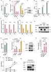
RNF5 expression is altered in HBV infection and correlates with host response. (A,B) RNF5 mRNA levels are upregulated in vitro. HepG2, Hep2.2.15, and HepAD38 cells were collected, and RNA was extracted for qPCR analysis of RNF5 expression (A). HepG2 cells were transfected with pHBV1.2 plasmids, and RNF5 mRNA levels were evaluated by qPCR at 0, 24, 48, and 72 h (B). (C–E) RNF5 mRNA levels are elevated in HBV patients and show age-related differences compared to healthy controls. (C) RNF5 mRNA levels were measured by qPCR in PBMCs from healthy controls (n = 19) and HBV patients (n = 44). (D,E) Correlation analysis of RNF5 mRNA levels with age in HBV patients and healthy controls. Data were obtained from patients at the First Hospital of Jilin University. Results are presented as mean ± SD from three independent experiments. *p < 0.05; ***p < 0.001.
FIGURE 2
FIGURE 2
RNF5 inhibits HBV replication. (A–J) RNF5 suppresses HBV replication. (A–C) HepG2 cells were co-transfected with pHBV1.2 and increasing amounts of Flag-RNF5 or an empty vector (EV). After 48 h, supernatants were analyzed for HBeAg levels via ELISA (A), RNA was extracted for qPCR to measure HBV DNA and pgRNA levels (B), and cell lysates were subjected to immunoblotting with the indicated antibodies (C). (D–G) Time-course analysis of RNF5’s effect on HBV replication. HepG2 cells were co-transfected with pHBV1.2 and Flag-RNF5 or EV. Samples were collected at 24, 48, and 72 h for analysis of HBeAg levels (D), HBV DNA and pgRNA levels (E,F), and protein expression by immunoblotting (G). (H–J) RNF5 suppresses HBV replication in HepAD38 cells. WT and RNF5 KO HepAD38 cells were seeded in 12-well plates, and after 48 h, samples were collected to analyze HBeAg levels (H), HBV DNA and pgRNA levels (I), and protein expression via immunoblotting (J). (K–M) RNF5 suppresses HBV infection. WT and RNF5 KO HepG2-NTCP cells were infected with HBV for 24 h. Supernatants were collected every 2 days, and the medium was changed after washing the cells five times with PBS. Cells were harvested on day 7 post-infection. Samples were analyzed for HBV DNA levels via qPCR (K), HBeAg levels via ELISA (L), and protein expression via immunoblotting (M). *p < 0.05; **p < 0.01; ***p < 0.001; ****p < 0.0001.
FIGURE 3
FIGURE 3
RNF5 inhibits HBV replication independent of its E3 enzyme activity. (A) Schematic representation of RNF5 wild-type (WT) and RNF5-C42S mutant structures. (B–D) Effect of RNF5 E3 ligase activity on HBV replication. HepG2 cells were co-transfected with pHBV1.2 and increasing amounts of Flag-RNF5-C42S or EV. After 48 h, HBV DNA and pgRNA levels were measured by qPCR (B), HBeAg and HBsAg levels were analyzed by ELISA (C), and HBx protein level was assessed by immunoblotting (D). ***p < 0.001; ****p < 0.0001.
FIGURE 4
FIGURE 4
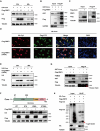
RNF5 mediates degradation of HBV core protein independent of the proteasome pathway. (A) Time-course analysis of RNF5-mediated degradation of Core. HepG2 cells were co-transfected with HA-Core and Flag-RNF5-WT or EV. Cell lysates were analyzed by Western blotting at 30 and 48 h post-transfection. (B,C) Interaction between RNF5 and Core. Co-IP confirmed interactions between RNF5 and Core (B) and between Core and endogenous RNF5 (C) in HepG2 cells 28 h post-transfection. (D) RNF5 colocalizes with Core protein. HepG2 cells co-transfected with HA-Core and Flag-RNF5-WT, C42S, or EV were subjected to immunofluorescence analysis using anti-HA and anti-Flag antibodies. (E) RNF5-mediated degradation of Core is independent of its E3 enzyme activity. HepG2 cells were co-transfected with HA-Core and Flag-RNF5-C42S or EV. Cell lysates were analyzed by Western blotting at 30 and 48 h. (F) RNF5 promotes degradation of Core lysine mutants (K7A, K96A, K7/96A). Schematic representation of Core and its lysine residues (upper panel). HepG2 cells were co-transfected with wild-type or mutant Core plasmids and RNF5 or EV, and protein levels were analyzed by immunoblotting (lower panel). (G) RNF5 interacts with endogenous proteasome 26S subunit non-ATPase 2 (PSMD2). Co-IP analysis confirmed this interaction in HepG2 cells co-transfected with Flag-RNF5 or EV, along with GST-Core or EV. (H) Effect of RNF5 on the ubiquitination of Core. 293T cells were transfected with HA-Core, Flag-ubiquitin (Flag-Ub), and Flag-RNF5 expression plasmids for 30 h, followed by collection and Co-IP analysis.
FIGURE 5
FIGURE 5
RNF5 degrades HBV core protein via the caspase-3 pathway. (A) Z-VAD inhibits RNF5-mediated Core degradation. HepG2 cells were co-transfected with HA-Core and Flag-RNF5 or EV, treated with Z-VAD (20 μM), MG132 (20 μM), or BafA1 (2 μM) for 6 h, and analyzed by immunoblotting. (B,C) Caspase-3 knockout (KO) prevents RNF5-mediated Core degradation. Caspase-1, -3, -4, and -8 were individually knocked out in HepG2 cells via CRISPR/Cas9. WT or KO cells were co-transfected with HA-Core and Flag-RNF5 or EV, and protein levels were analyzed by Western blotting. (D) Validation of Caspase-3 KO in HepG2 cells by immunoblotting. (E) RNF5 interacts with endogenous Caspase-3. Co-IP confirmed interaction in 293T cells transfected with Flag-RNF5 or EV. (F) 293T cells were co-transfected with HA-Core and Flag-Caspase-3 plasmids for 24 h, followed by treatment with or without MG132 (10 μM) or BafA1(1 μM) for an additional 24 h. The cells were then harvested, and whole-cell lysates were subjected to immunoblotting with the indicated antibodies. (G) Schematic representation of Core and its aspartate residues. (H) RNF5-Mediated Degradation of Core Is Inhibited by Alanine Substitutions at Aspartate Residues (D2/4A, D78A). HepG2 cells were co-transfected with wild-type or mutant Core plasmids and RNF5 or EV, and protein levels were analyzed by immunoblotting.

Similar articles

References

    1. Atani Z., Hosseini S., Goudarzi H., Faghihloo E. (2024). Human viral oncoproteins and ubiquitin-proteasome system. Glob. Med. Genet. 11 285–296. 10.1055/s-0044-1790210 - DOI - PMC - PubMed
    1. Chan Y., Liu N., Hsin F., Lu J., Lin J., Liu H. (2024). Temporal regulation of MDA5 inactivation by Caspase-3 dependent cleavage of 14-3-3η. PLoS Pathog. 20:e1012287. 10.1371/journal.ppat.1012287 - DOI - PMC - PubMed
    1. Collins J., Piscopio R., Reyland M., Johansen C., Benninger R., Farnsworth N. (2024). Cleavage of protein kinase c δ by caspase-3 mediates proinflammatory cytokine-induced apoptosis in pancreatic islets. J. Biol. Chem. 300:107611. 10.1016/j.jbc.2024.107611 - DOI - PMC - PubMed
    1. Didier C., Broday L., Bhoumik A., Israeli S., Takahashi S., Nakayama K., et al. (2003). RNF5, a RING finger protein that regulates cell motility by targeting paxillin ubiquitination and altered localization. Mol. Cell. Biol. 23 5331–5345. 10.1128/MCB.23.15.5331-5345.2003 - DOI - PMC - PubMed
    1. Fu J., Schroder K., Wu H. (2024). Mechanistic insights from inflammasome structures. Nat. Rev. Immunol. 24 518–535. 10.1038/s41577-024-00995-w - DOI - PMC - PubMed

LinkOut - more resources