. 2025 Apr 15;16(4):100113.
doi: 10.4239/wjd.v16.i4.100113.

MicroRNA-122-5p is upregulated in diabetic foot ulcers and decelerates the transition from the inflammatory to the proliferative stage

Affiliations

MicroRNA-122-5p is upregulated in diabetic foot ulcers and decelerates the transition from the inflammatory to the proliferative stage

Mei-Jie Yuan et al. World J Diabetes. .

Abstract

Background: Shifting from the inflammatory to the proliferative phase represents a pivotal step during managing diabetic foot ulcers (DFUs); however, existing medical interventions remain insufficient. MicroRNAs (miRs) highlight notable capacity for accelerating the repair process of DFUs. Previous research has demonstrated which miR-122-5p regulates matrix metalloproteinases under diabetic conditions, thereby influencing extracellular matrix dynamics.

Aim: To investigate the impact of miR-122-5p on the transition from the inflammatory to the proliferative stage in DFU.

Methods: Analysis for miR-122-5p expression in skin tissues from diabetic ulcer patients and mice was analyzed using quantitative real-time polymerase chain reaction (qRT-PCR). A diabetic wound healing model induced by streptozotocin was used, with mice receiving intradermal injections of adeno-associated virus -DJ encoding empty vector or miR-122. Skin tissues were retrieved at 3, 7, and 14 days after injury for gene expression analysis, histology, immunohistochemistry, and network studies. The study explored miR-122-5p's role in macrophage-fibroblast interactions and its effect on transitioning from inflammation to proliferation in DFU healing.

Results: High-throughput sequencing revealed miR-122-5p as crucial for DFU healing. qRT-PCR showed significant upregulation of miR-122-5p within diabetic skin among DFU individuals and mice. Western blot, along with immunohistochemical and enzyme-linked immunosorbent assay, demonstrating the upregulation of inflammatory mediators (hypoxia inducible factor-1α, matrix metalloproteinase 9, tumor necrosis factor-α) and reduced fibrosis markers (fibronectin 1, α-smooth muscle actin) by targeting vascular endothelial growth factor. Fluorescence in situ hybridization indicated its expression localized to epidermal keratinocytes and fibroblasts in diabetic mice. Immunofluorescence revealed enhanced increased presence of M1 macrophages and reduced M2 polarization, highlighting its role in inflammation. MiR-122-5p elevated inflammatory cytokine levels while suppressing fibrotic activity from fibroblasts exposed to macrophage-derived media, highlighting its pivotal role in regulating DFU healing.

Conclusion: MiR-122-5p impedes cutaneous healing of diabetic mice via enhancing inflammation and inhibiting fibrosis, offering insights into miR roles in human skin wound repair.

Keywords: Diabetic foot ulcer; Fibrosis; Inflammation; MicroRNA-122-5p; Wound healing.


Conflict of interest statement

Conflict-of-interest statement: The authors declare that they have no conflict of interest.

Figures
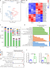
Figure 1
Figure 1
Sequencing and bioinformatics analysis of MicroRNAs. A: In diabetic ulcer mice wounds, wound tissues were harvested 14 days postinjury, and total RNA was extracted. Subsequently, MicroRNAs (miR)-seq was performed, followed by the generation of a volcano plot; B: Representative heatmap of differentially expressed miRs in the skin tissues of diabetic ulcer mice; C: The top 10 miRs with the highest expression levels in each group; D: Gene ontology annotations and enrichment analysis of the target genes of miRs; E: Kyoto encyclopedia of genes and genomes annotations and enrichment analysis of the target genes of miRs; F: Relative expression levels of miR-122-5p in wound tissue from streptozotocin-induced diabetic mice [2-∆∆Ct = 2.30, 95% confidence interval (CI): 1.81-2.72] showed a significant increase compared to normal mice (2-∆∆Ct = 1.12, 95%CI: 0.64-1.31, P < 0.001) by quantitative real-time polymerase chain reaction; Further experiments demonstrated that the miR-122-5p overexpression group (2-∆∆Ct = 3.82, 95%CI: 3.45-4.37, P < 0.001) exhibited significantly higher expression compared to the diabetic ulcer group; miR-122-5p levels were significantly increased in patients with diabetic foot ulcer (2-∆∆Ct = 1.86, 95%CI: 2.14-3.51) compared with healthy individuals (2-∆∆Ct = 1, 95%CI: 0.96-1.23, P < 0.001). cP < 0.001. miR: MicroRNA; FC: Fold change; DU: Diabetic ulcer; GO: Gene ontology; KEGG: Kyoto encyclopedia of genes and genomes; DFU: Diabetic foot ulcer; AAVDJ: Adeno-associated virus-DJ.
Figure 2
Figure 2
Adeno-associated virus-DJ-microRNA-122-5p up-decelerated wound healing in diabetic mice. A: Schematic diagram of the timeline of mice tests on the therapeutic effect of wound; B-D: A full-thickness skin wound was created on the dorsal area of the mice (1 cm × 1 cm). Animals were randomized into three groups and treated with phosphate-buffered saline. Optical pictures and related quantification of the wound closure rate in the control, diabetic ulcer (DU), and adeno-associated virus (AAV)-DJ-microRNA (miR)-122-5p groups at days 0, 3, 7, and 14 after the skin operation (n = 5); E: Gross view of wounds and wound area among the three groups of mice; F: Hematoxylin-eosin staining images of wound tissues in the control, DU, and AAVDJ-miR-122-5p groups at day 14 (n = 5; scale bar = 500 μm for 10 × and 100 μm for 40 ×); G: Masson trichrome staining at day 14 post operation (n = 5, scale bar = 500 μm for 10 × and 100 μm for 40 ×). aP < 0.05. bP < 0.01. DU: Diabetic ulcer; miR: MicroRNA; AAVDJ: Adeno-associated virus-DJ; STZ: Streptozotocin; HE: Hematoxylin-eosin.
Figure 3
Figure 3
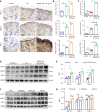
MicroRNA-122-5p upregulated matrix metalloproteinase 9 to target inflammatory factors during the transition from the inflammatory phase to the proliferative phase. A and B: Levels of matrix metalloproteinase (MMP) 9, tumor necrosis factor (TNF)-α, and hypoxia inducible factor (HIF)-1α in wound tissues of mice after 14 days were detected using immunohistochemistry (20 ×); C: The expression of MMP9, TNF-α, and HIF-1α in wound tissues of mice was assessed using quantitative real-time polymerase chain reaction; D and E: Expressions of MMP9, TNF-α, and HIF-1αproteins were tested in wound tissues (n = 3); F and G: Expressions of MMP9, TNF-α, and HIF-1α proteins were tested in NIH3T3 cells in different groups (n = 3). aP < 0.05. bP < 0.01. cP < 0.001. DU: Diabetic ulcer; miR: MicroRNA; AAVDJ: Adeno-associated virus-DJ; TNF-α: Tumor necrosis factor-α; HIF-1α: Hypoxia inducible factor-1α; MMP9: Matrix metalloproteinase 9; GAPDH: Glyceraldehyde-3-phosphate dehydrogenase; NC: Normal control.
Figure 4
Figure 4
MicroRNA-122-5p reduced vascular endothelial growth and delayed fibrosis during the transition from the inflammatory phase to the proliferative phase. A and B: Levels of vascular endothelial growth factor (VEGF), fibronectin (FN) 1, and α-smooth muscle actin (α-SMA) in wound tissues of mice after 14 days were detected using immunohistochemistry (20 ×); C: The expression of VEGF, FN1, and α-SMA in wound tissues of mice was assessed using quantitative real-time polymerase chain reaction; D and E: Expressions of VEGF, FN1, and α-SMA proteins were tested in wound tissues (n = 3); F and G: Expressions of VEGF, FN1, and α-SMA proteins were tested in NIH3T3 cells in different groups (n = 3). aP < 0.05. bP < 0.01. cP < 0.001. DU: Diabetic ulcer; miR: MicroRNA; AAVDJ: Adeno-associated virus-DJ; SMA: Smooth muscle actin; VEGF: Vascular endothelial growth factor; FN: Fibronectin; NC: Normal control.
Figure 5
Figure 5
Adeno-associated virus-DJ-microRNA-122-5p up-decelerated the transition from the inflammatory to the proliferative stage. A: In situ hybridization was performed using a microRNA (miR)-122-5p-specific probe. Green indicates miR-122-5p expression, and blue indicates 4’,6-diamidino-2-phenylindole (20 ×); B: Tissue immunofuorescence staining of F4/80, ARG1, and inducible nitric oxide synthase (20 ×); C and D: The migration capacity and quantitative analysis of NIH3T3 cultured with RAW264.7 media in different groups were assessed using a cell scratch assay (n = 3); E and F: Matrix metalloproteinase 9, vascular endothelial growth factor, inflammation factors, and fibrosis factors were detected in vivo by enzyme-linked immunosorbent assay assays (n = 3). aP < 0.05. bP < 0.01. cP < 0.001. miR: MicroRNA; DU: Diabetic ulcer; AAVDJ: Adeno-associated virus-DJ; DAPI: 4’,6-diamidino-2-phenylindole; iNOS: Inducible nitric oxide synthase; TNF-α: Tumor necrosis factor-α; HIF-1α: Hypoxia inducible factor-1α; MMP9: Matrix metalloproteinase 9; SMA: Smooth muscle actin; VEGF: Vascular endothelial growth factor; FN: Fibronectin; NC: Normal control.

Similar articles

References

    1. Armstrong DG, Boulton AJM, Bus SA. Diabetic Foot Ulcers and Their Recurrence. N Engl J Med. 2017;376:2367–2375. - PubMed
    1. Falanga V, Isseroff RR, Soulika AM, Romanelli M, Margolis D, Kapp S, Granick M, Harding K. Chronic wounds. Nat Rev Dis Primers. 2022;8:50. - PMC - PubMed
    1. Zhao L, Guo Z, Chen K, Yang W, Wan X, Zeng P, He H, Luo Y, Xiao Q, Mo Z. Combined Transplantation of Mesenchymal Stem Cells and Endothelial Colony-Forming Cells Accelerates Refractory Diabetic Foot Ulcer Healing. Stem Cells Int. 2020;2020:8863649. - PMC - PubMed
    1. Hashempour R, MirHashemi S, Mollajafari F, Damiri S, ArabAhmadi A, Raei B. Economic burden of diabetic foot ulcer: a case of Iran. BMC Health Serv Res. 2024;24:363. - PMC - PubMed
    1. Zhang S, Liu H, Li W, Liu X, Ma L, Zhao T, Ding Q, Ding C, Liu W. Polysaccharide-based hydrogel promotes skin wound repair and research progress on its repair mechanism. Int J Biol Macromol. 2023;248:125949. - PubMed

LinkOut - more resources