. 2025 Apr;21(4):e70086.
doi: 10.1002/alz.70086.

Lecanemab preferentially binds to smaller aggregates present at early Alzheimer's disease

Affiliations

Lecanemab preferentially binds to smaller aggregates present at early Alzheimer's disease

Emre Fertan et al. Alzheimers Dement. 2025 Apr.

Abstract

Introduction: The monoclonal antibodies Aducanumab, Lecanemab, Gantenerumab, and Donanemab were developed for the treatment of Alzheimer's disease (AD).

Methods: We used single-molecule detection and super-resolution imaging to characterize the binding of these antibodies to diffusible amyloid beta (Aβ) aggregates generated in-vitro and harvested from human brains.

Results: Lecanemab showed the best performance in terms of binding to the small-diffusible Aβ aggregates, affinity, aggregate coating, and the ability to bind to post-translationally modified species, providing an explanation for its therapeutic success. We observed a Braak stage-dependent increase in small-diffusible aggregate quantity and size, which was detectable with Aducanumab and Gantenerumab, but not Lecanemab, showing that the diffusible Aβ aggregates change with disease progression and the smaller aggregates to which Lecanemab preferably binds exist at higher quantities during earlier stages.

Discussion: These findings provide an explanation for the success of Lecanemab in clinical trials and suggests that Lecanemab will be more effective when used in early-stage AD.

Highlights: Anti amyloid beta therapeutics are compared by their diffusible aggregate binding characteristics. In-vitro and brain-derived aggregates are tested using single-molecule detection. Lecanemab shows therapeutic success by binding to aggregates formed in early disease. Lecanemab binds to these aggregates with high affinity and coats them better.

Keywords: Alzheimer's disease; amyloid beta; diffusible aggregate; monoclonal antibody; single‐molecule detection; super‐resolution microscopy; therapeutic success.


Conflict of interest statement

B.D.S. has been a consultant for Eli Lilly, Biogen, Janssen Pharmaceutica, Eisai, and AbbVie and other companies, but not on their antibody programs. He is consultant to Muna Therapeutics. B.D.S. is a scientific founder of Augustine Therapeutics and a scientific founder and stockholder of Muna Therapeutics. Author disclosures are available in the Supporting Information.

Figures

FIGURE 1
FIGURE 1
Visual summary of the experiments: mAbs were produced in‐house from publicly available sequences and tested with silica nanoparticles coated with Aβ, in‐vitro aggregates, and human post‐mortem Alzheimer's disease brain homogenates using SiMoA and dSTORM. The SiMoA platform utilizes paramagnetic beads coated with capture antibodies, binding to targets of interest at ultra‐low concentrations, forming a small number of immunocomplexes on the beads, which are then detected using streptavidin beta‐galactosidase and resorufin beta‐D‐galactopyranoside interactions; this provides a binary (digital) readout for aggregate quantification. The signal is quantified as average number of enzymes or in this case, AEBs. dSTORM along with SiMPull uses a glass surface optimized to capture targets of interest with high sensitivity, which are then imaged with a resolution limit of 30 nm (under the diffraction limit of light), allowing the morphological characterization of soluble aggregates (scale bars are 50 nm). Aβ, amyloid beta; AEB, aggregate per bead; dSTORM, direct stochastic optical reconstruction microscopy; mAbs, monoclonal antibodies; SiMPull, single‐molecule pulldown; SiMoA, single‐molecule array.
FIGURE 2
FIGURE 2
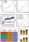
Characterization of the mAbs using synthetic aggregates: Signal on SiMoA was compared between Aducanumab, Lecanemab, Gantenerumab, and Donanemab using Aβ‐ 42 (A) and pyroglutamate Aβ– (B) coated glass nanoparticles at different concentrations (x‐axis shows concentration in nM). Each antibody–bead concentration combination was tested at least on four independent wells. For Aβ42‐coated beads, a significant antibody by concentration interaction was determined (AIC = 470.4, F = 306.6, p < 0.001, ηp2 = 0.855). While Donanemab did not show a dose dependent increase in signal, Aducanumab, Lecanemab, and Gantenerumab all showed a significant increase with increased bead concentration, with the greatest increase seen in Lecanemab (A). For pyroglutamate Aβ coated beads, once again a significant antibody by concentration interaction was present (AIC = 101.3, F = 264.4, p < 0.001, ηp2 = 0.860). All four antibodies showed a concentration‐dependent increase in signal, with the greatest signal acquired from Lecanemab and Donanemab, whereas the weakest signal was measured from gantenerumab (B). In addition to coated silica‐nanoparticles, in‐vitro aggregates of Aβ were also prepared by incubating Aβ42 and pyroglutamate Aβ for different durations. A ThT assay was performed to characterize the aggregation dynamics of different species, revealing the formation of ThT‐positive aggregates over time in the samples containing pure Aβ42 and a mixture of Aβ42 and pyroglutamate Aβ; however, the signal did not change for the pure pyroglutamate Aβ signal, showing a lack of aggregation (C; x‐axis shows time in minutes). Then these samples were tested on SiMoA on three independent wells, revealing a three‐way interaction between the antibody, aggregate type, and concentration (AIC = 584.2, F = 23.7, p < 0.001, ηp2 = 0.797). Overall, the signal with the mixed aggregates was higher for all antibodies than the pure Aβ aggregates, and the signal increased with aggregate concentration. The highest signal was detected by Lecanemab, followed by Aducanumab, Gantenerumab, and Donanemab (D; x‐axis shows concentration in nM). The limit of detection was in parallel to the signal at high concentrations (E). The performance of Aducanumab, Lecanemab, and Gantenerumab with in‐vitro aggregates at different sizes was also compared using single‐molecule pulldown (FG), with imaging the aggregates on two independent wells for at least nine fields of view per well. For the number of aggregates detected, there was an antibody by aggregate type interaction (AIC = 800.0, F = 4.9, p = 0.002, ηp2 = 0.214). The greatest number of aggregates were detected with gantenerumab, followed by aducanumab and lecanemab. Although Aducanumab and Lecanemab showed a strong preference for early and late oligomers, the difference was smaller for gantenerumab (F). Aggregate brightness was also measured, with controlling for the number of dyes per antibody, providing a measure for numbers of antibodies binding to unit area of each aggregate (i.e., the aggregate‐coating ability). Again, an antibody by aggregate type interaction (AIC = 703317.0, F = 91.8, p < 0.001, ηp2 = 0.006) was found. Lecanemab achieved the highest brightness of all antibodies when tested with early oligomers, which was significantly higher than the signal it provided with fibrils and least with late oligomers. On the other hand, Gantenerumab and Aducanumab provided the brightest signal with fibrils, with the signal from Aducanumab being significantly dimmer than Gantenerumab, collectively showing a strong preference for early oligomers by lecanemab, and a preference for fibrillar aggregates by gantenerumab, and to a lesser extent aducanumab (G). Aβ, amyloid beta; AIC, Akaike information criterion; mAbs, monoclonal antibodies; nM, nanomolar; 𝞰p2, partial eta squared; SiMoA, single‐molecule array; ThT, thioflavin T.
FIGURE 3
FIGURE 3
Characterization of the mAbs using post‐mortem human Alzheimer's disease brain homogenates: Signal on SiMoA was compared between Aducanumab, Lecanemab, Gantenerumab, and Donanemab using human post‐mortem brain homogenates from patients with Braak stages 0, 3, or 5 Alzheimer's disease, with two independent brain samples tested in two independent wells per condition (A). A significant antibody by Braak stage interaction was observed (AIC = 63.19, F = 128.1, p < 0.001, ηp2 = 0.964); whereas Aducanumab and Gantenerumab signal correlated positively with Braak stage, Lecanemab showed the highest detection with the Stage 3 samples. Meanwhile, Donanemab signal was at the baseline for the brain homogenates containing soluble Aβ aggregates. Brain homogenates were also tested on SiMPull with dSTORM, to measure the length (B) and area (C) of the aggregates detected by the antibodies on two independent wells with at least two fields of view each. There was an mAb by Braak stage interaction for both aggregate length (AIC = 231669.0, F = 54.9, p < 0.001, ηp2 = 0.008) and area (AIC = 497262.0, F = 61.7, p < 0.001, ηp2 = 0.008). All mAbs showed a size preference and mostly bound to the aggregates 90 nm in length; yet the average length of the detected aggregates increased at Braak Stage 5 for Aducanumab and Gantenerumab, whereas Lecanemab remained preferentially binded to the smaller aggregates at this stage. Average aggregate brightness as also measured during diffraction‐limited imaging (same brain samples imaged in two independent wells with nine fields of view), with controlling for the number of dyes per antibody, provided a measure for number of antibodies binding to unit area of each aggregate (D). Although the brightness signal was provided by Lecanemab overall and on average the Stage 5 brains were brighter, Lecanemab actually provided the brightest signal with the Stage 0 samples, followed by Stage 3, showing a preference for aggregates formed at early disease stages, unlike Gantenerumab and Aducanumab, which had a brighter signal with the Stage 5 samples, resulting in an antibody by Braak stage interaction (AIC = 466760.0, F = 98.6, p < 0.001, ηp2 = 0.007). Finally, an immuno‐pulldown was performed using the antibodies to identify the amount of available binding regions on the aggregates they detect (E). The greatest signal reduction was seen in aducanumab, followed by gantenerumab and lecanemab, once again showing the superior aggregate coating ability of lecanemab. Aβ, amyloid beta; AIC, Akaike information criterion; dSTORM, direct stochastic optical reconstruction microscopy; mAbs, monoclonal antibodies; 𝞰p2, partial eta squared; SiMoA, single‐molecule array; SiMPull, single‐molecule pulldown.

Similar articles

Cited by

References

    1. Selkoe DJ. Amyloid protein and Alzheimer's disease. Scientific American. 1991;265(5):68‐71. - PubMed
    1. Hardy J, Allsop D. Amyloid deposition as the central event in the aetiology of Alzheimer's disease. Trends Pharmacol Sci. 1991;12(10):383‐388. - PubMed
    1. Arndt JW, Qian F, Smith BA, et al. Structural and kinetic basis for the selectivity of aducanumab for aggregated forms of amyloid‐β. Sci Rep. 2018;8(1):6412. doi:10.1038/s41598-018-24501-0 - DOI - PMC - PubMed
    1. Swanson CJ, Zhang Y, Dhadda S, et al. A randomized, double‐blind, phase 2b proof‐of‐concept clinical trial in early Alzheimer's disease with lecanemab, an anti‐Aβ protofibril antibody. Alzheimers Res Ther. 2021;13:80. doi:10.1186/s13195-021-00813-8 - DOI - PMC - PubMed
    1. Bohrmann B, Baumann K, Benz J, et al. Gantenerumab: a novel human anti‐Aβ antibody demonstrates sustained cerebral amyloid‐β binding and elicits cell‐mediated removal of human amyloid‐β. J Alzheimers Dis. 2012;28(1):49‐69. doi:10.3233/JAD-2011-110977 - DOI - PubMed

MeSH terms