. 2025 May 14;16(5):e0060225.
doi: 10.1128/mbio.00602-25. Epub 2025 Apr 16.

AUP1 and UBE2G2 complex targets STING signaling and regulates virus-induced innate immunity

Affiliations

AUP1 and UBE2G2 complex targets STING signaling and regulates virus-induced innate immunity

Xin-Tao Wang et al. mBio. .

Abstract

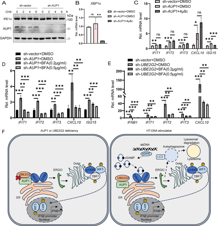
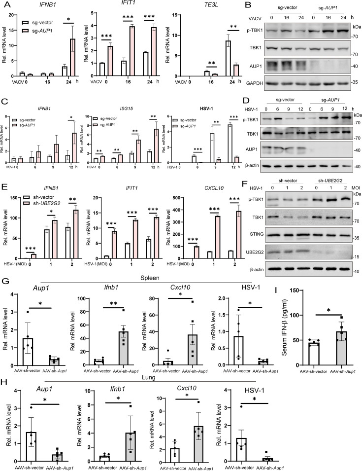
Stimulator of interferon genes (STING) is an endoplasmic reticulum (ER) signaling adaptor that is essential for the host immune response triggered by DNA pathogens. Precise regulation of STING is crucial for maintaining a balanced immune response and preventing harmful autoinflammation. Activation of STING requires its translocation from the ER to the Golgi apparatus. However, the mechanisms that maintain STING in its resting state remain largely unclear. Here, we find that deficiency of the ancient ubiquitous protein 1 (AUP1) causes spontaneous activation of STING and enhances the expression of type I interferons (IFNs) under resting conditions. Furthermore, deficiency of UBE2G2, a cofactor of AUP1, also promotes the abnormal activation of STING. AUP1 deficiency significantly enhances STING signaling induced by DNA virus, and AUP1 deficiency exhibits increased resistance to DNA virus infection in vitro and in vivo. Mechanistically, AUP1 may form a complex with UBE2G2 to interact with STING, preventing its exit from the ER membrane. Notably, infection with the RNA virus vesicular stomatitis virus (VSV) promotes the accumulation of lipid droplets (LDs) and AUP1 proteins. Additionally, AUP1 deficiency markedly inhibits the replication of VSV because AUP1 deficiency reduces lipid accumulation and alters the expression of lipid metabolism genes, such as carnitine palmitoyltransferase 1A (CPT1A), monoglyceride lipase (MGLL), and sterol regulatory element-binding transcription factor 1 (SREBF1). This study uncovers the essential roles of AUP1 in the STING signaling pathway and lipid metabolism pathway, highlighting its dual role in regulating virus replication.IMPORTANCEThe stimulator of interferon genes (STING) signaling cascade plays an essential role in coordinating innate immunity against DNA pathogens and autoimmunity. Precise regulation of the innate immune response is essential for maintaining homeostasis. In this study, we demonstrate that ancient ubiquitous protein 1 (AUP1) and UBE2G2 act as negative regulators of the innate immune response by targeting STING. Notably, AUP1 interacts with STING to retain STING in the endoplasmic reticulum (ER), preventing STING translocation and thereby limiting STING signaling in the resting state. In addition, deficiency of AUP1 markedly inhibits the replication of DNA virus and RNA virus. Our findings provide new insights into the regulation of STING signaling and confirm AUP1 has a dual role in regulating virus replication.

Keywords: AUP1; STING; UBE2G2; innate immunity; virus infection.


Conflict of interest statement

The authors declare no conflict of interest.

Figures

Fig 1
Fig 1
AUP1 deficiency promotes the cGAS-STING signaling pathway. (A) Control cells or stable AUP1-deficient HeLa cells were transfected with HT-DNA (1 µg/mL) for indicated time points. The levels of IFNB1, IFIT1, ISG15, and CXCL10 mRNA were examined by qRT-PCR. (B) Control cells or AUP1-knockdown HeLa cells were transfected with HT-DNA (1 µg/mL) for indicated time points. The levels of IFNB1, IFIT1, ISG15, and CXCL10 mRNA were examined by qRT-PCR. (C) qRT-PCR analysis of ISG15 and IFIT1 mRNA abundance in AUP1-deficient HeLa cells and cells transfected with different doses of HT-DNA for 6 h. (D) qRT-PCR analysis of IFNB1 and IFIT1 mRNA abundance in wild-type and AUP1-deficient HeLa cells transfected with poly(I:C) for indicated time points. (E) Immunoblot analysis of the indicated proteins in wild-type and AUP1-deficient HeLa cells transfected with the HT-DNA for indicated time points. *, p-STING. (F) Immunoblot analysis of the indicated proteins in wild-type and AUP1 stable knockdown HeLa cells transfected with the HT-DNA for indicated time points. (G) Immunoblot analysis of the indicated proteins in wild-type and AUP1-deficient HeLa cells transfected with poly(I:C) for indicated time points. Data are representative of three experiments with similar results. Bar graphs show the means ± standard deviation (SD). *P < 0.05, **P < 0.01, and ***P < 0.001.
Fig 2
Fig 2
AUP1 interacts with STING. (A) HEK293T cells were transfected with the indicated plasmids. Twenty-four hours after transfection, the cell lysates were immunoprecipitated with an anti-Flag antibody or normal immunoglobulin G (IgG) and then immunoblotted with the indicated antibodies. (B and C) HeLa cells were transfected with the indicated plasmids. Twenty-four hours after transfection, the cell lysates were immunoprecipitated with an anti-Flag antibody or IgG and then immunoblotted with the indicated antibodies. (D) HeLa cells were stained with the indicated antibodies and imaged by confocal microscopy. Scale bar represents 5 µm. (E) HeLa cells were transfected with the indicated plasmids for 24 h and then transfected with HT-DNA for the indicated times, and the cell lysates were subjected to immunoprecipitation with the indicated antibodies. (F) Schematic of AUP1 and its truncation mutant protein domains. TM, transmembrane; CUE, coupling of ubiquitin conjugation to endoplasmic reticulum degradation; G2BR, UBE2G2 binding region. (G) Flag-tagged AUP1 or its mutants were individually transfected into HEK293T cells along with the HA-tagged STING. The cell lysates were immunoprecipitated with an anti-Flag antibody or IgG and then immunoblotted with the indicated antibodies. (H) Schematic of STING and its truncation mutant protein domains. TM, transmembrane; LBD, ligand-binding domain; CTT, carboxy-terminal tail. (I) Flag-tagged STING or its mutants were individually transfected into HEK293T cells along with V5-tagged AUP1. The cell lysates were immunoprecipitated with an anti-Flag antibody or IgG and then immunoblotted with the indicated antibodies. (J) 3D structures of AUP1/STING. Human STING crystal structure derived from PDB (6NT5). The structure of human AUP1 protein was derived from AlphaFold prediction.
Fig 3
Fig 3
AUP1 restricts STING signaling. (A) qRT-PCR analysis of IFNB1 and ISGs (IFIT1 and ISG15) mRNA expression in wild-type and AUP1 knockout HeLa cells after stimulation with diABZI (2.5 µM) for indicated time points. (B and C) qRT-PCR analysis of IFNB1 and ISGs mRNA expression in wild-type and AUP1 knockdown THP1 cells after stimulation with diABZI or 2′,3′-cGAMP (1 µg/mL) for indicated time points. (D) Immunoblot analysis of the indicated proteins in wild-type and AUP1 knockout HeLa cells stimulated with diABZI for indicated time points. (E) Immunoblot analysis of the indicated proteins in wild-type and AUP1 knockdown THP1 cells stimulated with diABZI for indicated times. Data are representative of three experiments with similar results. Bar graphs show the means ± SD. *P < 0.05, **P < 0.01, and ***P < 0.001.
Fig 4
Fig 4
UBE2G2 is required for AUP1 to regulate STING signaling. (A) Cells were treated with CHX (50 µg/mL) for the indicated times to inhibit protein synthesis and UBE2G2 degradation monitored by Western blot in wild-type and AUP1 knockdown HeLa cells. (B and C) HEK293T cells were transfected with the indicated plasmids. Twenty-four hours after transfection, the cell lysates were immunoprecipitated with an anti-Flag antibody or IgG and then immunoblotted with the indicated antibodies. (D) qRT-PCR analysis of the baseline IFIT1, ISG15, and CXCL10 expression in AUP1 knockdown cells, wild-type cells, and AUP1 knockdown cells stably expressing wild-type UBE2G2. (E) qRT-PCR analysis of IFNB1 and CXCL10 mRNA expression in wild-type and UBE2G2 knockdown THP1 cells after stimulation with HT-DNA for indicated times. (F) qRT-PCR analysis of IFNB1 and ISGs (IFIT1, ISG15, and CXCL10) mRNA expression in wild-type and UBE2G2 knockdown HeLa cells after stimulation with HT-DNA for indicated times. (G) qRT-PCR analysis of IFNB1, IFIT1, and CXCL10 expression in wild-type, UBE2G2 knockdown, and UBE2G2 knockdown with knockdown of STING HeLa cells after stimulation with HT-DNA for indicated times. (H and I) Immunoblot analysis of the indicated proteins in wild-type and UBE2G2 knockdown THP1 or HeLa cells after stimulation with HT-DNA for indicated times. (J) Immunoblot analysis of the indicated proteins in wild-type, UBE2G2 knockdown, and UBE2G2 knockdown with knockdown of STING HeLa cells after stimulation with HT-DNA for indicated times. Data are representative of three experiments with similar results. Bar graphs show the means ± SD. *P < 0.05, **P < 0.01, and ***P < 0.001.
Fig 5
Fig 5
Knockdown of UBE2G2 activates STING signaling. (A) qRT-PCR analysis of the baseline ISGs (CXCL10, IFIT1, ISG15, MX1, and OAS1) expression in wild-type cells and UBE2G2 knockdown cells. (B) qRT-PCR analysis of the baseline IFNB1 and ISGs (IFIT1, CXCL10) expression in wild-type cells, UBE2G2 knockdown cells, and UBE2G2 knockdown cells stably expressing wild-type or mutant UBE2G2. (C) qRT-PCR analysis of IFNB1, IFIT1, and CXCL10 expression in wild-type, UBE2G2 knockdown, and UBE2G2 knockdown with knockdown of STING HeLa cells after stimulation with diABZI for indicated times. (D) Immunoblot analysis of the indicated proteins in wild-type and UBE2G2 knockdown HeLa cells after stimulation with diABZI for indicated times. (E) Immunoblot analysis of the indicated proteins in wild-type, UBE2G2 knockdown, and UBE2G2 knockdown with knockdown of STING HeLa cells after stimulation with diABZI for indicated times. (F) qRT-PCR analysis of IFNB1, IFIT1, and CXCL10 expression in wild-type and UBE2G2 knockdown HT1080 cells after stimulation with diABZI for indicated times. (G) Immunoblot analysis of the indicated proteins in wild-type and UBE2G2 knockdown HT1080 cells after stimulation with diABZI for indicated times. Data are representative of three experiments with similar results. Bar graphs show the means ± SD. *P < 0.05, **P < 0.01, and ***P < 0.001.
Fig 6
Fig 6
Knockdown of Ube2g2 activates STING signaling in L929 cells. (A) Immunoblot analysis of Ube2g2 protein (left) and qRT-PCR analysis of Ube2g2 mRNA level (right) in Ube2g2 stably knockdown L929 cells. (B) qRT-PCR analysis of baseline ISGs (Ifit1, Isg15, Oas1, and Cxcl10) in wild-type Ube2g2 and two independent Ube2g2 knockdown L929 cell lines. (C and D) qRT-PCR analysis of Ifnb1, Ifit1, Isg15, and Cxcl10 mRNA expression in wild-type and Ube2g2 knockdown L929 cells after stimulation with HT-DNA (C) or cGAMP (1 µg/mL) (D) for indicated times. (E and F) Immunoblot analysis of the indicated proteins in wild-type and Ube2g2 knockdown L929 cells after stimulation with HT-DNA (E) or cGAMP (1 µg/mL) (F) for indicated times. Data are representative of three experiments with similar results. Bar graphs show the means ± SD. *P < 0.05, **P < 0.01, and ***P < 0.001.
Fig 7
Fig 7
AUP1 and UBE2G2 retain STING at the ER. (A) Cells were treated with CHX (50 µg/mL) for the indicated times to inhibit protein synthesis and IRE1α degradation monitored by Western blot in wild-type and AUP1 knockdown HeLa cells. (B) qRT-PCR analysis of spliced XBP1 mRNA expression in wild-type and AUP1 knockdown HeLa cells treated with DMSO or 4μ8c (50 µM) for 24 h. (C) qRT-PCR analysis of the baseline ISGs (IFIT1, IFIT2, IFIT3, CXCL10, and ISG15) in wild-type and AUP1 knockdown HeLa cells treated with DMSO or 4μ8C (50 µM) for 24 h. (D) qRT-PCR analysis of the baseline ISGs (IFIT1, IFIT2, IFIT3, CXCL10, and ISG15) in wild-type and AUP1 knockdown HeLa cells treated with DMSO or BFA for 6 h. (E) qRT-PCR analysis of the baseline IFNB1 and ISGs (IFIT1, IFIT2, IFIT3, and CXCL10) in wild-type and UBE2G2 knockdown THP1 cells treated with DMSO or BFA for 6 h. (F) A proposed model of AUP1 and UBE2G2 complex controls STING-mediated innate immunity. On the one hand, AUP1 and UBE2G2 interact with STING in the resting state, but when AUP1 or UBE2G2 deficiency occurs, STING exits from the ER and begins trafficking, inducing activation of downstream signaling. On the other hand, HT-DNA stimulation can disrupt the interaction of AUP1 and UBE2G2 complex with STING and promote the normal signaling of STING. Data are representative of three experiments with similar results. Bar graphs show the means ± SD. *P < 0.05, **P < 0.01, and ***P < 0.001.
Fig 8
Fig 8
AUP1 deficiency enhances DNA virus-triggered signaling and restricts viral infection in vitro and in vivo. (A) qRT-PCR analysis of IFNB1, IFIT1, and TE3L mRNA expression in wild-type and AUP1 knockout HeLa cells infected with VACV (MOI = 0.1) for the indicated times. (B) Immunoblot analysis of the indicated proteins in wild-type and AUP1 knockout HeLa cells infected with VACV (MOI = 0.1) for indicated times. (C) qRT-PCR analysis of mRNA abundance of the indicated genes in wild-type and AUP1 knockout HeLa cells infected with HSV-1 (MOI = 1) for indicated times. (D) Immunoblot analysis of the indicated proteins in wild-type and AUP1 knockout HeLa cells infected with HSV-1 (MOI = 1) for indicated times. (E) qRT-PCR analysis of mRNA abundance of the indicated genes in wild-type and UBE2G2 knockdown THP1 cells infected with HSV-1 for 4 h. (F) Immunoblot analysis of the indicated proteins in wild-type and UBE2G2 knockdown THP1 cells infected with HSV-1 for 4 h. (G and H) Two sets of mice were injected intravenously with AAV-sh-vector (1 × 10¹² vg/mouse) or AAV-sh-Aup1 (1 × 10¹² vg/mouse) and kept under the same conditions for 4 weeks. Mice injected with AAV-sh-vector or AAV-sh-Aup1 (n  =  5) were intravenously infected with HSV‐1 (0.5  ×  107 PFU) for 24 h and then the lungs and spleens of the mice were subjected to real‐time PCR assays. (I) ELISA analyses of IFN-β from the sera of control and Aup1 knockdown mice (n  =  5) after intravenous injection with HSV-1 (0.5  ×  107 PFU) for 24 h. (G–I) Each symbol represents one independent biological replicate. Bar graphs show the means ± SD. *P < 0.05, **P < 0.01, and ***P < 0.001.
Fig 9
Fig 9
VSV infection induces AUP1 protein accumulation, and knockdown of AUP1 alters lipid metabolism genes. (A) Immunoblot analysis of the indicated proteins at different time intervals upon VSV (MOI = 1) infection in HT1080 cells (left) and densitometric analyses to quantitate increased expression of AUP1 (right). GAPDH was used as an internal control. (B and C) LDs (stained with Nile Red) visualized using confocal imaging. Scale bar represents 50 µm. The fluorescence intensity of lipid was analyzed with ImageJ. (D) qRT-PCR analysis of mRNA expression levels of various lipid metabolism-associated factors in wild-type and AUP1 knockdown HeLa cells. (E and F) HeLa cells were transfected with vector or Flag-CPT1A for 24 h, followed by infection with VSV (MOI = 0.1) for indicated time. The mRNA and protein levels of VSV were determined by qRT-PCR and Western blot. (G and H) HeLa cells were transfected with vector or Flag-MGLL for 24 h, followed by infection with VSV (MOI = 0.1) for indicated times. The mRNA and protein levels of VSV were determined by qRT-PCR and Western blot. (I and J) HeLa cells were infected with VSV (MOI = 1) and then treated with DMSO or AM580 (20 µM) for indicated times. The mRNA and protein levels of VSV were determined by qRT-PCR and Western blot. Data are representative of three experiments with similar results. Bar graphs show the means ± SD. *P < 0.05, **P < 0.01, and ***P < 0.001.

References

    1. Luo Y, Chang L, Ji Y, Liang T. 2024. ER: a critical hub for STING signaling regulation. Trends Cell Biol 34:865–881. doi:10.1016/j.tcb.2024.02.006 - DOI - PubMed
    1. Decout A, Katz JD, Venkatraman S, Ablasser A. 2021. The cGAS-STING pathway as a therapeutic target in inflammatory diseases. Nat Rev Immunol 21:548–569. doi:10.1038/s41577-021-00524-z - DOI - PMC - PubMed
    1. Ishikawa H, Barber GN. 2008. STING is an endoplasmic reticulum adaptor that facilitates innate immune signalling. Nature 455:674–678. doi:10.1038/nature07317 - DOI - PMC - PubMed
    1. Jeremiah N, Neven B, Gentili M, Callebaut I, Maschalidi S, Stolzenberg M-C, Goudin N, Frémond M-L, Nitschke P, Molina TJ, Blanche S, Picard C, Rice GI, Crow YJ, Manel N, Fischer A, Bader-Meunier B, Rieux-Laucat F. 2014. Inherited STING-activating mutation underlies a familial inflammatory syndrome with lupus-like manifestations. J Clin Invest 124:5516–5520. doi:10.1172/JCI79100 - DOI - PMC - PubMed
    1. Gui X, Yang H, Li T, Tan X, Shi P, Li M, Du F, Chen ZJ. 2019. Autophagy induction via STING trafficking is a primordial function of the cGAS pathway. Nature 567:262–266. doi:10.1038/s41586-019-1006-9 - DOI - PMC - PubMed

LinkOut - more resources