. 2025 Jul 30;232(1):e116-e125.
doi: 10.1093/infdis/jiaf192.

Increased Atherosclerosis in HIV-Infected Humanized Mice Is Caused by a Single Viral Protein, Nef

Affiliations

Increased Atherosclerosis in HIV-Infected Humanized Mice Is Caused by a Single Viral Protein, Nef

Yongsen Wang et al. J Infect Dis. .

Abstract

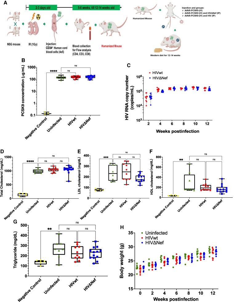
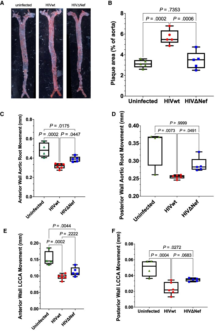
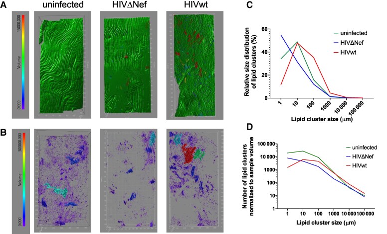
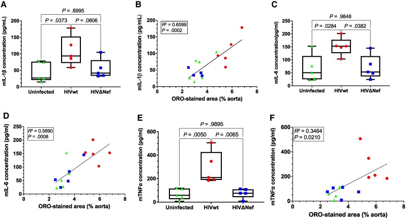
Antiretroviral therapy suppresses human immunodeficiency virus (HIV) replication, reverses immunodeficiency, and reduces AIDS-related symptoms, but non-AIDS comorbidities like cardiovascular diseases remain a major challenge for people with HIV (PWH). The pathogenic mechanisms driving these comorbidities are poorly understood. We previously showed that the HIV protein Nef contributes to chronic inflammation in PWH. Here, we explored Nef's role in HIV-associated atherosclerosis using a novel model: HIV-infected humanized mice expressing a gain-of-function mutant of proprotein convertase subtilisin/kexin type 9 (PCSK9) and fed a high-fat diet. Comparing atherosclerosis in uninfected mice to those infected with Nef-positive or Nef-deficient HIV-1, we found that Nef exacerbates atherosclerotic changes by increasing inflammation. These results identify Nef as a key driver of HIV-related atherosclerosis and provide a platform for testing therapeutic interventions targeting Nef to mitigate cardiovascular risks in PWH.

Keywords: HIV; Nef; atherosclerosis; humanized mice; inflammation.


Conflict of interest statement

Potential conflicts of interest. All authors: No reported conflicts of interest. All authors have submitted the ICMJE Form for Disclosure of Potential Conflicts of Interest. Conflicts that the editors consider relevant to the content of the manuscript have been disclosed.

Figures

Figure 1.
Figure 1.
Mouse model of HIV-associated atherosclerosis. A, Experimental procedure schematic. B, PCSK9 concentrations at euthanasia. Results shown as box plots (mean ± SD). Groups compared using 1-way ANOVA with Tukey test. C, HIV RNA quantified every 2 weeks by qRT-PCR. D, Total cholesterol measured using Wako kit. Results analyzed as in (B). Negative control was humanized mice fed a high-fat diet without AAV-PCSK9. E, LDL-C levels measured and analyzed as in (B). F, HDL-C levels measured using Crystal Chem kit. Results are presented as in (B). Results analyzed using Kruskal-Wallis test with Dunn correction. G, Triglycerides measured using Wako kit. Results analyzed as in (B). H, Body weight measured every 2 weeks. Box plots show the median (line), interquartile range (box), and minimum/maximum values (whiskers). ****P < .0001, ***P < .001, **P < .01, *P < .05. Abbreviations: HDL-C, high-density lipoprotein cholesterol; HIVwt, HIV wild type; HIVΔNef, Nef-deficient HIV; IP, intraperitoneal; IR, irradiation; IV, intravenous; LDL-C, low-density lipoprotein cholesterol; ns, not significant; qRT-PCR, quantitative reverse transcription polymerase chain reaction.
Figure 2.
Figure 2.
Vascular atherosclerosis analysis. A, En face ORO staining of aorta. B, Quantitative staining analysis. Results shown as box plots (mean ± SD). Groups compared using 1-way ANOVA with Tukey test. C–F, Vascular ultrasound analysis. Imaging performed in M Mode using a 40-MHz transducer. Wall displacement averaged over 3 cardiac cycles. Error bars indicate SD. C, Anterior aortic root displacement. Groups compared using 1-way ANOVA with Tukey test. D, Posterior aortic root displacement. Analysis by Kruskal-Wallis test with Dunn correction. E, Anterior LCCA displacement. Analysis as in (C). F, Posterior LCCA displacement. Analysis as in (C). n = 4 (uninfected), n = 6 (HIVwt), n = 5 (HIVΔNef). Abbreviations: HIVwt, HIV wild type; HIVΔNef, Nef-deficient HIV; LCCA, left common carotid artery. Box plots show the median (line), interquartile range (box), and minimum/maximum values (whiskers).
Figure 3.
Figure 3.
Two-photon imaging of aorta. Aortas imaged using 25×/0.8 oil immersion objective. A, ORO labeling surface model. B, ORO plus elastin autofluorescence. Lipid cluster size is color coded by volume (red, largest; blue, smallest). Image volume stitched from 2 × 3 (uninfected, HIVΔNef) or 4 × 3 (HIVwt) fields. C, ORO-stained lipid cluster size distribution. D, ORO-stained lipid clusters per media volume. Abbreviations: HIVwt, HIV wild type; HIVΔNef, Nef-deficient HIV; ORO, Oil Red O.
Figure 4.
Figure 4.
Cytokine levels in blood. A, Murine IL-1β levels. Results shown as box plots (mean ± SD). Groups compared using 1-way ANOVA with Tukey test. B, IL-1β versus ORO-stained area correlation. C, Murine IL-6 levels. D, IL-6 versus ORO-stained area correlation. E, Levels of murine TNF-α. Results are presented as in (A). F, TNF-α versus ORO-stained area correlation. All comparisons were made as in (A). Color and shape of the symbols in (B, D, and F) match (A, C, and E). Abbreviations: HIVwt, HIV wild type; HIVΔNef, Nef-deficient HIV; IL, interleukin; ORO, Oil Red O; TNF-α, tumor necrosis factor-α. Box plots show the median (line), interquartile range (box), and minimum/maximum values (whiskers).

Similar articles

References

    1. Bai R, Li Z, Lv S, et al. Persistent inflammation and non-AIDS comorbidities during ART: coming of the age of monocytes. Front Immunol 2022; 13:820480. - PMC - PubMed
    1. Hsue PY, Waters DD. HIV infection and coronary heart disease: mechanisms and management. Nat Rev Cardiol 2019; 16:745–59. - PMC - PubMed
    1. Singh RB, Mengi SA, Xu YJ, Arneja AS, Dhalla NS. Pathogenesis of atherosclerosis: a multifactorial process. Exp Clin Cardiol 2002; 7:40–53. - PMC - PubMed
    1. Wang T, Yi R, Green LA, Chelvanambi S, Seimetz M, Clauss M. Increased cardiovascular disease risk in the HIV-positive population on ART: potential role of HIV-Nef and tat. Cardiovasc Pathol 2015; 24:279–82. - PMC - PubMed
    1. Cui HL, Ditiatkovski M, Kesani R, et al. HIV protein Nef causes dyslipidemia and formation of foam cells in mouse models of atherosclerosis. FASEB J 2014; 28:2828–39. - PMC - PubMed

Substances