. 2025 Sep 8;27(7):1801-1812.
doi: 10.1093/neuonc/noaf076.

FOXR2 activation is not exclusive of CNS neuroblastoma

Affiliations

FOXR2 activation is not exclusive of CNS neuroblastoma

Alexa N Siskar et al. Neuro Oncol. .

Abstract

Background: FOXR2 activation is regarded as pathognomonic for CNS neuroblastoma (NB). However, a comprehensive understanding of the landscape for CNS tumors exhibiting FOXR2 activation is lacking.

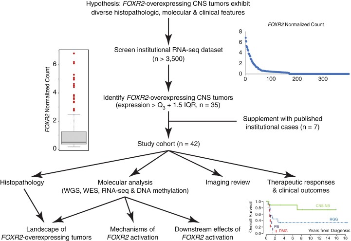
Methods: Histopathologic, molecular, imaging, and clinical data of 42 CNS tumors with FOXR2 overexpression identified through screening institutional datasets and published institutional cases were analyzed.

Results: Among the 42 tumors, 21 (50.0%) were high-grade gliomas (HGGs), and 18 (42.9%) were embryonal tumors. The HGGs included ten H3 K27M-mutant diffuse midline gliomas (DMGs) and eight radiation-associated tumors. The embryonal tumors included 11 CNS NBs and six pineoblastomas (PBs). FOXR2 expression was similar between CNS NB and other tumor types (P = 0.82). HGGs with FOXR2 overexpression, unlike NBs and PBs, displayed diverse concomitant genetic alterations. The most common mechanisms of FOXR2 activation involved structural alterations causing promoter donation and enhancer hijacking from active genes essential for brain development, followed by alternative promoter activation or truncated LINE-1 retrotransposition. The preferential activation mechanism varied by tumor type. All but two aberrant FOXR2 transcripts incorporated non-canonical, non-coding exons. Gene set enrichment analysis demonstrated shared downstream effects of FOXR2 activation at the epigenome and transcriptome levels across tumor types. DMGs and PBs with FOXR2 overexpression were aggressive, with 0% 2-year overall survival, whereas CNS NBs responded well to combined chemotherapy and radiation.

Conclusions: CNS tumors with FOXR2 overexpression manifest significant histological, molecular, imaging, and clinical diversity. While HGGs and PBs with FOXR2 overexpression demonstrated inferior prognosis, CNS NBs showed favorable outcomes. Integrating histologic and molecular diagnostic approaches is imperative for accurate prognostication and optimal therapeutic decision-making.

Keywords: CNS tumors with FOXR2 overexpression; LINE-1 retrotransposition; alternative promoter; patient outcomes; promoter donation and enhancer hijacking.


Conflict of interest statement

The authors declare no competing interests.

Figures

Figure 1.
Figure 1.
Study design. The Institutional RNA-sequencing (RNA-seq) dataset was screened for tumors that showed significantly elevated, more than 1.5 interquartile range (IQR), FOXR2 expression. Tumors that showed no apparent exonic structure of the FOXR2 transcript, likely indicating processing artifacts, were excluded. The 35 tumors identified, supplemented with seven previously published institutional cases, were subjected to comprehensive histologic, molecular, imaging, and clinical data review.
Figure 2.
Figure 2.
Study cohort characteristics and patient outcomes. (A) Oncoplot summarizing the study cohort’s demographic, histologic, and molecular characteristics. Event-free (B) and overall survival (C) of the four major tumor types in the study cohort. (D) Swimmer plot showing the detailed treatment response and patient outcomes of CNS NB in our cohort. CNS NB: CNS neuroblastoma with FOXR2 activation. DMG: histone H3 K27M-mutant diffuse midline glioma. HGG: high-grade glioma. PB: Pineoblastoma.
Figure 3.
Figure 3.
Histologic spectrum of CNS NB-FOXR2. (A–C) The characteristic embryonal-looking areas of CNS NB-FOXR2 showed substantial variation. Areas resembling high-grade glioma may be found (D). Diffusely infiltrating tumor cells may masquerade as a glial neoplasm (E). Some tumors contained low-grade appearing regions (F), but there were nodules of embryonal cells (G). There may be genuine GFAP expression among the tumor cells (H). The typical diffuse Olig2 (I) and synaptophysin (Synap, K) expression could be patchy (J, L) instead. Scale bar: 100 microns
Figure 4.
Figure 4.
Imaging features of CNS tumors with FOXR2 overexpression. Baseline axial diffusion-weighted (A), Apparent Diffusion Coefficient (ADC) map (B), and CT (C) images showing a hypercellular left frontal CNS NB-FOXR2 with coarse calcifications and cortical remodeling, suggestive of a CNS NB-FOXR2 (C, arrow). Axial FLAIR (D) and post-contrast T1 (E) images showing an infiltrative left frontal high-grade glioma (HGG) with enhancement several years after radiation therapy for a CNS NB-FOXR2 at the same location. Axial T2 (F), and post-contrast T1 (G) images showing a heterogeneously enhancing left pontine diffuse midline glioma (DMG). (H) Four months post-RT fluid-attenuated inversion recovery (FLAIR) image showing extensive new subependymal metastases. (I) Axial T2 and post-contrast T1 (J) images showing a circumscribed and heterogeneously enhancing left dorsal thalamic DMG. (K) Baseline diffusion-weighted images and (L) ADC map showing a hypercellular pineal mass, suggestive of a pineoblastoma.
Figure 5.
Figure 5.
Mechanisms of FOXR2 activation. Summary of structural variant (SV) promoter donation and FOXR2 promoter activation events observed per tumor type. SV events occur as gene fusions to exons-6, -2, or 1, while promoter activation events occur at exon-3 or -7. FOXR2 transcripts 1–3 are based on the splicing pattern seen in our cohort of activated tumors, and the exons are annotated as in Tsai et al. The RefSeq FOXR2 transcript has only one exon that starts > 200 bp downstream of the exon 1 splice site in activated transcripts 1–3. The coding region of FOXR2 occurs within exon 1 and is identical for all transcripts. Exon 1 coordinates are for transcripts 1–3, not RefSeq.
Figure 6.
Figure 6.
Gene set enrichment analysis. (A) Results of FOXR2-activated histone H3 K27M-mutant diffuse midline glioma (n = 10) vs non-activated tumors (n = 29). (Left) Results with regulatory target gene sets with a total of 337/3160 gene sets showing upregulation in the activated group and a total of 2 gene sets are significant at FDR < 5%. (Right) Results with hallmark gene set show that a total of 35/50 gene sets are upregulated in the activated group, and no gene sets are significant at FDR < 5%. (B) Results of FOXR2-activated CNS tumors (n = 32) vs non-activated tumors (n = 303). (Left) Results with regulatory target gene sets with a total of 401/3160 gene sets show upregulation in the activated group, and a total of 18 gene sets are significant at FDR < 5%. (Right) Results with hallmark gene set show that a total of 11/50 gene sets are upregulated in the activated group, and two gene sets are significant at FDR < 5%.

References

    1. Gharbaran R. Insights into the molecular roles of FOXR2 in the pathology of primary pediatric brain tumors. Crit Rev Oncol Hematol. 2023;192:104188. - PubMed
    1. Tsai JW, Cejas P, Wang DK, et al. FOXR2 is an epigenetically regulated pan-cancer oncogene that activates ETS transcriptional circuits. Cancer Res. 2022;82(17):2980–3001. - PMC - PubMed
    1. Sturm D, Orr BA, Toprak UH, et al. New brain tumor entities emerge from molecular classification of CNS-PNETs. Cell. 2016;164(5):1060–1072. - PMC - PubMed
    1. Louis DN, Perry A, Wesseling P, et al. The 2021 WHO classification of tumors of the central nervous system: a summary. Neuro Oncol. 2021;23(8):1231–1251. - PMC - PubMed
    1. Korshunov A, Okonechnikov K, Schmitt-Hoffner F, et al. Molecular analysis of pediatric CNS-PNET revealed nosologic heterogeneity and potent diagnostic markers for CNS neuroblastoma with FOXR2-activation. Acta Neuropathol Commun. 2021;9(1):20. - PMC - PubMed

MeSH terms