. 2025 Apr 16;14(4):356-367.
doi: 10.1302/2046-3758.144.BJR-2024-0327.R1.

Carpaine ameliorates synovial inflammation by promoting p65 degradation and inhibiting the NF-κB signalling pathway

Affiliations

Carpaine ameliorates synovial inflammation by promoting p65 degradation and inhibiting the NF-κB signalling pathway

Hongbo Zhang et al. Bone Joint Res. .

Abstract

Aims: Osteoarthritis (OA) is a chronic and debilitating joint disease. Despite its prevalence, especially in ageing and obese populations, effective treatments targeting the molecular mechanisms of OA are limited. This study aimed to investigate the role of carpaine (CP), a major alkaloid from the Carica papaya leaf, in inhibiting articular cartilage destruction and synovitis during OA progression, and to elucidate the underlying molecular mechanisms.

Methods: CP (purity > 98%) was dissolved in dimethyl sulfoxide (DMSO). Various antibodies and reagents were sourced from Sigma-Aldrich, Abcam, and other suppliers. Peritoneal macrophages (pMACs) were cultured in Dulbecco's Modified Eagle Medium (DMEM) and treated with CP to assess its effects on inflammatory cytokine production and nuclear factor-kappa B (NF-κB) signalling. A total of 40 ten-week-old male C57/BL6 mice underwent destabilization of the medial meniscus (DMM) surgery to induce OA. Post-surgery, mice were treated with CP (0.5 or 3 mg/kg) or vehicle via intra-articular injections for up to ten weeks. Cartilage degradation and synovitis were evaluated using Safranin O, Fast Green staining, haematoxylin and eosin (H&E) staining, immunohistochemistry, and quantitative polymerase chain reaction (PCR).

Results: CP treatment significantly reduced cartilage degeneration and maintained hyaline cartilage thickness compared to the vehicle group. Indicators of cartilage degeneration, such as collagen X (Col X) and matrix metallopeptidase 13 (MMP13), were markedly decreased in the CP-treated group. CP-treated mice exhibited significantly lower synovitis scores at both five and ten weeks post-DMM surgery. CP prominently decreased the production of proinflammatory cytokines (interleukin (IL)-1β, IL-6) in M1 polarized macrophages both in vitro and in vivo. CP impeded NF-κB signalling by promoting p65 degradation through the E3 ubiquitin ligase LRSAM1. The defensive effect of CP was reversed by Lrsam1 small interfering RNA (siRNA), confirming the role of LRSAM1 in CP-mediated NF-κB inhibition.

Conclusion: CP acts as a 'physiological brake' on NF-κB activation, thereby mitigating synovial inflammation and cartilage destruction in OA. These findings suggest that targeting synovitis via CP could be a promising therapeutic strategy for OA.


Conflict of interest statement

H. Zhang reports funding from the state-funded postdoctoral researcher program of Southern Medical University (No. GZC20231062 (CN)), related to this study. D. Cai reports funding from the National Natural Science Foundation of China (82172491), related to this study. C. Zeng reports funding from the Natural Science Foundation of Guangdong Province (2022A1515011101 and 2024A1515011231), related to this study.

Figures

Fig. 1
Fig. 1
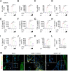
Carpaine (CP) alleviates osteoarthritis (OA) by decreasing articular cartilage degeneration and synovitis inflammation. a) and b) Representative images of Safranin O/Fast Green staining and quantitative analysis of the Osteoarthritis Research Society International (OARSI) scale in OA mice treated with vehicle or 3 mg/kg CP at five or ten weeks post-surgery. Higher magnification images are shown at the bottom of panel A, with dotted lines indicating the tide line. Scale bars: 100 µm. N ≥ 4 per group. c) to h) Representative images of immunohistochemistry (IHC) staining for collagen X (Col X) and ADAM metallopeptidase with thrombospondin type 1 motif 5 (ADAMTS5), immunofluorescence for matrix metallopeptidase 13 (MMP13), and quantitative analysis of positive chondrocytes in the articular cartilage of OA mice treated with vehicle or CP at five or ten weeks post-surgery. Scale bars: 50 µm. N ≥ 4 per group. i) and j) Representative images of haematoxylin and eosin (HE) staining and quantitative analysis of synovium inflammation scores in OA mice treated with vehicle or CP at five or ten weeks post-surgery. Scale bars: 50 µm. N ≥ 4 per group. *p < 0.05, **p < 0.01. CC, calcified cartilage; DMM, destabilization of the medial meniscus; HC, hyaline cartilage.
Fig. 2
Fig. 2
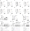
Carpaine (CP) decreases M1 macrophage polarization and inhibits inflammatory cytokines in vitro and in vivo. a) to d) Quantitative polymerase chain reaction (PCR) analysis of Il1b, Il6, Tnfa, and Inos expression in lipopolysaccharide (LPS)-stimulated bone marrow-derived macrophages (BMDMs) treated with or without 1 μM or 4 μM CP. N ≥ 4 per group. e) to g) Enzyme-linked immunosorbent assay (ELISA) analysis of interleukin (IL)-1β, IL-6, and TNF-α levels in the supernatant of LPS-induced BMDMs treated with or without 1 μM or 4 μM CP. N ≥ 4 per group. h) to j) ELISA analysis of IL-1β, IL-6, and TNF-α levels in the serum of destabilization of the medial meniscus (DMM) mice treated with vehicle or CP (0.5 mg/kg or 3 mg/kg) at five weeks post-surgery. N ≥ 4 per group. k) to m) Representative images of immunofluorescence for F4/80 (green) and iNOS (red), and quantitative analysis of positive cells in the synovium of osteoarthritis (OA) mice treated with vehicle or 3 mg/kg CP at five weeks post-surgery. N ≥ 4 per group. Scale bar: 50 µm. **p < 0.01. IL, interleukin; iNOS, inducible nitric oxide synthase; mRNA, messenger RNA; TNF-α, tumour necrosis factor-α.
Fig. 3
Fig. 3
Carpaine (CP) inhibits nuclear factor-kappa B (NF-κB) signalling activation during osteoarthritis (OA). a) and d) Western blot analysis and densitometry of p-p65 and p-IKKα/β in lipopolysaccharide (LPS)-induced bone marrow-derived macrophages (BMDMs) treated with 1 μM or 4 μM CP. N = 3 per group. b) and e) Western blot analysis and densitometry of p-p65 and p-IKKα/β in LPS-induced BMDMs treated with vehicle or 4 μM CP for various durations. N = 3 per group. c) and f) Western blotting and densitometry analysis of p-p65 and p-IKKα/β in the synovial tissue of OA mice treated with or without 0.5 or 3 mg/kg CP. N = 3 per group. **p < 0.01. ns, not significant.
Fig. 4
Fig. 4
Carpaine (CP) suppresses the nuclear factor-kappa B (NF-κB) pathway by promoting ubiquitin-proteasomal degradation of p65. a) and b) NF-κB luciferase activity was measured in 293T cells stably expressing the NF-κB luciferase reporter construct after exposure to interleukin (IL)-1β or tumour necrosis factor-α (TNF-α) with or without 1 μM or 4 μM CP for six hours. Luciferase activity is presented as relative light units (RLUs) normalized to Renilla luciferase activity. N = 3 per group. c) to h) NF-κB luciferase activity was measured in 293T cells stably expressing the NF-κB luciferase reporter construct after overexpression of MyD88, IRAK1, TRAF6, IKKα, IKKβ, or p65 with or without 1 μM or 4 μM CP. N = 3 per group. i) and j) Western blotting and densitometry analysis of Flag-p65 in 293T cells transfected with Flag-p65 and increasing doses of CP for 12 hours. N = 3 per group. k) Quantitative polymerase chain reaction (PCR) analysis of p65 in 293T cells transfected with Flag-p65 and increasing doses of CP for 12 hours. n = 3 per group. l) Western blotting and densitometry analysis of Flag-p65 in 293T cells treated with varying doses of cycloheximide (CHX) with or without 4 μM CP for 12 hours. N = 3 per group. m) and n) Western blotting and densitometry analysis of Flag-p65 in 293T cells treated with dimethyl sulfoxide (DMSO), MG132 (10 μM), chloroquine (CQ) (50 μM), 3-methyladenine (3-MA) (10 mM), or MLN7243 (0.2 μM) for six hours, with or without 4 μM CP. N = 3 per group. *p < 0.05, **p < 0.01. mRNA, messenger RNA; ns, not significant.
Fig. 5
Fig. 5
Carpaine (CP) enhances the interaction between p65 and LRSAM1. a) Western blotting and densitometry analysis of Flag-p65 in 293T cells transfected with Flag-p65 and Lrsam1 siRNA, with or without 4 μM CP for 12 hours. N = 3 per group. b) Western blotting and densitometry analysis of Flag-p65 in 293T cells transfected with Flag-p65 and varying concentrations of Myc-LRSAM1 plasmids for 24 hours. N = 3 per group. c) and d) Immunoprecipitation of p65 from 293T cells pre-treated with MG132 (10 μM) for six hours, followed by stimulation with lipopolysaccharide (LPS) or Myc-LRSAM1 plasmids and CP for 24 hours. Western blot analysis was performed to detect the protein levels of p65 and LRSAM1 in both immunoprecipitated (IP) and whole cell lysate (WCL) samples. **p < 0.01. IB, immunoblotting; ns, non-significant; siRNA, small interfering RNA.
Fig. 6
Fig. 6
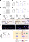
Carpaine (CP) inhibits the nuclear factor-kappa B (NF-κB) pathway by recruiting LRSAM1 to degrade p65 during osteoarthritis (OA). a) and b) Western blotting and densitometry analysis of p-p65, p65, and LRSAM1 in synovial tissue from destabilization of the medial meniscus (DMM) mice treated with or without 0.5 μM or 3 μM CP. N = 3 per group. c) and d) Western blotting and densitometry analysis of p-p65, p65, and LRSAM1 in bone marrow-derived macrophages (BMDMs) induced by lipopolysaccharide (LPS) and treated with or without CP and Lrsam1 siRNA. e) Quantitative polymerase chain reaction (PCR) analysis of Il1b, Il6, Tnfa, and Inos in samples described in c) and d). N = 3 per group. f) to h) Representative images of immunohistochemistry (IHC) staining for collagen X (Col X), immunofluorescence for matrix metallopeptidase 13 (MMP13), and quantitative analysis of positive chondrocytes in the cartilage explants cultured with conditioned medium (CM) from the M1-like macrophage administrated with or without CP and Lrsam1 siRNA. Scale bars: 50 μm. N ≥ 4 per group. i) CP decreases synovial inflammation and inhibits NF-κB signalling by recruiting LRSAM1 to promote ubiquitin-proteasomal degradation of p65. *p < 0.05, **p < 0.01. DAMPs, danger-associated molecular patterns; IB, immunoblotting; mRNA, messenger RNA; ns, non-significant; siRNA, small interfering RNA; TNF-α, tumour necrosis factor-α.

Similar articles

References

    1. Bijlsma JWJ, Berenbaum F, Lafeber F. Osteoarthritis: an update with relevance for clinical practice. Lancet. 2011;377(9783):2115–2126. doi: 10.1016/S0140-6736(11)60243-2. - DOI - PubMed
    1. Glyn-Jones S, Palmer AJR, Agricola R, et al. Osteoarthritis. Lancet. 2015;386(9991):376–387. doi: 10.1016/S0140-6736(14)60802-3. - DOI - PubMed
    1. Moseng T, Vliet Vlieland TPM, Battista S, et al. EULAR recommendations for the non-pharmacological core management of hip and knee osteoarthritis: 2023 update. Ann Rheum Dis. 2024;83(6):730–740. doi: 10.1136/ard-2023-225041. - DOI - PMC - PubMed
    1. Katz JN, Arant KR, Loeser RF. Diagnosis and treatment of hip and knee osteoarthritis: a review. JAMA. 2021;325(6):568–578. doi: 10.1001/jama.2020.22171. - DOI - PMC - PubMed
    1. Hunter DJ, Bierma-Zeinstra S. Osteoarthritis. Lancet. 2019;393(10182):1745–1759. doi: 10.1016/S0140-6736(19)30417-9. - DOI - PubMed