. 2025 Apr 18;11(16):eadu2159.
doi: 10.1126/sciadv.adu2159. Epub 2025 Apr 16.

Earlier onset of chemotherapy-induced neuropathic pain in females by ICAM-1-mediated accumulation of perivascular macrophages

Affiliations

Earlier onset of chemotherapy-induced neuropathic pain in females by ICAM-1-mediated accumulation of perivascular macrophages

Li Chen et al. Sci Adv. .

Abstract

Sex differences in the pathogenesis of a variety of diseases have drawn increasing attention. However, it remains unclear whether such differences exist in chemotherapy-induced neuropathic pain. Here, we conducted a retrospective analysis of clinical case data and found that peripheral sensory disorders occurred earlier in females than in males following bortezomib (BTZ) treatment in patients with multiple myeloma. BTZ treatment led to an early elevation of intercellular adhesion molecule-1, which triggered the infiltration of peripheral monocytes into the perivascular region of the spinal cord in female mice. The CC-chemokine ligand 1 released by infiltrating macrophages directly activated neurons or indirectly activated neurons by enhancing the astrocyte activity, ultimately leading to the earlier onset of BTZ-induced neuropathic pain in females. Together, clarifying the mechanism underlying the earlier onset of BTZ-induced neuropathic pain will contribute to the precise treatment of multiple myeloma in females.


Figures

Fig. 1.
Fig. 1.. Females exhibit early sensory disorders following BTZ treatment in the patients with MM.
(A to C) The data were derived from the case records of the First Affiliated Hospital of Sun Yat-sen University from 1 January 2014 to 30 June 2021. (A) Flow chart of patient selection in this study. (B) The proportion of sensory disorders occurred in the first cycle among the patients with sensory disorders (**P < 0.01, chi-square test). (C) The number of BTZ therapy for the initial occurrence of sensory disorder (**P < 0.01, Mann-Whitney test). (D and E) The data including 23 females and 17 males were derived from the case records of the Third Affiliated Hospital of Sun Yat-sen University from 2010 to 2020. (D) The proportion of sensory disorders occurred in the first cycle among patients with sensory disorders (**P < 0.01, chi-square test). (E) The number of BTZ therapy for the initial occurrence of sensory disorders (**P < 0.01, Mann-Whitney test).
Fig. 2.
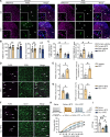
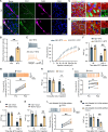
Fig. 2.. BTZ induced an earlier onset of mechanical allodynia and elevated the level of CD45high/CD11b+ cells in the spinal cord of female mice.
(A) The hind paw withdraw (PWT) threshold was examined in female and male mice following BTZ treatment. n = 10. (B) BTZ treatment produced mechanical allodynia in female, but not male, mice on days 3 and 4. n = 10. BL, baseline. (C) Presentative trace of action potential induced by an 80-pA current injection (left). A line chart showed the number of action potentials evoked by inward currents with different genders on day 3 following BTZ treatment (right). n = 5 mice. (D) Top 10 biological pathways (BP) of differential cytokines in BTZ treatment from GO analysis (top: female, bottom: male). (E) Flow cytometry analysis of macrophage population in the L4-L6 spinal cord. Gated cells that were positive in CD11b and higher in CD45 were selected as macrophages. (F) Percentages of CD45highCD11b+ cells in the CD11b+ cell population. n = 4 to 5. Significance: *P < 0.05, #P < 0.05, **P < 0.01, and ##P < 0.01. d, days; veh, vehicle.
Fig. 3.
Fig. 3.. Circulating monocytes contribute to the increased perivascular macrophages in the spinal cord following BTZ treatment in female mice.
(A) Representative images showing TMEM119+ (purple) and F4/80+ (green) cells within the dorsal horn of the spinal cord in mice. Scale bars, 100 μm. (B) Quantification of TMEM119+ microglial including the number of cells (left) and the soma area (right) in the dorsal horn of the spinal cord in mice. n = 5 to 6. (C) The number (left) and fluorescence area (right) of F4/80high/TMEM119 cells in the dorsal horn of the spinal cord in mice. n = 5 to 6. (D) Immunostaining for F4/80 (purple) and CD31 (green) in the spinal cords of female mice on day 3 after BTZ treatment. Scale bar, 50 μm. (E) Quantification showed the number of F4/80high cells (left) and the percentage of F4/80high area within CD31+ area (right) in the dorsal horn. n = 4. (F) Immunostaining for F4/80 (purple) and CD31 (green) in the spinal cords of female mice after tail vein injection of clodronate liposomes (CLLs) before BTZ treatment. Scale bar, 50 μm. (G) Quantification showed the number of F4/80high cells (left) and the percentage of F4/80high area within CD31+ area (right) in the dorsal horn. n = 4. (H) Representative traces (top) and statistical data (bottom) for the action potential firing recorded in spinal cord lamina II neurons of female mice after tail vein injection of CLLs before BTZ treatment. n = 4 mice. (I) Preintravenous injection of CLLs attenuated the mechanical allodynia induced by BTZ. n = 5 to 8. Significance: not significant (n.s.), *P < 0.05, #P < 0.05, and **P < 0.01.
Fig. 4.
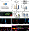
Fig. 4.. ICAM-1 increases in the early stage of BTZ treatment and promotes macrophage pass through the vascular wall in female mice.
(A) Venn diagram showed the overlap of cytokines between the female BTZ group versus the female vehicle group and the female BTZ group versus the male BTZ group. TGFβ1, transforming growth factor–β1; TWEAK, TNF-related weak inducer of apoptosis; Fas, tumor necrosis factor receptor superfamily member 6; TremL1, Trem-like transcript 1 protein. VEGF-B, vascular endothelial growth factor B. (B) The mRNA expression of Icam1 in the dorsal horn of mice. n = 3 to 4. (C) The protein levels of ICAM-1 in the dorsal horn of mice were measured 3 days after BTZ treatment. n = 5. (D) Immunofluorescence staining of ICAM-1 (green) was colocalize with CD31+ signal (the endothelial marker, red). Scale bars, 100 μm (low) or 5 μm (high). (E) Preoral application of A205804 attenuated the mechanical allodynia induced by BTZ in female mice. n = 5 to 8. (F) Preoral application of A205804 did not affect the mechanical allodynia in male mice on day 3 or 4 following BTZ treatment. n = 5 to 8. (G) Immunofluorescence representative image showed the localization of ICAM-1 (turquoise), macrophages (green), and vascular endothelial cells (red). Images are maximum projections of confocal z-stacks. Scale bar, 10 μm. DAPI, 4′,6-diamidino-2-phenylindole. (H) The localization of the macrophage and vascular wall. The image showed F4/80high monocyte (green) passing through the blood vessel wall (red) at 1-μm intervals (Ha). Images are maximum projections of confocal z-stacks (Hb). The magnifications showed the corresponding three-dimensional (3D) reconstructions viewed from different sides (Hc). Scale bars, 20 μm. MIP, macrophage inflammatory protein. Significance: *P < 0.05, #P < 0.05, **P < 0.01, and ##P < 0.01.
Fig. 5.
Fig. 5.. Up-regulation of ICAM-1 in the dorsal horn vascular contributes to neuropathic pain in female mice.
(A and B) Immunostaining for F4/80 (green) and CD31 (red) in the spinal dorsal horn from female mice injected with ICAM-1 siRNA before BTZ treatment. Scale bar, 200 μm (A). The number of F4/80high cells in the dorsal horn of the spinal cord [(B), left]. The percentage of F4/80high area among CD31+ area [(B), right]. n = 5. (C) Representative traces (left) and statistical data (right) for the action potential firing recorded in spinal cord lamina II neurons of female mice after injection of ICAM-1 siRNA before BTZ treatment. n = 3 mice. (D) Intrathecal injection of ICAM-1 siRNA attenuated the mechanical allodynia induced by BTZ on days 3 and 4. n = 5. (E and F) Immunostaining for F4/80 (green) and CD31 (red) in the spinal dorsal horn from female mice intravenously injected with AAV-CAG–ICAM-1 or its control. Scale bar, 200 μm (E). The number of F4/80high cells in the dorsal horn of the spinal cord in female mice [(F), left]. The percentage of F4/80high area among CD31+ area [(F), right]. n = 5. (G) Representative traces (left) and statistical data (right) for the action potential firing recorded in spinal cord lamina II neurons of female naïve mice after intravenous injection of AAV-CAG–ICAM-1. n = 3 mice. (H) Intravenous injection of AAV-CAG–ICAM-1 significantly reduced the withdrawal threshold 21 days after injection in the female naïve mice. n = 6. Significance: *P < 0.05, #P < 0.05, and **P < 0.01.
Fig. 6.
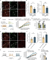
Fig. 6.. ICAM-1 and IL-1α released by endothelial cells synergistically promote macrophage infiltration in female mice following BTZ.
(A) Il1a, Ccl5, and Ccl1 mRNA levels in the dorsal horn of female mice. n = 5 to 8. (B) Immunofluorescence image of IL-1α (green) in vascular endothelial cells (red). Scale bars, 50 μm (low) or 20 μm (high). (C) Double immunofluorescence staining of CCL1 (green) and vascular endothelial cells (red). Scale bars, 50 μm (low) or 20 μm (high). (D and E) Coimmunostaining image of F4/80 (green) and CD31 (red) in the spinal dorsal horn and the statistical graph. Scale bar, 200 μm. n = 5. (F) Intrathecal injection of IL-1α siRNA attenuated the mechanical allodynia induced by BTZ. n = 5. (G) Immunofluorescence image shows IL-1R1 (red) expression in macrophages (green). Scale bar, 10 μm. (H and I) Intrathecal IL-1R1 injection inhibited the up-regulation of F4/80 (green) and CD31 (red) in the near vascular of the spinal dorsal horn. Scale bar, 200 μm (H). The number of F4/80high cells in the dorsal horn of the spinal cord [(I), left]. The percentage of F4/80high area among CD31+ area [(I), right]. n = 4 to 5. (J and K) Immunostaining shows F4/80 (green) and CD31 (red) expression in the spinal dorsal horn. Scale bar, 200 μm (J). The number of F4/80high cells in the dorsal horn of the spinal cord [(K), left]. The percentage of F4/80high area among CD31+ area [(K), right]. n = 5. (L) Intrathecal injection of IL-1α siRNA attenuated the mechanical allodynia induced by BTZ. n = 5. Significance: *P < 0.05, #P < 0.05, **P < 0.01, and ##P < 0.01.
Fig. 7.
Fig. 7.. The infiltrated macrophage-released CCL1 enhances the neuron excitability and contributes to the neuropathic pain in female mice.
(A) Coimmunofluorescence of CCL1 (green) and macrophages (purple) on day 3 following BTZ treatment in female mice. Scale bar, 50 μm. (B) Quantification of CCL1 fluorescence intensity in macrophages. n = 5. (C) Coimmunofluorescence of CCR8 (red) and NeuN (marker of neuron, green). Scale bars, 20 μm. (D) Coimmunofluorescence of CCR8 (red) and glial fibrillary acidic protein (GFAP; a marker of astrocyte, green). Scale bars, 20 μm. (E) Representative traces (left) and statistical data (right) for the action potential firing recorded in spinal cord lamina II neurons. n = 4 mice. (F) Intrathecal injection of CCL1-neutralizing antibody attenuated the mechanical allodynia induced by BTZ. n = 5 to 7. (G) CCL1 increased the action potentials of neurons in female mouse spinal slices. n = 6. (H) Intraspinal CCL1 injection reduced the mechanical withdrawal threshold. n = 5. (I) R243 preincubation blocked the CCL1-induced action potential increase in female mouse spinal slices. n = 6. (J) Intrathecal injection of R243 attenuated the mechanical allodynia induced by BTZ. n = 5. (K) Intrathecal injection of R243 attenuated the mechanical allodynia induced by recombinant CCL1. n = 5. (L) Recombinant CCL1 increased the action potentials in astrocyte CCR8 knockdown mouse spinal slices. n = 5. (M) Intraspinal CCL1 injection reduced the mechanical withdrawal threshold in astrocyte CCR8 knockdown female mice. n = 5. Significance: not significant (n.s.), *P < 0.05 and **P < 0.01.
Fig. 8.
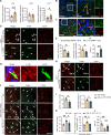
Fig. 8.. CCL1 is involved in the neuronal excitability and mechanical allodynia by activating astrocytes.
(A) Immunofluorescence imaging showed BTZ-activated astrocytes (turquoise) in close proximity to macrophages (green) and vascular endothelial cells (red). Scale bars, 20 μm. (B) Schematic of two-photon imaging configuration. (C) Representative image showed the increased GCaMP6s (green) in spinal cord astrocytes (SR101, red) following CCL1 incubation. Scale bar, 150 μm. (D) Example signal trace (left) and histogram of area under the curve (AUC) (right) of calcium signaling value from the spinal dorsal horn astrocyte in mice treated with CCL1. n = 3. (E to G) R243 incubation prevented CCL1-induced calcium signaling increase in GCaMP6s+ astrocytes. Representative image (E) and signal trace (F) show R243 blocked CCL1-induced calcium up-regulation. Scale bar, 20 μm. AUC quantification (G). n = 3. (H to J) Response of astrocytes to recombinant CCL1 during bath application of TTX (1 μM). Representative image (H) and signal trace (I) showed that TTX did not inhibit the increase in intracellular calcium signaling induced by CCL1. Scale bar, 20 μm. AUC quantification (J). n = 3. (K to M) Application of CNO increased the astrocyte intracellular calcium signaling in the female with injection of AAV2/5-GfaABC1D-CRE and AAV-DIO-hM3Dq-mCherry. Scale bar, 20 μm. (N) The action potential number in female mouse spinal cord slice was increased by CNO application. Representative trace (left) and statistical data (right) were shown. n = 3. (O) Application with CNO decreased the paw withdrawal threshold in female mice with intraspinal injection of AAV2/5-GfaABC1D-CRE and AAV-DIO-hM3Dq-mCherry. n = 5. Significance: not significant (n.s.) and *P < 0.05.
Fig. 9.
Fig. 9.. The mechanism underlying the earlier pain induced by BTZ in females.
(A) Increased ICAM-1 promotes monocyte adhesion to vascular endothelial cells following BTZ treatment. (B) IL-1α/ICAM-1 collaboratively promote monocyte transmigration across the vascular wall. (C) Infiltrated macrophage-released CCL1 enhances neuron excitability directly or indirectly via the activated astrocytes.

References

    1. Beijers A. J., Oerlemans S., Mols F., Eurelings M., Minnema M. C., Vreugdenhil A., van de Poll-Franse L. V., The magnitude of neurotoxicity in patients with multiple myeloma and the impact of dose modifications: Results from the population-based PROFILES registry. Ann. Hematol. 96, 653–663 (2017). - PubMed
    1. Cowan A. J., Green D. J., Kwok M., Lee S., Coffey D. G., Holmberg L. A., Tuazon S., Gopal A. K., Libby E. N., Diagnosis and management of multiple myeloma: A review. JAMA 327, 464–477 (2022). - PubMed
    1. Rajkumar S. V., Multiple myeloma: 2022 update on diagnosis, risk stratification, and management. Am. J. Hematol. 97, 1086–1107 (2022). - PMC - PubMed
    1. Midavaine É., Côté J., Marchand S., Sarret P., Glial and neuroimmune cell choreography in sexually dimorphic pain signaling. Neurosci. Biobehav. Rev. 125, 168–192 (2021). - PubMed
    1. Mogil J. S., Sex differences in pain and pain inhibition: Multiple explanations of a controversial phenomenon. Nat. Rev. Neurosci. 13, 859–866 (2012). - PubMed

LinkOut - more resources