. 2025 Jun 4;15(6):jkaf085.
doi: 10.1093/g3journal/jkaf085.

An RNAi screen of Rab GTPase genes in Caenorhabditis elegans reveals that morphogenesis has a higher demand than stem cell niche maintenance for rab-1 in the somatic cells of the reproductive system

Affiliations

An RNAi screen of Rab GTPase genes in Caenorhabditis elegans reveals that morphogenesis has a higher demand than stem cell niche maintenance for rab-1 in the somatic cells of the reproductive system

Noor Singh et al. G3 (Bethesda). .

Abstract

Membrane trafficking is a crucial function of all cells and is regulated at multiple levels from vesicle formation, packaging, and localization to fusion, exocytosis, and endocytosis. Rab GTPase proteins are core regulators of eukaryotic membrane trafficking, but developmental roles of specific Rab GTPases are less well characterized, potentially because of their essentiality for basic cellular function. Caenorhabditis elegans gonad development entails the coordination of cell growth, proliferation, and migration-processes in which membrane trafficking is known to be required. Here, we take an organ-focused approach to Rab GTPase function in vivo to assess the roles of Rab genes in reproductive system development. We performed a whole-body RNAi screen of the entire Rab family in C. elegans to uncover Rabs essential for gonad development. Notable gonad defects resulted from RNAi knockdown of rab-1, the key regulator of ER-Golgi trafficking. We then examined the effects of tissue-specific RNAi knockdown of rab-1 in somatic reproductive system and germline cells. We interrogated the dual functions of the distal tip cell as both a leader cell of gonad organogenesis and the germline stem cell niche. We find that rab-1 functions cell-autonomously and non-cell-autonomously to regulate both somatic gonad and germline development. Gonad migration, elongation, and gamete differentiation-but surprisingly not germline stem niche function-are highly sensitive to rab-1 RNAi.

Keywords: C. elegans; Animalia; RNAi; Rab GTPase; gamete differentiation; germline; gonad; stem cell.


Conflict of interest statement

Conflicts of interest: The authors declare no conflicts of interest.

Figures

Fig. 1.
Fig. 1.
Whole-body rab-1 knockdown leads to large-scale defects in the gonad. a) Cartoon showing position of 2 gonad arms inside of worm body (above) and representative gonad shapes of control (lower left) and rab-1 RNAi gonads (center and right). Vulva shown with V, and location where proximal gonad fails to form in RNAi samples shown with asterisks. b)–d) Representative images of germ cells (yellow; mex-5p::H2B::mCherry::nos-2 3′UTR) (b′–d′) and somatic gonad DTC and sheath cells (magenta; GFP::INX-9) (b″–d″). Adults that escaped severe embryonic and larval defects after hatching on whole-body rab-1 RNAi (imaged 4 days post-maternal exposure) (c–c″, d–d″) have fewer germ cells and smaller gonads in n = 4/16 worms compared with adults hatched on L4440 empty RNAi vector (b-b″) and display turning and gonad growth defects (c-c″) as well as catastrophic gonad defects (d–d″). e)–g) Representative images of strain expressing markers for the DTC (cyan; lag-2p::mNeonGreen::PLCδPH) (e′–g′) and gonadal sheath cells (magenta; mKate::INX-8) (e″–g″). Adults that escaped severe embryonic and larval defects after hatching on rab-1 RNAi (f–f″, g–g″) have DTC and sheath cells in the correct relative positions, but with aberrant morphology in n = 6/12 worms compared to L4440 treated controls (e-e″). Phenotypes range from growth defects (f–f″) to catastrophic gonad defects (g–g″). Gonads outlined in white dashed lines. h) Representative image of gonad morphology in unmarked N2 animals hatched on rab-1 RNAi (imaged 4 days post-maternal exposure) showing growth arrest and gonad morphology defects. Visible portion of the affected gonad in DIC is outlined in white dashed line. All scale bars = 20 μm. i) Graph showing percentage lethality and percentage body growth defects in larvae exposed to rab-1 RNAi from the L1 stage for 72 h in both the marked strain shown in b)–d) (GFP::1NX-9; mex-5p::H2B::mCherry::nos-2 3′UTR) and the unmarked N2 strain. Error bars represent SE of the sample proportion (see Methods).
Fig. 2.
Fig. 2.
C. elegans with germline-specific rab-1 RNAi knockdown develop gonads with embryo defects. Representative DIC images of MAH23 (rrf-1(pk1417)) worms treated with L4440 control (a) or rab-1 RNAi (b and c) for 72 h starting at the L1 stage. b) A portion of germline-specific rab-1 RNAi-treated gonads have largely normal gonad morphology and gametes, but have disorganized, clumped, or misshapen embryos (classified as mild embryo defect, n = 12/38 gonads). c) Another portion of germline-specific rab-1 RNAi-treated gonads have more severe gamete and embryo defects (severe embryo defect, n = 16/38 gonads). c′ shows the inset indicated by yellow box with abnormal gametes and no embryos. d) Quantification of gonad and vulva defects in germline-specific rab-1 RNAi- and L4440 control-treated animals for 72 h from the L1 stage. n = 3/20 worms had a mild vulval defect (delay and pvl); n = 11/38 gonads had gonad defects (see Methods for scoring criteria). Error bars represent SE of the sample proportion. e). Quantification of embryo and gamete defects in germline-specific rab-1 RNAi- and L4440 control-treated animals for 72 h from the L1 stage. n = 16/38 gonads had severe embryo defects, n = 12/38 had mild embryo defects, and n = 10/38 had normal/fertilized embryos. n = 9/38 gonads had severe gamete defects (see Methods for scoring criteria). Error bars represent SE of the sample proportion. Representative DIC images of MAH23 (rrf-1(pk1417)) worms treated with L4440 control (f) or rab-1 RNAi (g) imaged 4 days post-maternal exposure. g′) shows the inset indicated by yellow box with the absence of gametes and embryos. h) 100% of maternal rab-1 germline-specific RNAi-treated gonads (n = 12/12 gonads) had severe gonad, gamete, and embryo defects. Yellow arrows mark the vulva, yellow arrowheads mark embryos, blue arrowheads mark sperm (spermatheca), and pink arrowheads mark oocytes. All scale bars = 20 μm. All gonads outlined in black dashed lines.
Fig. 3.
Fig. 3.
Somatic gonad-specific RNAi knockdown of rab-1 causes numerous gonad defects. Representative images of rde-1(ne219) mutants rescued with a lag2p::mNG::PLCδPH::F2A::rde-1 transgene restoring RNAi function and driving membrane-localized fluorescence protein mNeonGreen in cells that express the lag-2 promoter on empty L4440 vector control (a) and rab-1 RNAi (b) for 48 h after L1 arrest. Fluorescence merged with a single DIC z-slice (left), fluorescence alone (right, 8 μm maximum intensity projection displayed on a log scale to show DTC and dimmer VPCs, yellow arrows). Scale bars = 20 μm. Gonads outlined in dashed lines; control uterine lumen outlined in solid black line. Unbroken basement membrane in rab-1 RNAi-treated (b) animal bracketed in yellow. c) Quantification of vulval (left) and DTC migration (right) defects in L4440 control and rab-1 RNAi-treated animals following lag-2p+-specific RNAi for 48 h post-L1 exposure. (Left) Tissue-specific rab-1 RNAi-treated worms (n = 17) have delayed vulva formation compared with controls (n = 16). For controls, n = 1/16 had not yet completed vulva formation, but n = 17/17 rab-1 RNAi-treated worms had such a delay. (Right) Tissue-specific rab-1 RNAi-treated worms (n = 50) have defective DTC migration compared with controls (n = 31). One gonad arm scored per worm. Growth of both gonad arms was typically affected, but the deeper DTC under the gut was difficult to score for orientation of turning. All control DTCs completed migration. For rab-1 RNAi, n = 17/50 had no turn, n = 7/50 had just a first turn, n = 9/50 had a misdirected second turn, n = 4/50 had no extension after the second turn, and n = 13/50 completed migration. Error bars show SE of the sample proportion in c). Representative DIC images of uterine-specific (fos-1ap::rde-1) gonads treated with L4440 control (d) and rab-1 RNAi (e) for 72 h post-L1 exposure. Yellow arrows mark vulva, yellow arrowheads mark embryos, blue arrowheads mark sperm (spermatheca), and pink arrowheads mark oocytes. Scale bars = 20 μm. Gonads outlined in black dashed lines. f) Quantification of gonad, vulval, gamete, and embryo defects in L4440 control and rab-1 RNAi-treated animals represented in d) and e). In L4440 controls, n = 1/10 gonads had both severe embryo and gamete defect, n = 3/12 gonads had gonad defects, and n = 1/10 worms had a vulval defect. The variation in the total gonads scored is due to some gonads not having one of the scored features in frame. For rab-1 RNAi-treated animals, n = 17/17 gonads had both severe gamete and embryo defects, n = 18/21 gonads had gonad defects, and n = 4/11 worms had vulval defects. Not every phenotype could be scored in every sample, causing variations in sample size within a treatment group. Error bars show SE of the sample proportion in f).
Fig. 4.
Fig. 4.
Reproductive system defects persist after prolonged rab-1 RNAi knockdown in lag-2 promoter-expressing somatic gonad cells. Representative images of reproductive age (72 h post-L1) animals with tissue-specific RNAi in cells expressing a lag2p::mNG::PLCδPH::F2A::rde-1 transgene restoring RNAi function and driving membrane-localized fluorescence protein mNeonGreen on empty L4440 vector control (a) and rab-1 RNAi (b, c) for 72 h after L1 exposure. Fluorescence merged with a single DIC z-slice (left), fluorescence alone (middle, maximum intensity projection through slices with mNeonGreen signal). a′) and b′) show insets indicated by yellow boxes. Scale bars = 20 μm. Gonads outlined in black or white dashed lines. Dashed yellow lines in a′) and b′) indicate length of DTC as measured for d). Yellow arrows (a–c) indicate vulva or site of expected vulval formation, showing normal vulva (a), protruding vulva (b), and vulvaless (c) phenotypes. Yellow arrowhead in a) indicates embryonic mNG expression. Quantification of rab-1 tissue-specific RNAi-treated worm defects in DTC growth (d), vulva formation (e), and DTC migration (f) compared with L4440 control-treated animals following 72 h of RNAi exposure (from the L1 stage). Both gonad arms in the same worm were scored if both were visible. d) rab-1 RNAi-treated worms (n = 21) have significantly shorter DTCs than controls (n = 18). Welch's t-test t(36.11) = 5.368, P < 0.0001, error bars show SEM. e) While n = 6/6 control samples had normal vulvas, only n = 1/17 rab-1 RNAi samples had normal vulva, n = 5/17 had a missing vulva, n = 10/17 had a protruding vulva, and n = 1/17 ruptured through its protruding vulva on the slide. Error bars show SE of the sample proportion. f) While n = 18/18 control samples had complete DTC migration, only n = 5/25 rab-1 RNAi samples completed migration, n = 10/25 failed to make any turns or elongate, n = 4/25 made the second turn in the wrong direction, and n = 6/25 made both turns and then failed to extend. Error bars show SE of the sample proportion.
Fig. 5.
Fig. 5.
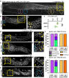
RNAi knockdown of rab-1 in lag-2 promoter-expressing somatic gonad cells causes germline proliferation and differentiation defects. Representative images of DAPI-stained reproductive age adult rde-1(ne219) mutants rescued with a lag2p::mNG::PLCδPH::F2A::rde-1 transgene restoring RNAi function in cells that express the lag-2 promoter on empty L4440 control (a) and the range of defects observed after tissue-specific rab-1 RNAi for 72 h after L1 arrest (b, d, e). b′) shows inset indicated by yellow box (b). Dividing/mitotic germ cells are marked by yellow circle. Gametes, when present, are circled in pink (oocytes) and blue (spermatids). Embryos circled in orange. Dashed green line shows length of progenitor zone as measured for c). c) lag-2p+ rab-1 RNAi-treated worms after 72 h following L1 RNAi exposure (n = 14) have shorter progenitor zones than control (n = 12), Welch's t-test, t(23.52) = 4.106, P = 0.0004; error bars show median with interquartile range. d) Pachytene arrest after 72 h on rab-1 RNAi. d′) Same specimen in d) acquired with 1.6× optical zoom on region boxed in d). Yellow arrowheads indicate exemplar pachytene germ cells. Yellow arrow indicates site of expected vulva formation. e) Spermatogenesis after 72 h on rab-1 RNAi. e′) Same specimen in e) acquired with 1.6× optical zoom of region boxed in e). Arrowheads indicate exemplar pachytene germ cells; spermatids circled in blue. Gonads outlined in dashed white lines. Yellow arrows indicate vulva or position of expected position of vulva formation, whenever in frame. Scale bars = 20 μm. f) Quantification of gametogenesis and embryo formation phenotypes in 72-h L4440 control- and rab-1 RNAi-treated lag-2p+ cell-specific RNAi animals represented in d) and e). f) Gamete (left) and embryo (right) defects observed for tissue-specific rab-1 RNAi (n = 52 gonads) and L4440 control (n = 20 gonads). Error bars show SE of the sample proportion. 100% of L4440 control-treated gonads formed both gametes and embryos. For rab-1 tissue-specific RNAi-treated animals, n = 28/52 gonads lacked gametes (germ cells in pachytene arrest), n = 12/52 gonads had both spermatids and oocytes, and n = 12/52 gonads had produced either spermatids or spermatocytes (developing sperm). n = 0/52 had formed embryos. g), h) Representative images of DAPI-stained lag-2p+ cell-specific rab-1 RNAi-treated worms after 96 h following L1 RNAi exposure. g) Pachytene arrest after 96 h on rab-1 RNAi. g′) shows inset indicated by yellow box in g). Yellow arrowheads indicate exemplar pachytene germ cells. Yellow arrow indicates site of expected vulva formation. h) Spermatogenesis after 96 h on rab-1 RNAi. h′) shows inset indicated by yellow box in h). Yellow arrow indicates site of expected vulva formation. Spermatids circled in blue. Gonads outlined in white dashed lines. i) Quantification of gametogenesis and embryo formation phenotypes in 96-h L4440 control- and rab-1 RNAi-treated lag-2p+ cell-specific RNAi animals represented in g) and h). Gamete (left) and embryo (right) defects observed for tissue-specific rab-1 RNAi (n = 28 gonads) and L4440 control (n = 9 gonads). 100% of L4440 control-treated gonads had formed both gametes and embryos. For rab-1 tissue-specific RNAi-treated animals, n = 15/28 gonads lacked gametes (germ cells in pachytene arrest), n = 4/28 gonads had both spermatids and oocytes, and another n = 9/28 gonads had produced either spermatids or spermatocytes (developing sperm). n = 0/28 had formed embryos. Error bars show SE of the sample proportion.

Update of

Similar articles

References

    1. Agarwal P, Shemesh T, Zaidel-Bar R. 2022. Directed cell invasion and asymmetric adhesion drive tissue elongation and turning in C. elegans gonad morphogenesis. Dev Cell. 57(17):2111–2126.e6. doi:10.1016/J.devcel.2022.08.003. - DOI - PubMed
    1. Arur S. 2017. Signaling-mediated regulation of meiotic prophase I and transition during oogenesis. Results Probl Cell Differ. 59:101–123. doi:10.1007/978-3-319-44820-6_4. - DOI - PMC - PubMed
    1. Ashrafi K, Chang FY, Watts JL, Fraser AG, Kamath RS, Ahringer J, Ruvkun G. 2003. Genome-wide RNAi analysis of Caenorhabditis elegans fat regulatory genes. Nature. 421(6920):268–272. doi:10.1038/nature01279. - DOI - PubMed
    1. Audhya A, Desai A, Oegema K. 2007. A role for Rab5 in structuring the endoplasmic reticulum. J Cell Biol. 178(1):43–56. doi:10.1083/jcb.200701139. - DOI - PMC - PubMed
    1. Blelloch R, Anna-Arriola SS, Gao D, Li Y, Hodgkin J, Kimble J. 1999. The gon-1 gene is required for gonadal morphogenesis in Caenorhabditis elegans. Dev Biol. 216(1):382–393. doi:10.1006/dbio.1999.9491. - DOI - PubMed

MeSH terms

Substances

LinkOut - more resources