. 2025 Apr 15;6(4):102051.
doi: 10.1016/j.xcrm.2025.102051.

Herpes simplex virus 1 encodes a STING antagonist that can be therapeutically targeted

Affiliations

Herpes simplex virus 1 encodes a STING antagonist that can be therapeutically targeted

An Wang et al. Cell Rep Med. .

Abstract

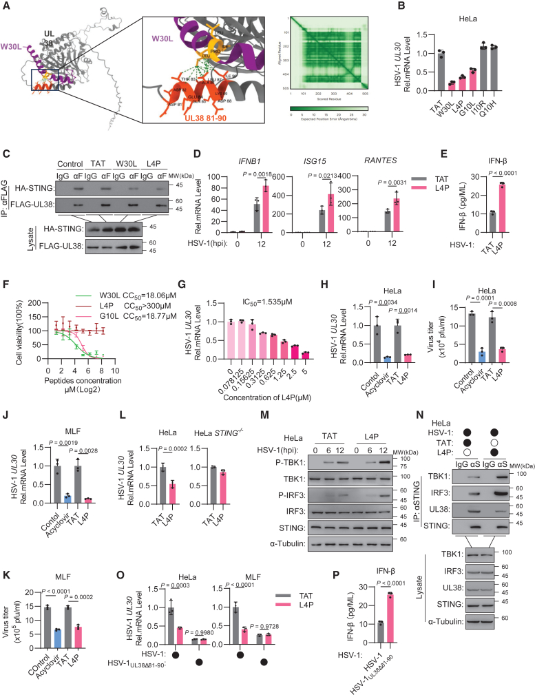
Herpes simplex virus 1 (HSV-1) is a ubiquitous human pathogen that causes serious symptoms and is known for its strong interactions with host immunity. Here, we revealed that the HSV-1-encoded UL38 is a stimulator of interferon genes (STING) antagonist that interacts with STING to abrogate the STING-TANK-binding kinase 1 (TBK1)-interferon regulatory factor 3 (IRF3) interaction, thereby suppressing cyclic GMP-AMP synthase (cGAS)-STING-dependent immune signaling. Losing UL38's STING antagonist activity made HSV-1 incapable of immune evasion and less replicable and pathogenic in vivo. Moreover, on the basis of the UL38-interacting sequence within STING, we rationally designed a series of peptides to target the STING-UL38 interface of UL38 specifically. Among them, a peptide effectively disrupts the STING-UL38 interaction, which unlocks the UL38-suppressed immune response and shows potent therapeutic efficacy against HSV-1 infection in vivo. Therefore, our findings demonstrate that HSV-1 UL38 is a STING antagonist, and targeting the activity of UL38 is a promising strategy for the development of antivirals against this notorious virus.

Keywords: STING; UL38; antiviral innate immunity; antivirals; drug target; herpes simplex virus 1.


Conflict of interest statement

Declaration of interests Wuhan Institute of Virology on behalf of Y.R., X.Z., and A.W. has filed a patent application for the use of antiviral peptide (Chinese patent application 202510237389.X).

Figures

None
Graphical abstract
Figure 1
Figure 1
HSV-1 UL38 inhibits DNA- or virus-triggered innate immune signaling (A) A luciferase reporter assay analyzing ISRE or NF-κB promoter activity was conducted in 293 cells transfected with a plasmid encoding FLAG-cGAS plus FLAG-STING or an empty vector together with increasing amounts of FLAG-UL38 expression plasmid for 24 h. The lower blots show the expression levels of these transfected proteins. (B) HeLa cells were transfected with a FLAG-UL38 plasmid or an empty vector. After 24 h, the cells were left untreated (control) or were transfected with poly(dA:dT) (1 μg/mL). At 6 hr post transfection (h.p.t.), total RNA was extracted and subjected to quantitative reverse-transcription PCR (qRT-PCR) of the indicated antiviral genes. (C) HeLa cells were transfected with an FLAG-UL38 plasmid or an empty vector. After 24 h, the cells were transfected with poly(dA:dT). The cell lysates were prepared and subjected to immunoblotting with the indicated antibodies. (D–F) HeLa cells were transfected with the FLAG-UL38 plasmid or an empty vector, followed by infection with HSV-1. At the indicated h.p.i., the transcription of the indicated antiviral genes was measured via qRT-PCR (D), ELISA was performed to measure the secretion of IFN-β (E), and the cell lysates were prepared and subjected to immunoblotting with the indicated antibodies (F). (G and H) HeLa cells were transfected with the FLAG-UL38 plasmid or empty vector and then infected with HSV-1 (MOI = 0.1). At 12 or 24 h.p.i., the mRNA levels of the HSV-1 UL30 gene were measured via qRT-PCR (G), and the virus titer was detected by the plaque assay (H). All experiments were independently conducted in triplicate and repeated at least twice with reproducible results. The graph shows the means ± SD, n = 3. p values were calculated via one-way or two-way ANOVA. See also Figure S1.
Figure 2
Figure 2
UL38 binds to STING to inhibit cGAS-STING-mediated immune signaling (A) A luciferase reporter assay analyzing ISRE or NF-κB promoter activity was conducted in 293 cells transfected with the ISRE reporter and plasmids encoding FLAG-tagged cGAS plus STING, TBK1, IRF3, or an empty vector for 24 h. The lower blots show the expression levels of these transfected proteins. (B) 293 cells were transfected with plasmids encoding FLAG-tagged cGAS, STING, TBK1, or IRF3 together with HA-UL38. After 24 h, the cells were prepared and subjected to immunoprecipitation (IP) with an anti-HA monoclonal antibody (ɑH) or IgG, followed by immunoblotting with anti-FLAG and anti-HA antibodies. (C) 293 cells were transfected with HA-tagged STING or STINGΔ161–190 together with the FLAG-UL38 expression plasmid. After 24 h, the cells were subjected to IP with an anti-FLAG monoclonal antibody (ɑF) or IgG, followed by immunoblotting with anti-FLAG and anti-HA antibodies. (D) 293 cells were transfected with plasmids containing FLAG-UL38 or the indicated truncations together with HA-STING. After 24 h, the cells were subjected to IP with an anti-FLAG monoclonal antibody or IgG, followed by immunoblotting with anti-FLAG and anti-HA antibodies. (E) HeLa cells were transfected with plasmids encoding FLAG-UL38, FLAG-UL38Δ81–90 or an empty vector. After 24 h, the cells were infected with HSV-1 (MOI = 0.1); at 24 h.p.i., qRT-PCR was performed to measure the transcription of the HSV-1 UL30 gene. (F and G) HeLa STING−/− cells were transfected with the ISRE reporter plasmid together with the indicated plasmids. After 24 h, the cell lysates were subjected to a luciferase assay. (H and I) HeLa cells were infected with HSV-1 (MOI = 1) or HSV-1UL38Δ81–90 (MOI = 1). At 0, 6, and 12 h.p.i., the cell lysates were subjected to immunoblotting with the indicated antibodies (H), and qRT-PCR was performed to measure the transcription of antiviral genes (I). (J–M) HeLa (J and K) or HeLa STING−/− (L and M) cells were infected with HSV-1 or HSV-1UL38Δ81–90 (MOI = 0.1) as indicated. At 24 h.p.i., qRT-PCR was performed to measure the expression of HSV-1 UL30 (J and L), and the virus titer was detected by the plaque assay (K and M). All experiments were independently conducted in triplicate and repeated at least twice with reproducible results. The graph shows the means ± SD, n = 3. p values were calculated via one-way or two-way ANOVA. See also Figure S2.
Figure 3
Figure 3
UL38 abrogates the interactions of STING with TBK1 and IRF3 (A) In vitro competition assay involving biotin-2′,3′-cGAMP, FLAG-STING, and His-UL38. Biotin-2′3'-cGAMP was incubated with affinity-purified FLAG-STING and His-UL38. After incubation, the reaction mixture was subjected to pull-down using Strep-beads (Str), and empty beads were used in controls (Ctrl). The mixture was then subjected to immunoblotting analysis with indicated antibodies. (B) HeLa cells were transfected with FLAG-UL38 for 24 h, followed by stimulating with 2′3′-cGAMP for 30 min. Cells were harvested and analyzed by immunoblotting to detect phosphorylated STING (P-STING). (C–F) 293 cells were transfected with the indicated plasmids. After 24 h, the cell lysates were prepared and subjected to IP with an indicated monoclonal antibody or IgG, followed by immunoblotting with indicated antibodies. (G) HeLa cells were transfected with the FLAG-UL38 plasmid; after 24 h, the cell lysates were subjected to IP with an anti-STING monoclonal antibody (ɑS) or IgG, followed by immunoblotting with the indicated antibodies. (H) HeLa cells were transfected with the HA-UL38 plasmid or an empty vector; at 24 h.p.t., the cells were untreated (control) or transfected with poly(dA:dT) (1 μg/mL). After 6 h, the cell lysates were prepared and subjected to IP with an anti-STING monoclonal antibody or IgG, followed by immunoblotting with the indicated antibodies. (I–K) 293 cells were transfected with the indicated plasmids. After 24 h, the cell lysates were subjected to IP with an anti-HA monoclonal antibody or IgG, followed by immunoblotting with anti-FLAG and anti-HA antibodies. (L) HeLa cells were infected with HSV-1 (MOI = 1) or HSV-1UL38Δ81–90 (MOI = 1). At 24 h.p.i., the cell lysates were subjected to IP with an anti-STING monoclonal antibody or IgG, followed by immunoblotting with the indicated antibodies. All experiments were independently conducted in triplicate and repeated at least twice with reproducible results.
Figure 4
Figure 4
HSV-1-expressed UL38 inhibits immune signaling and promotes viral replication and pathogenesis (A–E) Eight-week-old female C57BL/6J mice (n = 6) were infected with 1 × 107 plaque formation units (PFUs) of HSV-1 or 1 × 107 PFU of HSV-1UL38Δ81–90 by intraperitoneal injection (i.p.). Mouse survival was observed and recorded daily until 14 days post infection (d.p.i.) (A). At 4 d.p.i., total RNA was extracted from the lungs and spleen, and qRT-PCR was performed to measure the transcription of the indicated antiviral genes (B and C) and the HSV-1 UL30 gene (D and E). (F–H) Eight-week-old female C57BL/6J mice (n = 6) were infected with HSV-1 (1 × 104 PFU) or HSV-1UL38Δ81–90 (1 × 104 PFU) via intracerebroventricular injection (i.c.v.). Mouse survival was observed and recorded daily until 14 d.p.i (F). At 0.5 d.p.i., total RNA was extracted from the brain, and the mRNA levels of the antiviral genes (G) and the HSV-1 UL30 gene (H) were measured via qRT-PCR. (I) 8-week-old female C57BL/6J mice were i.c.v. infected with HSV-1 (1 × 104 PFU) or HSV-1UL38Δ81–90 (1 × 104 PFU). At 0.5 d.p.i., the murine brain was subjected to IP with anti-STING antibody or IgG, followed by immunoblots with indicated antibodies. All experiments were independently conducted in triplicate and repeated at least twice with reproducible results. The graph shows the means ± SD. p values were calculated via one-way ANOVA.
Figure 5
Figure 5
A STING-derived designer peptide targets the STING-UL38 interface and shows antiviral efficacy in cells (A) Left: the interaction sites between UL38 (gray) and the peptide W30L (purple) were predicted via AlphaFold3. The binding sites in W30L are shown in yellow, those in UL38 (amino acids 81–90) are shown in orange, and the interaction links are shown in green. Right: predicted aligned error (PAE) per token pair for the prediction on the left with rows and columns labeled by the chain ID and green gradient indicating PAE. (B) HeLa cells were incubated with the indicated peptides (10 μM). After 24 h, the cells were infected with HSV-1 (MOI = 0.1); at 24 h.p.i., qRT-PCR was performed to measure the transcription of HSV-1 UL30. (C) HeLa cells were transfected with FLAG-UL38 and HA-STING plasmids. At 24 h.p.t., the cells were incubated with the indicated peptide (10 μM) for 24 h. IP was performed using an anti-FLAG monoclonal antibody or IgG, followed by immunoblotting with anti-FLAG and anti-HA antibodies. (D and E) HeLa cells were incubated with TAT (10 μM) or L4P (10 μM). After 24 h, the cells were infected with HSV-1 (MOI = 1); at 0, 6, or 12 h.p.i., qRT-PCR was performed to measure the transcription of the indicated genes (D), and ELISA was performed to measure the secretion of IFN-β (E). (F) Increasing concentrations of L4P in DMEM supplemented with 2% FBS were added to HeLa cells for 12 h at 37°C. The viability of L4P cells was determined via a CCK-8 assay, and the absorbance at 450 nm was measured with a microplate reader (Infinite M200PRO). (G) HeLa cells were incubated with increasing concentrations of L4P as indicated for 24 h, after which the cells were infected with HSV-1 (MOI = 0.1). At 24 h.p.i., qRT-PCR was performed to measure the transcription of the HSV-1 UL30 gene. (H–K) HeLa cells (H and I) and MLFs (J and K) were incubated with acyclovir (10 μM) or buffer as a control and L4P (10 μM) or TAT as a control for 24 h. Then, the cells were infected with HSV-1 (MOI = 0.1), and at 24 h.p.i., qRT-PCR was performed to measure the expression of the HSV-1 UL30 gene (H and J), and the virus titer was detected by the plaque assay (I and K). (L) HeLa and HeLa STING−/− cells were incubated with TAT (10 μM) or L4P (10 μM) for 24 h, after which the cells were infected with HSV-1 (MOI = 0.1). At 24 h.p.i., qRT-PCR was performed to measure the expression of the HSV-1 UL30 gene. (M) HeLa cells were incubated with TAT (10 μM) or L4P (10 μM) for 24 h, after which the cells were infected with HSV-1 (MOI = 1). At 0, 6, or 12 h.p.i., the cell lysates were subjected to immunoblotting with the indicated antibodies. (N) HeLa cells were incubated with L4P (10 μM) for 24 h and then infected with HSV-1 (MOI = 1). At 24 h.p.i., the cells were subjected to IP with an anti-STING monoclonal antibody or IgG, followed by immunoblotting with the indicated antibodies. (O and P) HeLa cells and MLFs were incubated with L4P (10 μM) for 24 h and then infected with HSV-1 (MOI = 0.1) or HSV-1UL38Δ81–90 (MOI = 1). At 24 h.p.i., qRT-PCR was performed to measure the expression of the HSV-1 UL30 gene (O), and ELISA was performed to measure the secretion of IFN-β in HeLa cells (P). All experiments were independently conducted in triplicate and repeated at least twice with reproducible results. The graph shows the means ± SD, n = 3. p values were calculated via one-way or two-way ANOVA. See also Figure S3.
Figure 6
Figure 6
Peptide L4P shows potent in vivo antiviral efficacy against lethal HSV-1 infection (A–D) Model of the experiment (left of A and C). Groups (n = 10) of 8-week-old female C57BL/6J mice were treated with L4P or TAT at 10 mg/kg twice a day at 2 h post HSV-1 (1 × 107 PFU) infection via i.p. injection (A and B) or 1 day before HSV-1 infection (1 × 107 PFU) (C and D). Mouse survival (right of A and C) and body weight (B and D) were observed and recorded daily until 14 d.p.i. (E–H) Groups (n = 6) of 8-week-old female C57BL/6J mice were challenged with HSV-1 (1 × 107 PFU) via i.p. injection and treated with L4P or TAT at 10 mg/kg twice a day. At 0, 4, or 7 d.p.i., total RNA was extracted from the lungs (E, G) and spleen (F, H). qRT-PCR was performed to measure the transcription of HSV-1 UL30 (E and F) or the indicated antiviral genes (G and H). All experiments were independently conducted in triplicate and repeated at least twice with reproducible results. The graph shows the means ± SD. p values were calculated via two-way ANOVA. See also Figures S4 and S5.
Figure 7
Figure 7
L4P treatment protects mice from HSV-1 cerebral infection and mitigates its pathogenesis (A–D) Model of the experiment (left of A and C). Groups (n = 10) of 8-week-old female C57BL/6J mice were treated with L4P or TAT at 10 mg/kg twice a day via i.p. injection at 2 h post HSV-1 (1 × 104 PFU) i.c.v. infection (A and B) or 1 day before HSV-1 (1 × 104 PFU) i.c.v. infection (C and D). Mouse survival (right of A and C) and body weight (B and D) were observed and recorded daily until 14 d.p.i. (E–G) Groups (n = 6) of 8-week-old female C57BL/6J mice were i.c.v. infected with HSV-1 (1 × 104 PFU) and treated with L4P or TAT at 10 mg/kg twice a day via i.p. injection. At 0, 0.5, 3, or 5 d.p.i., total RNA was extracted from the brain, and qRT-PCR was performed to measure the transcription of HSV-1 UL30 (E), ELISA was performed to measure the secretion of IFN-β (F) and the indicated antiviral genes (G). (H and I) 8-week-old female C57BL/6J mice were i.c.v. infected with HSV-1 (1 × 104 PFU). At 2 h.p.i., these mice were treated with L4P or TAT at 10 mg/kg twice a day via i.p. injection. At 0 and 5 d.p.i., the murine brain was subjected to immunofluorescence staining, which revealed 2× views of the striatum sections, views of the piriform cortex, and views of the olfactory tubercle (H). The murine striatum brain slices were subjected to H&E staining, which revealed 20× views of the piriform cortex and olfactory tubercle (I). All experiments were independently conducted in triplicate and repeated at least twice with reproducible results. Graph shows mean ± SD. p values were calculated via one-way ANOVA or two-way ANOVA. See also Figures S6 and S7.

References

    1. Yin Y., Favoreel H.W. Herpesviruses and the Type III Interferon System. Virol. Sin. 2021;36:577–587. doi: 10.1007/s12250-020-00330-2. - DOI - PMC - PubMed
    1. Lee S., Ives A.M., Bertke A.S. Herpes Simplex Virus 1 Reactivates from Autonomic Ciliary Ganglia Independently from Sensory Trigeminal Ganglia To Cause Recurrent Ocular Disease. J. Virol. 2015;89:8383–8391. doi: 10.1128/jvi.00468-15. - DOI - PMC - PubMed
    1. Antony F., Kinha D., Nowińska A., Rouse B.T., Suryawanshi A. The immunobiology of corneal HSV-1 infection and herpetic stromal keratitis. Clin. Microbiol. Rev. 2024;37 doi: 10.1128/cmr.00006-24. - DOI - PMC - PubMed
    1. Gupta R., Warren T., Wald A. Genital herpes. Lancet. 2007;370:2127–2137. doi: 10.1016/s0140-6736(07)61908-4. - DOI - PubMed
    1. Yin D., Ling S., Wang D., Dai Y., Jiang H., Zhou X., Paludan S.R., Hong J., Cai Y. Targeting herpes simplex virus with CRISPR-Cas9 cures herpetic stromal keratitis in mice. Nat. Biotechnol. 2021;39:567–577. doi: 10.1038/s41587-020-00781-8. - DOI - PMC - PubMed

MeSH terms

LinkOut - more resources