Review
. 2025 May:106:101356.
doi: 10.1016/j.preteyeres.2025.101356. Epub 2025 Apr 14.

Animal models for the evaluation of retinal stem cell therapies

Affiliations
Review

Animal models for the evaluation of retinal stem cell therapies

Biju B Thomas et al. Prog Retin Eye Res. 2025 May.

Abstract

Retinal degeneration (RD) diseases leading to severe vision loss can affect photoreceptors (PRs) that are responsible for phototransduction, or retinal pigmented epithelium (RPE) providing support for PRs. Human pluripotent stem cell (hPSC)-based therapies are a potential approach for restoration of retinal structure in patients with currently incurable RD diseases. Currently, there are two targeted hPSC therapeutics: PR rescue and PR replacement. PR rescue involves the transplantation of RPE or other neural progenitors into the subretinal space to slow down or prevent further RD. RPE transplantation plays a critical role in preserving photoreceptors by providing trophic support and maintaining retinal integrity, particularly in diseases like age-related macular degeneration (AMD). Advances in RPE transplantation methods, such as polarized monolayer cultures and scaffold-based approaches, have shown promise in enhancing graft survival and integration. However, limitations include inconsistent integration, variable neurotrophic factor secretion, and immune rejection risks in non-autologous transplants. In PR replacement, stem cell-derived photoreceptor-like cells or photoreceptor progenitors (PRP) obtained are transplanted into the eye. While PRPs are commonly obtained from retinal organoids (ROs), alternative sources, such as early differentiation stages or direct differentiation protocols, are also utilized to enhance the efficiency and scalability of PRP generation. Challenges include achieving proper integration, forming outer segments, rosette formation, and avoiding immune rejection or tumorigenicity. Various animal models that simulate human RD diseases are being used for establishing surgical feasibility, graft survival and visual functional recovery but fail to replicate clinical immune challenges. Rodent models lack macula-like structures and have limited reliability in detecting subtle functional changes, while larger animal models pose ethical, logistical, and financial challenges. Immunocompromised models have been developed for minimizing xenograft issues. Visual functional testing for efficacy includes optokinetic testing (OKN), electroretinography (ERG), and electrophysiological recordings from the retina and brain. These tests often fail to capture the complexity of human visual recovery, highlighting the need for advanced models and improved functional testing techniques. This review aims to aggregate current knowledge about approaches to stem cell transplantation, requirements of animal models chosen for validating vision benefits of transplantation studies, advantages of using specific disease models and their limitations. While promising strides have been made, addressing these limitations remains essential for translating stem cell-based therapies into clinical success.

Keywords: Pluripotent stem cells; Retinal degeneration; Retinal disease models; Retinal organoids; Vision testing.


Conflict of interest statement

Declaration of interests BBT, DRN, MR, AKH, and TT have no financial interests to disclose. MJS (co-inventor): patent # 12,247,220: “3D printed micro-millifluidic bioreactors for long-term retinal organoid maintenance”, issued in March 2025.

Figures
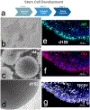
Fig. 1.
Fig. 1.. HESC-derived retinal organoids.
a) Schematic diagram of stages of retinal organoid development; b) image of CRX-GFP hESC colony; c) embryoid bodies on d4; d) retinal organoids on d152; e-g) retinal organoids stained for NRL (green), rhodopsin (red), and recoverin (white).
Fig. 2.
Fig. 2.. Self-organizing culture of human iPSCs to generate the 3D retina and dissected retinal sheet.
Adapted from (Watari et al., 2023), Fig. 1(b-e, f) under a Creative Commons Attribution 4.0 International License (http://creativecommons.org/licenses/by/4.0/). a) Bright-field view of iPSC-S17-derived cell aggregate containing retinal tissue on days 14, 42, 91, and 180 (upper). Scale bar in bright-field view: 100 μm. Immunostaining of iPSC-S17-derived retinal tissue on days 14, 42, 91, and 180 (middle and lower). Crx (green) and Chx10 (red) in middle panels. Pax 6 (green) and Recoverin (red) in lower panels. Blue: nuclear staining with DAPI. Scale bar in immunostaining: 20 μm. b) Bright-field view of iPSC-S17-derived retinal sheet. Scale bar: 100 μm. c) Immunostaining of iPSC-S17-derived retinal sheet on day 87. Crx (green) and Chx10 (red) in left panel. Rx (green) and Recoverin (red) in right panel. Blue: nuclear staining with DAPI. Scale bar: 100 μm.
Fig. 3.
Fig. 3.. Engraftment and photoreceptor maturation of iPSC-retinal sheets after subretinal transplantation in RD-nude rats.
Adapted from (Watari et al., 2023), Fig. 6 (part) under a Creative Commons Attribution 4.0 International License (http://creativecommons.org/licenses/by/4.0/). aj’ Immunostaining of rat eyes transplanted with iPSC-S17-derived retinal sheets. The retinal sheets were transplanted in the subretinal space of RD-nude rats. The rat retinas were fixed at 263 days after transplantation (341 days after initiation of differentiation). a, a’ Immunostaining for human Ku80 (green), Recoverin (red), and PKCalpha (white). Control non-transplanted area (a). Engrafted area (a’). Arrowheads in (a): human Ku80 , Recoverin+ and PKCalpha+ rat bipolar cells. bb” Immunostaining for human Ku80 (green), S-arrestin (red), and Cone-arrestin (white). Boxed area in (b) corresponds to (b’). High magnification in (b’) and higher magnification in (b”). ch’ Immunostaining for photoreceptor markers. NRL (green), Recoverin (red), and RXRG (white) in (c, c’). S-opsin (green) and L/M-opsin (red) in (d). GNAT1 (green) and PRPH2 (red) in (e, e’). GNAT2 (green) and PNA (red) in (f). Human Ku80 (green), Synaptophysin (red), and PKCalpha (white) in (gg”). Arrowheads in (g’, g”): Synaptophysin-positive neurites in no nuclear space. Human Ku80 (green), Calbindin (red), and S-arrestin (white) in (h, h’). Arrowheads in (h’): Calbindin-positive neurites. DAPI staining (blue) in (a, a’, bb”, c, d, e, e’, f, g, g’, h). Scale bars: 100 μm in (a, a’, b), 10 μm in (b’, b”, ch’). INL inner nuclear layer, GCL ganglion cell layer.
Fig. 4.
Fig. 4.. Immunohistochemical Examination of Photoreceptor Precursor Integration and Differentiation.
Adapted from (Aboualizadeh et al., 2020), Fig. 6, under a Creative Commons Attribution 4.0 International License (http://creativecommons.org/licenses/by/4.0/). (A) Some migratory transplanted photoreceptor precursors matured in vivo to express M/L opsin. (B) Neurites from transplanted cells were in contact with host second-order neurons, including G0α+ ON bipolar cell dendrites. (C and D) Some donor photoreceptor precursors extended axons toward host bipolar cells (PKCα+, C) and expressed the presynaptic protein marker (synaptophysin) (D). While expression of synaptophysin in transplanted cells was disorganized in cells away from the host OPL, processes close to the host INL had pronounced expression of synaptophysin in putative axonal terminals.(EH) Some photoreceptor precursors contacted host second-order neurons, including CALB1+ horizontal and bipolar cells (E and F) and expressed synaptophysin (G and H). The white squares in (E) and (G) are shown magnified in (F) and (H), respectively.
Fig. 5.
Fig. 5.. Instruments used for training rodents for vision testing.
A. Light dark box, apparatus. Dark/Light Box for Visual Activity Testing: The apparatus capitalizes on rodents’ natural aversion to brightly lit spaces and their exploratory behavior when faced with mild stressors, such as a novel environment and exposure to light. It consists of a small dark ‘safe’ compartment (one-third) and a larger illuminated ‘aversive’ compartment (two-thirds), enabling the assessment of visual function and anxiety-related responses in rodents."-B. Visual discrimination box, schematic drawing. Rats are introduced through a bend tube (1) that leads to the introduction chamber (2) from where the rats can move into one of the two escape alleys (3) by passing the transparent swing doors (dotted lines). At the far end of the escape alleys, a second set of swing doors are present which open into the home cage. The rats are trained to find the unlocked exit door based on the ‘positive’ visual stimulus displayed on one of the computer monitors (4), whereas a ‘negative’ visual cue is displayed on the other side B. Vision discrimination apparatus The open-field test box consisted of a dark compartment (one third of the floor area) and a larger illuminated compartment (two thirds). A small opening located at floor level in the center of the dividing wall allowed mice to freely move between the lit and dark chambers. C. Morris Water Maze. This set up-assesses visual function and spatial learning in rodents by requiring them to locate a hidden platform using visual cues around the pool. Impairments in vision are indicated by increased time or difficulty in finding the platform.
Fig. 6.
Fig. 6.. OKN Testing Instruments for Vision Assessments in Rodents.
A. Traditional set-up utilizing a rotating drum. The image depicts a top-down view of the drum, including the video camera used to record head movements, the stationary black wall surrounding the drum, and the rat holder positioned at the center. (Reproduced from (Thomas et al., 2004b), Thomas et al. Optokinetic test to evaluate visual acuity of each eye independently, Journal of Neuroscience Methods 138 (1–2):7–13)). B. Schematic representation of the optomotor testing apparatus. (A) Side view. A mouse is placed on a platform positioned in the middle of an arena created by a quad-square of computer monitors. Sine wave gratings drawn on the screens are extended vertically with floor and ceiling mirrors. A video camera is used to monitor the animal’s behavior from above. (B) Top view. The mouse is surrounded by 360° of gratings and is allowed to move freely on the platform. (Reproduced from (Prusky et al., 2004): Prusky et al. Rapid quantification of adult and developing mouse spatial vision using a virtual optomotor system, Invest Ophthalmol Vis Sci 45 (12):4611–6).

References

    1. Aboualizadeh E, Phillips MJ, McGregor JE, DiLoreto DA Jr., Strazzeri JM, Dhakal KR, Bateman B, Jager LD, Nilles KL, Stuedemann SA, Ludwig AL, Hunter JJ, Merigan WH, Gamm DM, Williams DR, 2020. Imaging transplanted photoreceptors in living nonhuman primates with single-cell resolution. Stem Cell Rep. 15, 482–497. 10.1016/j.stemcr.2020.06.019. - DOI - PMC - PubMed
    1. Acland GM, Aguirre GD, Ray J, Zhang Q, Aleman TS, Cideciyan AV, Pearce-Kelling SE, Anand V, Zeng Y, Maguire AM, Jacobson SG, Hauswirth WW, Bennett J, 2001. Gene therapy restores vision in a canine model of childhood blindness. Nat. Genet 28, 92–95. 10.1038/ng0501-92. - DOI - PubMed
    1. Ahmed F, Rajendran Nair DS, Thomas BB, 2022. A new optokinetic testing method to measure rat vision. J. Vis. Exp 185. 10.3791/63357, 10.3791/63357. - DOI - DOI - PMC - PubMed
    1. Ai X, Yu P, Luo L, Sun J, Tao H, Wang X, Meng X, 2022. Berberis dictyophylla F. inhibits angiogenesis and apoptosis of diabetic retinopathy via suppressing HIF-1alpha/VEGF/DLL-4/Notch-1 pathway. J. Ethnopharmacol 296, 115453. 10.1016/j.jep.2022.115453. - DOI - PubMed
    1. Ail D, Nava D, Hwang IP, Brazhnikova E, Nouvel-Jaillard C, Dentel A, Joffrois C, Rousseau L, Degardin J, Bertin S, Sahel JA, Goureau O, Picaud S, Dalkara D, 2023. Inducible nonhuman primate models of retinal degeneration for testing end-stage therapies. Sci. Adv 9, eadg8163. 10.1126/sciadv.adg8163. - DOI - PMC - PubMed

MeSH terms