Review
. 2025 Jul;31(3):771-795.
doi: 10.3350/cmh.2025.0048. Epub 2025 Apr 16.

Role of amino acids in the regulation of hepatic gluconeogenesis and lipogenesis in metabolic dysfunctionassociated steatotic liver disease

Affiliations
Review

Role of amino acids in the regulation of hepatic gluconeogenesis and lipogenesis in metabolic dysfunctionassociated steatotic liver disease

Eiji Kakazu et al. Clin Mol Hepatol. 2025 Jul.

Abstract

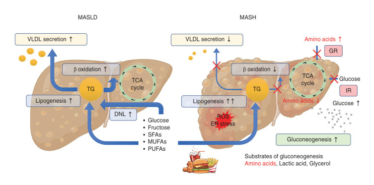
Metabolic dysfunction-associated steatotic liver disease (MASLD) and its relatively advanced form, metabolic dysfunction-associated steatohepatitis (MASH), are becoming increasingly prevalent worldwide, making their prevention and management an urgent global health priority. Central to their development are key metabolic defects, including abnormal concentrations of monosaccharides, fatty acids, and amino acids, but the complex relationships between these substances within the hepatic microenvironment remain only partially understood. Dysregulated glucose metabolism and selective insulin resistance (IR) promote hepatic gluconeogenesis, glycolysis, and de novo lipogenesis; and excessive concentrations of free fatty acids from the diet and adipose tissue drive steatosis. Emerging evidence also implies that amino acid metabolism affects mitochondrial function and redox balance. Dysfunctional mitochondrial oxidative phosphorylation and the associated increase in reactive oxygen species production further exacerbate the cellular stress, inflammation, and fibrosis. However, compared with monosaccharide and fatty acid metabolism, the role of amino acid metabolism in MASLD/MASH remains less well understood. A better understanding of the role of such metabolic dysfunction in liver pathobiology should aid the identification of more useful biomarkers and precision therapies for MASLD/MASH.

Keywords: Amino acid; Fatty acid; Gluconeogenesis; Glucose; Lipogenesis.


Conflict of interest statement

Conflicts of Interest

The authors have no conflicts to disclose.

Figures

Figure 1.
Figure 1.
Overview of the metabolic changes in the liver during the progression of MASLD to MASH. The excessive influx of nutrients from the portal circulation affects liver metabolism. In early MASLD, the liver responds to this nutrient overload by upregulating lipogenesis, fatty acid oxidation, and VLDL secretion. However, if this condition persists long term, lipotoxicity leads to ER stress, oxidative stress, and mitochondrial dysfunction. The reduction in hepatocyte numbers caused by apoptosis increases the lipotoxic burden on the surviving hepatocytes, leading to the formation of a vicious cycle. Selective IR develops where lipogenesis and gluconeogenesis are upregulated, while VLDL secretion and fatty acid oxidation are suppressed. Glucogenic amino acids, lactate, and glycerol are used as substrates for gluconeogenesis. As the disease progresses, GR develops, together with insulin resistance, leading to a suppression of amino acid uptake in the liver, resulting in lower hepatic amino acid concentrations and higher circulating concentrations. DNL, de novo lipogenesis; ER, endoplasmic reticulum; GR, glucagon resistance; IR, insulin resistance; MASH, metabolic dysfunction-associated steatohepatitis; MASLD, metabolic dysfunction-associated steatotic liver disease; MUFAs, monounsaturated fatty acids; PUFAs, polyunsaturated fatty acids; ROS, reactive oxygen species; SFAs, saturated fatty acids; TCA, tricarboxylic acid; TG, triglyceride; VLDL, very low-density lipoprotein.
Figure 2.
Figure 2.
Overview of the roles of gluconeogenesis and DNL in MASLD/MASH. In MASLD, and particularly in MASH, selective IR develops, and opposing metabolic processes, such as gluconeogenesis and lipogenesis, occur simultaneously. Gluconeogenesis is activated in the periportal region (zone 1), whereas glycolysis and DNL are activated in the pericentral region (zone 3) near the central vein. Proteins such as AMPK and FoxO1 modulate these metabolic pathways. AMPK suppresses G6PC and PEPCK activity, whereas FoxO1 increases this and reduces the activities of HK and PKM in the fasting state. These effects are impaired in MASLD/MASH. Fructose is converted to fructose-1-phosphate (F1P) by fructokinase, then metabolized to dihydroxyacetone phosphate (DHAP) and glyceraldehyde, which enter the glycolysis pathway. In general, DNL starts with acetyl-CoA, which is produced from glucose under aerobic conditions. However, in MASLD/MASH, lipogenesis enzymes, such as ACC, FASN, ELOVL, and SCD1, are activated by transcription factors, such as SREBP-1, even in the hypoxic environment of zone 3, leading to an increase in lipogenesis. Free fatty acids, MUFAs, and SFAs activate SREBP-1, but PUFAs reduce its activity. In the final stage of lipogenesis, triglycerides can be stored as lipid droplets or secreted as VLDL into the extracellular space via DGATs. The early stage of VLDL formation occurs in the ER. ApoB is synthesized in the rough ER, where MTTP facilitates the transfer of diacylglycerol and other lipids to ApoB. The immature VLDL formed in the ER is transferred to the Golgi apparatus, where it matures, before being secreted into the extracellular space. Genetic polymorphisms associated with MASLD/MASH, such as TM6SF2, influence the VLDL maturation process. The TM6SF2 risk allele is associated with less production of the larger VLDL1, leading to greater secretion of the smaller VLDL2 into the extracellular space. 1,3-BPG, 1,3-bisphosphoglycerate; 3PG, 3-phosphoglycerate; ACC, acetyl-CoA carboxylase; AMPK, AMP-activated protein kinase; ATGL, adipose triglyceride lipase; CV, central vein; DGAT, diacylglycerol acyltransferase; DNL, de novo lipogenesis; ELOVL, elongation of very long-chain fatty acids; ER, endoplasmic reticulum; F-1,6-BP, fructose 1,6-bisphosphate; F-6-P, fructose 6-phosphate; FASN, fatty acid synthase; FoxO1, forkhead box O1; G-6-P, glucose 6-phosphate; G6PC, glucose-6-phosphatase; G-A-P, glyceraldehyde 3-phosphate; HK, hexokinase; IR, insulin resistance; IRS, insulin receptor substrate; LD, lipid droplet; MASH, metabolic dysfunction-associated steatohepatitis; MASLD, metabolic dysfunction-associated steatotic liver disease; MTTP, microsomal triglyceride transfer protein; MUFA, monounsaturated fatty acid; OAA, oxaloacetate; PEP, phosphoenolpyruvate; PEPCK, phosphoenolpyruvate carboxykinase; PKM, pyruvate kinase muscle isozyme; PNPLA3, patatine-like phospholipase domain containing 3; PUFA, polyunsaturated fatty acid; PV, portal vein; SCD1, stearoyl-CoA desaturase 1; SFA, saturated fatty acid; SREBP-1, sterol regulatory element-binding protein 1; Star, target molecules under clinical investigation; TM6SF2, transmembrane 6 superfamily member 2; VLDL, very low-density lipoprotein.
Figure 3.
Figure 3.
Differences in metabolism in the various liver lobule zones and the distribution of insulin receptor substrates in MASLD. In the liver lobule, zone 1 is characterized by higher oxygen levels, gluconeogenesis and glycogen storage, fatty acid oxidation, and glutamine utilization and breakdown by glutaminase 2 (GLS2). In contrast, zone 3 is characterized by lower oxygen levels, and greater glycolysis, DNL, and glutamine synthesis. The activity of mTOR, an amino acid sensor, is also high, and this is involved in lipogenesis. These differences explain why zone 3 is the primary site of pathology in MASH. IRS1 is predominantly expressed in zone 3, whereas IRS2 is predominantly expressed in zones 1 and 3. In diabetes and obesity, IRS2 expression is low in both zones 1 and 3. Conversely, the phosphorylation of IRS1 remains relatively intact, and this leads to greater gluconeogenesis in zone 1 and lipogenesis in zone 3, which contributes to hepatic steatosis, causing selective IR. CV, central vein; DNL, de novo lipogenesis; FFA, fatty acid; IRS, insulin receptor substrate; MASH, metabolic dysfunction-associated steatohepatitis; MASLD, metabolic dysfunction-associated steatotic liver disease; mTOR, mechanistic target of rapamycin; PV, portal vein.
Figure 4.
Figure 4.
Role of the TCA cycle and entry points of FAAs. Twenty FAAs are used as substrates for protein synthesis or energy metabolism. FAAs are metabolized and the products enter the TCA cycle in mitochondria, and this occurs alongside glucose metabolism under aerobic conditions. The primary role of the TCA cycle is to generate NADH and FADH2 from NAD+ and FAD, which drive ATP synthesis through oxidative phosphorylation. In MASH, gluconeogenesis is often activated in zone 1, owing to selective IR in the liver (Fig. 2). Glucogenic amino acids, except for Leu and Lys, are used as substrates for glucose production. Ketone bodies are primarily produced from fatty acids via β-oxidation. However, ketogenic amino acids, such as Leu and Lys, also contribute to ketogenesis through their metabolism to acetyl-CoA and acetoacetyl-CoA, which are key intermediates in ketone body synthesis. This pathway becomes particularly important during prolonged fasting or in the presence of an energy deficit. In contrast, glycolysis and DNL are activated in zone 3, owing to mitochondrial dysfunction, and one-carbon metabolism is activated, alongside glycolysis. Glutamine that is produced by cytosolic glutamine synthetase (GS) is converted back to glutamate in the mitochondria by glutaminase (GLS2) and enters the lipogenesis pathway via α-ketoglutarate (αKG). Ala, alanine; Arg, arginine; Asn, asparagine; Asp, aspartate; ChREBP, carbohydrate-responsive element-binding protein; Cys, cysteine; DNL, de novo lipogenesis; FAAs, free amino acids; FoxO1, forkhead box O1; GDH, glutamate dehydrogenase; Gln, glutamine; GLS, glutaminase; Glu, glutamate; Gly, glycine; GS, glutamine synthetase; His, histidine; Ile, isoleucine; IR, insulin resistance; Leu, leucine; Lys, lysine; MASH, metabolic dysfunction-associated steatohepatitis; Met, methionine; PEP, phosphoenolpyruvate; Phe, phenylalanine; PPAR, peroxisome proliferator-activated receptor; Pro, proline; Ser, serine; SIRT1, sirtuin 1; SREBP-1, sterol regulatory element-binding protein 1; TCA, tricarboxylic acid; Thr, threonine; Trp, tryptophan; Tyr, tyrosine; Val, valine.
Figure 5.
Figure 5.
The role of one-carbon metabolism and related amino acids. One-carbon metabolism plays a pivotal role in the liver, supporting essential processes, such as redox control, ATP synthesis, VLDL synthesis, and epigenetics, with Gly, Ser, and Thr serving as key contributors. These amino acids provide one-carbon units that fuel the folate and methionine cycles, which are central to hepatocyte metabolism. In redox control, one-carbon metabolism generates NADPH and glutathione, a crucial reducing equivalent that defends against oxidative stress and maintains cellular redox balance. Glutathione synthesis is initiated by GCLC, which is regulated by Nrf2, and uses Gly, Cys, and Gly as substrates. For ATP synthesis, intermediates in these cycles contribute to purine and thymidylate biosynthesis, thereby supporting energy production through nucleotide metabolism. The methionine cycle, a key component of one-carbon metabolism, works in coordination with the folate cycle to facilitate VLDL synthesis. This interaction provides methyl groups, particularly from SAMe, for triglyceride methylation, which is critical for the assembly and secretion of VLDL, ensuring efficient lipid transport from the liver. Furthermore, the methyl groups generated are indispensable for DNA and histone methylation, driving epigenetic modifications that regulate gene expression and hepatic adaptation to metabolic demands. Ala, alanine; Cys, cysteine; GCLC, glutamate–cysteine ligase catalytic subunit; Glu, glutamate; Gly, glycine; Met, methionine; SAMe, S-adenosylmethionine; Ser, serine; Thr, threonine; VLDL, very low-density lipoprotein.
Figure 6.
Figure 6.
Abnormalities in branched-chain amino acid concentrations in metabolic dysfunction-associated steatotic liver disease (MASLD) and the effects of the host and gut microbiota. Although branched-chain amino acids (BCAAs) cannot be synthesized in the human body, Prevotella copri and Bacteroides vulgatus have been shown to synthesize BCAAs in the gut, contributing to insulin resistance. Bacteroides thetaiotaomicron is associated with high circulating concentrations of glutamate, BCAAs, and aromatic amino acids (AAAs). Clostridium sporogenes, Bacteroides ovatus, and Clostridium senegalense are associated with high circulating concentrations of BCAAs, tryptophan, arginine, and histidine in the host. Furthermore, a knockout of the BCAA transaminase gene (BCAT) in Clostridium sporogenes increases the serum BCAA concentrations and improves glucose tolerance. BCAAs bypass first-pass liver metabolism because of the low BCAT2 activity of hepatocytes. After being transaminated in extrahepatic tissues, branched-chain keto acids (BCKAs) are recirculated back to the liver for oxidation, where BCKA dehydrogenase activity is higher. As a result, in MASLD/metabolic dysfunction-associated steatohepatitis (MASH), the circulating concentrations of BCAAs are often high. The radar chart of 20 plasma FAAs in adults (right, lower panel) is reproduced from Mino et al. (Amino Acids 2024;57:3) [73], and shows fold differences from healthy adults. The light green, yellow, red, and blue lines represent healthy adults, individuals with cardiometabolic abnormalities but no steatotic liver disease (CC+SLD−), those with MASLD, and those with metabolic dysfunction-associated alcohol-related liver disease (MetALD), respectively. Ala, alanine; Arg, arginine; Asn, asparagine; Cys, cysteine; Gln, glutamine; Glu, glutamate; Gly, glycine; His, histidine; Ile, isoleucine; Leu, leucine; Lys, lysine; Met, methionine; Orn, ornithine; Phe, phenylalanine; Pro, proline; Ser, serine; Thr, threonine; Trp, tryptophan; Tyr, tyrosine; Val, valine.
Figure 7.
Figure 7.
Mechanisms of amino acid sensing and the regulation of energy metabolism. mTOR and GCN2 are critical amino acid sensors that regulate energy metabolism in the liver. mTORC1, which is activated by amino acids such as leucine via sestrin 2, arginine via CASTOR, and methionine via SAMTOR, promotes anabolic processes such as fatty acid and triglyceride synthesis through transcription factors such as SREBP-1, linking nutrient abundance to energy storage. GCN2 responds to amino acid starvation, activating ATF4. Leucine deficiency activates ATF4 to enhance FGF21 production. ATF4 is a transcription factor that plays a role in the ER stress response and cooperates with Nrf2, an oxidative stress sensor, to protect hepatocytes against lipotoxicity. AMPK and SIRT1 are key interacting regulators of cellular energy status. AMPK and SIRT1 activity is regulated by the AMP/ATP and NAD+/NADH ratios, respectively. AMPK and mTOR are mutually inhibitory. SIRT1 regulates lipid homeostasis by increasing Foxo1 activity. In MASLD/MASH, low PPARα activity leads to less fatty acid oxidation and greater lipid accumulation in the liver. Star, target molecules under clinical investigation. AMPK, AMP-activated protein kinase; Arg, arginine; ATF4, activating transcription factor; CASTOR1, cellular amino acid sensor for mTORC1; FGF21, fibroblast growth factor 21; FoxO1, forkhead box O1; GATOR, GTPase-activating protein toward Rags complex; GCN2, general control nonderepressible 2; Hcy, homocysteine; Leu, leucine; MASH, metabolic dysfunction-associated steatohepatitis; MASLD, metabolic dysfunction-associated steatotic liver disease; Met, methionine; mTORC1, mechanistic target of rapamycin complex 1; Nrf2, nuclear factor erythroid 2-related factor 2; PPAR, peroxisome proliferator-activated receptor; SAH, S-adenosylhomocysteine; SAMe, S-adenosylmethionine; SAMTOR, S-adenosylmethionine sensor upstream of mTORC1; SIRT1, sirtuin 1; SREBP-1, sterol regulatory element-binding protein 1.

Similar articles

Cited by

References

    1. Zhao Q, Deng Y. Comparison of mortality outcomes in individuals with MASLD and/or MAFLD. J Hepatol. 2024;80:e62–e64. - PubMed
    1. Kwak M, Kim HS, Jiang ZG, Yeo YH, Trivedi HD, Noureddin M, et al. MASLD/MetALD and mortality in individuals with any cardio-metabolic risk factor: A population-based study with 26.7 years of follow-up. Hepatology. 2025;81:228–237. - PMC - PubMed
    1. Mantovani A, Csermely A, Petracca G, Beatrice G, Corey KE, Simon TG, et al. Non-alcoholic fatty liver disease and risk of fatal and non-fatal cardiovascular events: an updated systematic review and meta-analysis. Lancet Gastroenterol Hepatol. 2021;6:903–913. - PubMed
    1. Rinella ME, Lazarus JV, Ratziu V, Francque SM, Sanyal AJ, Kanwal F, et al. A multisociety Delphi consensus statement on new fatty liver disease nomenclature. J Hepatol. 2023;79:1542–1556. - PubMed
    1. Hagström H, Vessby J, Ekstedt M, Shang Y. 99% of patients with NAFLD meet MASLD criteria and natural history is therefore identical. J Hepatol. 2024;80:e76–e77. - PubMed

LinkOut - more resources