. 2025 Jul;24(4):778-786.
doi: 10.1016/j.jcf.2025.04.003. Epub 2025 Apr 16.

Cystic Fibrosis-related neurodegenerative disease associated with tauopathy and cognitive decline in aged CF mice

Affiliations

Cystic Fibrosis-related neurodegenerative disease associated with tauopathy and cognitive decline in aged CF mice

Danica F Patton-Parfyonov et al. J Cyst Fibros. 2025 Jul.

Abstract

Background: Highly effective modulator therapies (HEMT) are increasing the lifespan for many people with cystic fibrosis (pwCF), making it necessary to identify and understand CF specific age-related consequences. In this study, we examine the impact of aging on cognitive function and age-related brain pathology in a CF mouse model focusing on phospho-Tau (pTau) pathology.

Methods: Cognitive function was measured by novel object recognition and spontaneous alternation behavior tests. Hippocampal neuronal function was assessed by measuring long-term potentiation (LTP) electrophysiology, the synaptic correlate of learning and memory. Tau pathology was assessed by immunohistochemical analyses and western blot assessment of pTau levels in CF mouse brain, as well as human nasal epithelial cells isolated from pwCF.

Results: Cognitive function declined progressively with age in Cftr (G542X/G542X) (G542X) mice, a model of CF, compared to wild-type (WT) littermate controls. LTP was also deficient in older G542X mice. Increased pTau was observed by staining and western blot analysis in the hippocampus of aged CF mice. Secondary impacts of tauopathy, including increased microglial uptake of cholesterol and reduced neuronal density were also observed. Lastly, human nasal epithelial cells from pwCF were found to display elevated pTau levels compared to non-CF controls.

Conclusions: Aging CF mice develop tauopathy, cognitive decline, LTP impairment, microglial activation, and neurodegeneration that is not experienced by age-matched WT littermates, a condition herein termed cystic fibrosis-related neurodegeneration (CFND). These findings suggest that pwCF may be at risk for tauopathy-related neurodegeneration and cognitive impairment with aging.

Keywords: Cognition; Cystic fibrosis; Dementia; Tauopathy.


Conflict of interest statement

Declaration of competing interest No authors have any conflicting interests with this work.

Figures

Fig. 1.
Fig. 1.
Spontaneous Alternation and Novel Object Recognition in CGX vs WT mice at 3, 7 and 15 months of age. Different cohorts of mice were run at 3 (n = 10 per genotype), 7 (n = 5 per genotype) and 15 (n = 18 per genotype). SA: mice of both genotypes explored the maze equally at all ages. No difference in total arm entries was observed across genotypes (Supplemental data) (Suppl Fig 1. A–B) At 3 months no difference on SA was observed between genotypes t(18)=1.229, p = 0.1174, 95 % (−5.684–21.72). Similarly no difference on NOR was observed between genotypes t(8)=0.23, p = 0.4119, 95 % (−0.3165 to 0.2591). (Fig 1. C–D) At 7 months, G542X mice were significantly impaired on SA with significantly fewer alternating arm entries observed in the G542X mice t(8)=3.056, p = 0.0078, 95 % (−35.13 to −4.912). However performance on NOR was preserved with no differences between genotypes t(8)=0.8286, p = 0.2144, 95 % (−0.8596 to 0.3987). (Fig 1. E–F) At 15 months, G542X were significantly impaired on SA t(33)=2.871, p = 0.0071, 95 % (−23.90 to −4.06) and NOR t(33)=3.813, p = 0.0006, 95 % (−0.5575 to −0.1698) compared to age matched WT controls. G542X engaged in significantly fewer alternating arm entries and exhibited a significantly reduced discrimination index compared to WT controls. Significance determined by student t-test: p > 0.05 NS, *p < 0.05, **p < 0.01, ***p < 0.0001.
Fig. 2.
Fig. 2.
Total and phosphorylated tau (AT8) in RIPA soluble and insoluble hippocampal fractions. (A) No significant changes were found in the RIPA soluble fraction for total Tau (A) t(6)=0.07294, p = 0.9442, 95 %(−0.2080 to 0.2208) or P-tau (B) t(6)=0.7845, p = 0.4626, 95 %(−0.5228 to 0.2689). In the RIPA insoluble fraction, total (C) t(6)=3.349, p = 0.0154, 95 % (0.1320 t0 0.8480) and p- tau (D) t(6)=3.851, p + 0.0085, 95 % (0.1483 to 0.6653) were significantly higher in G542X compared to WT. Significance determined by paired students t-test *p < 0.05, **p < 0.01.
Fig. 3.
Fig. 3.
Phosphorylated tau (AT8) in the dentate gyrus of the hippocampus in WT and G542X mice at 9 and 15 months. Mean fluorescence intensity of phosphorylated tau (AT8) in the anterior (A,C) and posterior (B,D) dentate gyrus of the hippocampus. AT8 intensity was significantly elevated in the anterior DG at 9 months t(11)=2.187, p = 0.0256, 95 %(−0.0655 to 19.99). At 15 months of age there is significantly more phosphorylated tau within the dentate gyrus in the G542X mice in both the anterior (C) t(7)=3.590, p = 0.0089, 95 % (11.46 to 55.71) portions of the hippocampus (D) t(7)=4.312, p = 0.0035, 95 % (4.955 to 16.99). Representative micrographs at 40X of the anterior dentate gyrus showing DAPI stained cell bodies and phosphorylated tau (AT8) in red anterior section from a WT (E) and G542X mouse (F).
Fig. 4.
Fig. 4.
Long-term potentiation in WT and CF (CGX) mouse brain. (A) Diagram of electrophysiological recordings from the schaffer collateral in ex vivo hippocampal sections (https://biorender.com) (B) Quantification of In-Out curves from 20-month-old female mice (two-way ANOVA, not significant, n = 28 slices from 5 WT, n = 32 slices from 5 CGX). (C) Quantification of Paired Pulse Facilitation from 20-month-old female mice. (two-way ANOVA, not significant, n = 28 slices from 5 WT, n = 32 slices from 5 CGX). (D) Quantification of Long-Term Potentiation after theta-burst stimulation from 20-month-old mice. (two-way ANOVA, F(79,4640)=2.515, P < 0.0001; posthoc Tukey main genotype effect: P < 0.0001, 95 % (21.84 to 26.36); posthoc Tukey main genotype effect: ****p < 0.0001; n = 28 slices from 5 WT, n = 32 slices from 5 CGX).
Fig. 5.
Fig. 5.
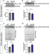
Cellular changes in the brain of G542X mice. Co-staining of the hippocampus for ionized calcium binding adaptor molecule (Iba1) and borondipyrromethene (BODIPY) suggests that macrophages are taking up more cholesterol in aged (15 month) G542X mice compared to controls t(12)=2.354, p = 0.0365, 95 % (2.405 to 62.28) (A-C) To assess if the observed changes in hyperphosphorylated tau and cholesterol accumulation and uptake by microglia in the hippocampus was associated with neuronal cell loss, NeuN staining was quantified in the hippocampus, were slight decreases in NeuN signal were observed in G542X mice in the dentate gyrus(D) t(12)=1.645, p = 0.0626, 95 % (−12.40 to 1.730), and CA1 (E) t(12)=2.283, p = 0.0207, 95 % (−8.487 to −0.197). Representative photomicrographs of NeuN staining in the anterior hippocampus from a WT (E) and G542X (F) mouse at 20X. Significance determined by t-test, *P < 0.05. (H) Western blot of pTau (AT8) in soluble fraction of HNE cells; lanes 1–3 non-CF subjects, lanes 4–7 F508del homozygous CF subjects, lane 8 G542X homozygous CF subject. (I) Densitometry analysis of AT8 and GAPDH. Significance determined by t-test; t(6)=2.815, p = 0.0306, 95 %(0.05661 to 0.8101). (J-K)Representative images of immunostain of pTau (AT8) in non-CF and F508del (CF) HNE cells showing nuclear accumulation.

References

    1. Drumm ML, Collins FS. Molecular biology of cystic fibrosis. Mol. Genet. Med. 1993;3:33–68. - PubMed
    1. Chun SW, Somers ME, Burgener EB. Highly effective cystic fibrosis transmembrane conductance (regulator) modulator therapy: shifting the curve for most while leaving some further behind. Curr. Opin. Pediatr. 2024;36(3):290–5. - PMC - PubMed
    1. Mulberg AE, Resta LP, Wiedner EB, Altschuler SM, Jefferson DM, Broussard DL. Expression and localization of the cystic fibrosis transmembrane conductance regulator mRNA and its protein in rat brain. J. Clin. Invest. 1995;96(1):646–52. - PMC - PubMed
    1. Roy B, Woo MS, Vacas S, Eshaghian P, Rao AP, Kumar R. Regional brain tissue changes in patients with cystic fibrosis. J. Transl. Med. 2021;19(1):419. - PMC - PubMed
    1. Chadwick HK, Abbott J, Hurley MA, Dye L, Lawton CL, Mansfield MW, et al. Cystic fibrosis-related diabetes (CFRD) and cognitive function in adults with cystic fibrosis. J. Cyst. Fibros. 2022;21(3):519–28. - PubMed