. 2025 Apr 16;16(1):3570.
doi: 10.1038/s41467-025-58876-2.

GRA12 is a common virulence factor across Toxoplasma gondii strains and mouse subspecies

Affiliations

GRA12 is a common virulence factor across Toxoplasma gondii strains and mouse subspecies

Francesca Torelli et al. Nat Commun. .

Abstract

Toxoplasma gondii parasites exhibit extraordinary host promiscuity owing to over 250 putative secreted proteins that disrupt host cell functions, enabling parasite persistence. However, most of the known effector proteins are specific to Toxoplasma genotypes or hosts. To identify virulence factors that function across different parasite isolates and mouse strains that differ in susceptibility to infection, we performed systematic pooled in vivo CRISPR-Cas9 screens targeting the Toxoplasma secretome. We identified several proteins required for infection across parasite strains and mouse species, of which the dense granule protein 12 (GRA12) emerged as the most important effector protein during acute infection. GRA12 deletion in IFNγ-activated macrophages results in collapsed parasitophorous vacuoles and increased host cell necrosis, which is partially rescued by inhibiting early parasite egress. GRA12 orthologues from related coccidian parasites, including Neospora caninum and Hammondia hammondi, complement TgΔGRA12 in vitro, suggesting a common mechanism of protection from immune clearance by their hosts.


Conflict of interest statement

Competing interests: The authors declare no competing interests.

Figures
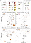
Fig. 1
Fig. 1. CRISPR screens in vivo identify GRA12 as the most important common secreted virulence factor for survival across clonal and atypical Toxoplasma strains.
a Scheme of the CRISPR screen pipeline. Two protospacers libraries targeting the putative Toxoplasma secretome were assembled and included either the DHFR-TS or the HXGPRT selection cassette to create pyrimethamine-resistant (PYRR) or mycophenolic acid/xanthine-resistant (M/XR) parasite KO pools. The wild-type strains VAND and VEG were transfected with the DHFR-TS library, while the ∆HXGPRT strains RH and PRU were transfected with the HXGPRT library. The resulting KO parasite pools were injected in the peritoneum of 5 mice/parasite strain, retrieved after 5 days and expanded for one lytic cycle in vitro before gDNA extraction and sgRNA amplification for sequencing. The relative abundance of each guide (L2FC) at day 5 versus inoculum indicates the importance for in vivo survival of the relative Toxoplasma gene. b Scatter plots of the median L2FC for each gene in vitro and in vivo in CRISPR screens of, in order from the top left: VAND in CAST/EiJ mice, RH ∆HXGPRT in PWD/PhJ mice, PRU ∆HXGPRT and VEG in C57BL/6J mice. The colour and size of each point reflects the Discordance/Concordance (DISCO) score, and the dashed grey line indicates equal L2FC. Displayed data are thresholded for L2FC in vitro > −1 according to published in vitro whole genome screens. Data points with a L2FC in vivo < −1.5 are labelled in addition to ROP18 as control.
Fig. 2
Fig. 2. GRA12 is a virulence factor in the highly virulent South American VAND strain in vivo.
a Immunofluorescence localisation of the C-terminal HA-tagged GRA12 in the VAND ∆GRA12::GRA12-HA strain. Scale bar represents 10 µm. b Verification of GRA12 expression by anti-HA detection in the VAND ∆GRA12::GRA12-HA strain via western blot. c Survival curve of PWD/PhJ mice infected with either VAND, ∆GRA12 or ∆GRA12::GRA12-HA strains, dose in parenthesis. Significance was tested using a two-sided Mantel-Cox test, N = 5 mice per group. d Number of brain cysts recovered from PWD/PhJ mice infected with VAND ∆GRA12 parasites, N = 5. (i) and immunofluorescence detection of a representative brain cyst stained with DBA. Scale bar represents 50 µm (ii). Source data are provided as a Source Data file.
Fig. 3
Fig. 3. GRA12 is the first identified virulence factor to survive the IFNγ-mediate clearance in different murine subspecies.
a Scheme of the CRISPR screen of type I RH secretome in PWD/PhJ BMDMs to identify Toxoplasma factors important to survive IFNγ-mediated restriction. The KO pool was used to infect BMDMs either untreated or pretreated with IFNγ for two consecutive lytic cycles. Surviving parasites were expanded in fibroblasts prior to gDNA extraction and sgRNA amplification for sequencing. The relative abundance of each guide (L2FC) in IFNγ-restricted versus untreated parasites indicates the importance for in vitro survival of the relative Toxoplasma gene. b Scatter plots of the median L2FCs for each gene in vitro in CRISPR screens of the RH KO pool in PWD/PhJ BMDMs, treated or not with IFNγ. The grey line indicates equal L2FCs. c Quantification of high content-automated imaging of parasite vacuoles in IFNγ-treated BMDMs relative to untreated controls. BMDMs of different mouse subspecies were infected with RH or VAND ∆UPRT, ∆GRA12, ∆GRA12::GRA12-HA and ∆ROP18 and vacuoles were quantified at 24 h after infection. The box-plot shows the median value ± SD and the whiskers show minimum and maximum values. Symbol shapes indicate biological repeats, N > = 3. Significance was tested using the One-way Anova test with the Benjamini, Krieger and Yekutieli FDR correction. Source data are provided as a Source Data file.
Fig. 4
Fig. 4. Lack of GRA12 results in IFNγ-mediated host cell death by necrosis.
a Time course of PWD/PhJ BMDMs cell death quantified via Propidium Iodide uptake. The graph shows the median value ± SEM, N = 4. Cells were pre-stimulated for 24 h with IFNγ (continuous line) or left untreated as control (dashed line) and then infected with VAND ∆UPRT, ∆GRA12, ∆GRA12::GRA12-HA or left uninfected. The number of dead cells is expressed as a percentage of the total at each time point. b Cell death quantification at 9 hpi of IFNγ-treated PWD/PhJ BMDMs infected with ∆UPRT, ∆GRA12, ∆GRA12::GRA12-HA in the PRU, RH and VAND strains, or left uninfected as control. The bar graph shows the median value ± SD, N > = 4. Significance was tested using the Tukey’s multiple comparisons test for the RH and VAND strains, and with a paired t test for the PRU strains. c Western blot analysis of the supernatant and cell lysate of C57BL/6J BMDMs pre-treated with IFNγ and infected for 8 h with RH ∆UPRT, ∆GRA12, ∆GRA12::GRA12-HA, or left untreated as control. d Live cell imaging of IFNγ-treated C57BL/6J BMDMs infected with RH ∆GRA12 parasites. Time post infection reported as hh:mm in the bottom right corner. Filled arrowheads indicate dying cells and an egressed parasite. Complete video is in Supplementary Movie 1. e Fluorescence microscopy detection of RH ∆UPRT parasites after 48 h growth in HFFs treated or not with 1 µM ML10. Scale bar represents 20 µm. f Time course of C57BL/6J BMDMs cell death quantified via Propidium Iodide uptake. Cells were pre-stimulated for 24 h with IFNγ, and infected with RH ∆UPRT, ∆GRA12, ∆GRA12::GRA12-HA before treatment with 1 µM ML10 (triangle symbol), or left untreated as control (continuous line). The number of dead cells is expressed as a percentage of the total at each time point. g Cell death quantification at 9 hpi of the ML10 treatment experiment. The bar graph shows the median value ± SD, N > = 4. Symbol shapes indicate biological repeats. Significance was tested using the One-way Anova test with the Benjamini, Krieger and Yekutieli FDR correction. Source data are provided as a Source Data file.
Fig. 5
Fig. 5. Parasites lacking GRA12 have a collapsed intravacuolar space and are less targeted by host immune proteins.
a Immunofluorescence localisation of GRA12 in partially permeabilised human fibroblasts infected with the N-terminally tagged line (upper panel) or the C-terminally tagged line (lower panel). Filled arrowheads indicate a permeabilised vacuole and empty arrowheads indicate a non-permeabilised vacuole. Scale bar represents 10 µm. b Example TEM images of IFNγ-treated PWD/PhJ BMDMs infected with RH ∆KU80, ∆GRA12, ∆GRA12::GRA12-HA for 2 h. Scale bar represents 0.5 µm. c Example of analysis for the vacuolar space quantification on a parasite infecting a BMDM. Scale bar represents 1 µm. d Quantification of the vacuolar area from the TEM images. Individual vacuoles were randomly imaged in a single biological experiment. Scatter plot of quantified area. When the PVM was not visible, the vacuolar area was imputed to 0.01 µm2. Significance was tested using the Kruskal-Wallis test. e Quantification of the recruitment of host proteins (IRGd, IRGb10 and GBP2) to the PVM after 90 min infection of IFNγ-treated or untreated C57BL/6J BMDMs with PRU ΔUPRT or ΔGRA12 parasites. The bar graph shows the median value ± SD, N = 3. Significance was tested using the Tukey’s multiple comparisons. f Representative images of anti-IRGd stained infections with PRU ΔUPRT (left panel) or PRU ΔGRA12 (right panel). Scale bar represents 50 µm. Source data are provided as a Source Data file.
Fig. 6
Fig. 6. GRA12 is an intravacuolar protein conserved across Coccidia.
a Neighbor-Joining tree of Coccidian parasites GRA12 homologues built with Bootstrap method and considering GRA12D as outgroup. The bootstrap support values, based on 100 replicates, are indicated above each node. b Structural overlay of Toxoplasma GRA12 (grey) and N. caninum GRA12 homologue (teal) as predicted by AlphaFold2. Root Main Square Deviation (RMSD) was calculated using PyMol. c Relative Toxoplasma growth in IFNγ-treated versus untreated BMDMs. BMDMs were infected with the RH ∆GRA12 strain, or the RH∆GRA12 strain complemented with Toxoplasma (Tg) GRA12 or the H. hammondi (Hh) or N. caninum (Nc) GRA12 homologues, or the RH ∆UPRT strain as control for 24 h before a plate reader quantification of the mCherry signal as proxy for parasite growth. The box-plot shows the median value ± SD and the whiskers show minimum and maximum values. Significance was tested using the One-way Anova test with the Benjamini, Krieger and Yekutieli FDR correction, N > = 4. Source data are provided as a Source Data file.

References

    1. Pappas, G., Roussos, N. & Falagas, M. E. Toxoplasmosis snapshots: global status of Toxoplasma gondii seroprevalence and implications for pregnancy and congenital toxoplasmosis. Int. J. Parasitol.39, 1385–1394 (2009). - PubMed
    1. Su, C., Howe, D. K., Dubey, J. P., Ajioka, J. W. & Sibley, L. D. Identification of quantitative trait loci controlling acute virulence in Toxoplasma gondii. Proc. Natl. Acad. Sci. USA99, 10753–10758 (2002). - PMC - PubMed
    1. Galal, L., Hamidović, A., Dardé, M. L. & Mercier, M. Diversity of Toxoplasma gondii strains at the global level and its determinants. Food Waterborne Parasitol.15, e00052 (2019). - PMC - PubMed
    1. Howe, D. K. & Sibley, L. D. Toxoplasma gondii comprises three clonal lineages: correlation of parasite genotype with human disease. J. Infect. Dis.172, 1561–1566 (1995). - PubMed
    1. Grigg, M. E., Ganatra, J., Boothroyd, J. C. & Margolis, T. P. Unusual abundance of atypical strains associated with human ocular toxoplasmosis. J. Infect. Dis.184, 633–639 (2001). - PubMed

LinkOut - more resources