. 2025 Apr 16;16(1):3612.
doi: 10.1038/s41467-025-58931-y.

Inflammatory conditions shape phenotypic and functional characteristics of lung-resident memory T cells in mice

Affiliations

Inflammatory conditions shape phenotypic and functional characteristics of lung-resident memory T cells in mice

Anna Schmidt et al. Nat Commun. .

Abstract

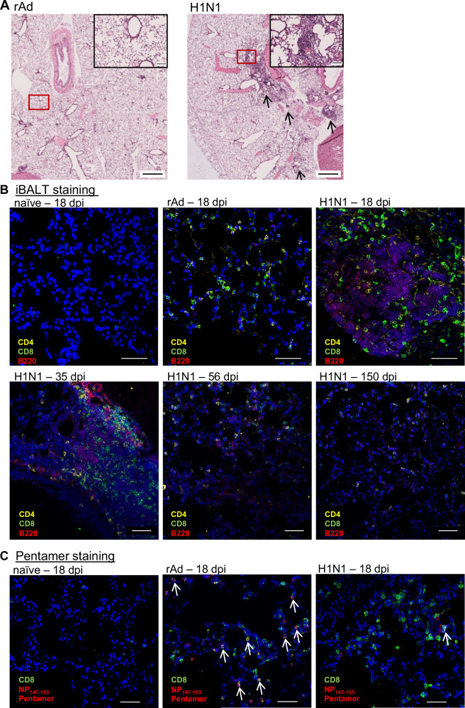
Lung tissue-resident memory T cells (TRM) are critical for the local control of respiratory tract infections caused by influenza A viruses (IAV). Here we compare TRM populations induced by intranasal adenoviral vector vaccines encoding hemagglutinin and nucleoprotein (NP) with those induced by an H1N1 infection in BALB/c mice. While vaccine-induced TRM express high levels of CD103 and persist longer in the lung parenchyma, short-lived, H1N1-induced TRM have a transcriptome associated with higher cytotoxic potential and distinct transcriptional profile as shown by single-cell RNA sequencing. In both the vaccine and H1N1 groups, NP-specific CD8+ T cells expand during heterologous influenza virus infection and protect the mice from disease. Meanwhile, lung inflammation in response to an infection with unrelated respiratory syncytial virus do not influence the fate of pre-existing TRM. Our preclinical work thus confirms that inflammatory conditions in the tissue shape the phenotypic and functional characteristics of TRM to serve relevant informations for optimizing mucosal vaccines.


Conflict of interest statement

Competing interests: The authors declare no competing interests.

Figures

Fig. 1
Fig. 1. Schedule of experimental treatments and kinetic phenotypic analysis of antigen-specific CD8+ TRM.
A On day 0, seven-weeks-old female BALB/c mice were either i.n. immunized with rAd-HA, rAd-NP, and rAd-IL-1β (each 2 × 108 particles) or infected with the H1N1 strain A/PR/8/34 (100 PFU). At the indicated time points, lymphocytes were isolated from lung tissues to evaluate T-cell phenotypes and functional responses. In addition, blood and BALF samples were collected at the specified times to assess systemic and local antibody responses. The localization and organization of different immune cells was examined by histological techniques. B At the indicated times, antigen-specific cells were identified by pentamer staining, and total numbers of HA533-541- and NP147-155-specific Pent+ cells are depicted. The distribution of antigen-specific T cells within the iv- and iv+ proportions are illustrated by different coloring. C Absolute numbers of circulating effector and memory T-cell subsets, and those of the TRM pool induced by mucosal vaccination or H1N1 infection are shown. Iv- TRM (KLRG1-) are colored in different shades of blue (regarding CD69 and CD103 expression), while effector T cells (Teff; iv+ KLRG1+ CD127-) are shown in black, and effector memory T cells (TEM; iv+ KLRG1 + CD127 + ), and central memory T cells (TCM; iv+ KLRG1- CD127 + ) are shown in gray. Each data set represents the mean + SEM of per group (n = 6 mice per group/timepoint, except 150 dpi in the H1N1 group represents only n = 4). Statistical significances were analyzed by two-way ANOVA followed by Šídák’s multiple comparison test (*, p < 0.1; **, p < 0.01; ***, p < 0.001 (gray line: iv-)). (A) Created in BioRender. Tenbusch, M. (2025) https://BioRender.com/o92n191.
Fig. 2
Fig. 2. Functional CD8+ T-cell responses measured by ICS.
Lymphocytes from rAd-immunized or H1N1-infected mice were isolated, and ICS was used to identify influenza-reactive T cells. HA- and NP-specific cytokine secretion was measured to determine functional CD8+ subpopulations, which are shown for day 35 as frequencies of the circulating (iv+) or tissue-resident (iv-) CD8+ T-cell compartment. Each data point represents an individual mouse, and the bars represent the mean of the group (n = 6 mice per group). Statistical significances were analyzed by a two-tailed Mann-Whitney test (*, p < 0.05; **, p < 0.01).
Fig. 3
Fig. 3. Histological analyses of lung tissue post immunization or H1N1 infection.
A Lungs from rAd-immunized or H1N1-infected mice were obtained 18 days post-treatment, and HE-stained lung sections were scanned at 40x magnification. Airways were generally intact and solely H1N1 infection caused lymphocytic infiltration reflected by areas of lymphocyte accumulations, without neutrophils, monocytes, and macrophages (arrows). One representative example of four mice per group is shown. Scale bars in whole slide lung images are 1000 µm and scale bars in magnified images are 100 µm. B At the indicated time points, lungs of naïve, immunized, and infected mice were removed, embedded into O.C.T. compound, and finally stained with anti-CD4, anti-CD8, and anti-B220. Nuclei are shown in blue (Hoechst33342), CD4+ T cells are shown in yellow, CD8+ T cells are shown in green, and B cells are shown in red. C To identify the localization of antigen-specific CD8+ T cells in the lung parenchyma, lung sections from day 18 p.i. were primary stained with anti-CD8, followed by MHC-I pentamer staining. The signal intensity of bound APC-labeled pentamers was amplified with anti-APC-AF647, and anti-mouse IgG2b-AF647. Nuclei are shown in blue (Hoechst33342), CD8+ T cells are shown in green, and MHC-I pentamer-specific T cells are shown in red. Images were acquired on a Leica SP5X laser scanning confocal microscope using a 40x oil objective and are representatives from a collection of different slices of four mice per group (n = 4). Scale bars in each image are 50 µm.
Fig. 4
Fig. 4. Experimental schedule, neutralizing capacity, bodyweight analysis, and blood oxygen saturation after lethal H3N2 challenge in the mid-memory phase.
A On day 0, seven-weeks-old female BALB/c mice were either i.n. immunized with rAd-HA, rAd-NP, and rAd-IL-1β or infected with the H1N1 strain A/PR/8/34 as described before. 56 days after the initial treatment, mice were challenged with H3N2 A/HK/68 (10,000 PFU) to determine the protective capacity of vaccine- or infection-induced immunity in the mid-phase of immune memory, as well as the fate of pre-existing TRM. B Serum samples were collected before the challenge as well as two and six weeks after the H3N2 challenge to analyze the neutralizing capacity against heterosubtypic H3N2. Each data point represents an individual mouse, and the bars represent the mean of the group (n = 8 mice per group). C Weight loss was monitored daily and is expressed as a percentage of the initial weight on day 0. Naïve mice served as control, but all of them reached the endpoint criteria on day eight after the challenge. Curves represent the mean with SEM (n = 4 mice for naïve, 8 mice for rAd and H1N1). D Pulse-oximetry was performed three, six, and eight days p.i., and percentages of blood oxygen saturation are presented. Each data point represents an individual mouse, and bars represent the mean of the group mean ± SEM per group (n = 4 mice for naïve, 8 mice for rAd and H1N1) (B). Statistical significances were analyzed by two-way ANOVA followed by Tukey’s multiple comparisons test (B; **, p < 0.01; ****, p < 0.0001)., by two-way ANOVA followed by Tukey’s multiple comparisons test (C; *, p < 0.05 H1N1 vs. rAd; #, p < 0.05 H1N1 vs. naïve; +, p < 0.05 rAd vs. naive) or by one-way ANOVA followed by Tukey’s multiple comparison test (D; ****, p < 0.0001). A Created in BioRender. Tenbusch, M. (2025) https://BioRender.com/z57x523.
Fig. 5
Fig. 5. Phenotypic and functional profile of CD8+ T cells after H3N2 challenge.
Previously, rAd-immunized or H1N1-infected mice were challenged with H3N2 (10,000 PFU) 56 days after the initial prime, and lymphocytes from lung tissues were isolated 44 days after the challenge (+ H3N2). One set of mice was not reinfected and served as the control group (- H3N2). Additionally, some lymphocytes were restimulated in vitro using MHC-II- or MHC-I-restricted peptides derived from HA and NP, and ICS was used for the functional identification. A, B Phenotypic differentiation between antigen-specific Teff, TEM, and TCM, and different subsets of TRM. The graphs show the total number of HA533-541- and NP147-155-specific Pent+ CD8+ T cells. C Frequencies of cytokine-specific CD8+ T cells investigated in secondary infected mice (+ H3N2) compared to only primed mice (- H3N2) are shown. AC Each data point represents an individual mouse, and bars represent the mean of the group (n = 6 mice for rAd and H1N1 (-H3N2), 7 mice for H1N1 (+ H3N2) and 8 mice for rAd (+ H3N2). To compare statistical effects between unchallenged (- H3N2) and challenged (+ H3N2) mice of one group, statistical significances were analyzed by two-tailed Mann-Whitney test (*, p < 0.05; **, p < 0.01; ***, p < 0.001).
Fig. 6
Fig. 6. Experimental schedule to analyze the fate of functional antigen-specific CD8+ TRM upon unrelated secondary virus infection.
A The survival of previously induced IAV-specific TRM was examined upon secondary inflammatory events by subsequent infection with the unrelated RSV (1 × 106 PFU) or by LPS treatment (10 µg) on day 56 after priming. Three weeks after the challenge, lymphocytes were isolated from lung tissues. One set of initially primed mice remained without secondary exposure (- RSV, - LPS, respectively) and served as the control group. B Body weight was measured for nine days following the RSV challenge, and values represent percentages of the initial weight on day 0. C Antigen-specific CD8+ T cells were identified by pentamer staining. Total numbers of HA533-541-, NP147-155-, and M282-90-specific tissue-resident CD8+ T cells are shown. D Frequencies of non-circulating CD8+ T cells that are at least positive for the single expression of either CD107a, IFNγ, IL-2, or TNFα were assessed after in vitro restimulation. E Immunofluorescence staining of lung tissues from initially rAd-immunized or H1N1-infected mice reinfected with RSV on day 35 and collected an additional 35 days after challenge. Tissue sections were stained with anti-B220 (green), RSV M282-90 pentamer (yellow), and IAV NP147-155 pentamer (red). Nuclei are shown in blue (Hoechst33342). Images were acquired on a Leica SP5X laser scanning confocal microscope using a 40x oil objective and are representatives from a collection of different slices of three mice per group (n = 3). Scale bars in each image are 50 µm. C, D Each data point represents an individual mouse, and bars represent the mean of the group mean ± SEM per group (n = 6 mice per group, only in (D) one sample in the group rAd (-RSV) was lost due to technical issues). Numbers above the columns show fold change between unchallenged (- RSV) and challenged (+ RSV) mice. Statistical significances were analyzed (B) by two-way ANOVA followed by Tukey’s multiple comparison test, or (C, D) by one-way ANOVA followed by Tukey’s multiple comparison test ((B): *, p < 0.05 H1N1 + RSV vs. naïve + RSV; (C, D); *, p < 0.1; ***, p < 0.001; ****, p < 0.001). (A) Created in BioRender. Tenbusch, M. (2025) https://BioRender.com/g83h450.
Fig. 7
Fig. 7. Experimental schedule to define the transcriptional profile of NP-specific TRM after mucosal immunization compared to H1N1 infection.
On day 0, seven-weeks-old female BALB/c mice were either i.n. immunized with rAd-NP and rAd-IL-1β (each 2 × 108 particles) or infected with the H1N1 strain A/PR/8/34 (100 PFU). On day 56, lymphocytes were isolated from the lung tissue, and NP147-155-specific CD8+ T cells were sorted and sequenced by scRNA-seq to determine potential differences between rAd-NP/IL-1β- and H1N1-induced TRM at the transcriptomic level. The analysis includes four mice per group (n = 4). A Leiden clustering of the dataset with a resolution of 0.5 revealed nine clusters of memory T cells. The clusters are circled separately according to the color code. In the table, the number of cells per cluster is provided for each of the two groups. B For selected characteristic genes of memory T cells, the number of transcripts per million and the relative expression levels for each assigned cluster were indicated divided into transcription factors, effector molecules, and surface receptor/migration. The absolute TMP is given as the number within each cell, and the relative expression level is indicated by the color of the cell. For each gene, the TMP of the cluster with the highest expression level was set as maximum, and the ratios were calculated accordingly, ranging from 0 to 1. C Volcano plot of a total of 15730 genes analyzed for differential expression. The solid, light gray line indicates the significance threshold. The p-value and log2fc was calculated in DESeq2 with default settings. Significant differentially expressed genes are highlighted in green (rAd) or red (H1N1), and selected gene names are indicated.

References

    1. WHO. Influenza (Seasonal). https://www.who.int/news-room/fact-sheets/detail/influenza-(seasonal) (2025).
    1. Wong, S.-S. & Webby, R. J. Traditional and new influenza vaccines. Clin. Microbiol Rev.26, 476–492 (2013). - PMC - PubMed
    1. Padilla-Quirarte, H. O., Lopez-Guerrero, D. V., Gutierrez-Xicotencatl, L. & Esquivel-Guadarrama, F. Protective antibodies against influenza proteins. Front. Immunol.10, 1677 (2019). - PMC - PubMed
    1. Tripp, R. & Tompkins, S. Virus-vectored influenza virus vaccines. Viruses6, 3055–3079 (2014). - PMC - PubMed
    1. Angeletti, D. et al. Defining B cell immunodominance to viruses. Nat. Immunol.18, 456–463 (2017). - PMC - PubMed

MeSH terms

LinkOut - more resources