

The prophylactic anti-aging effect of aspirin (acetylsalicylic acid) on oxidative stress-induced damage in the buccal mucosa of D-galactose-induced aged rats
- PMID: 40240423
- PMCID: PMC12003813
- DOI: 10.1038/s41598-025-94566-1
The prophylactic anti-aging effect of aspirin (acetylsalicylic acid) on oxidative stress-induced damage in the buccal mucosa of D-galactose-induced aged rats
Abstract
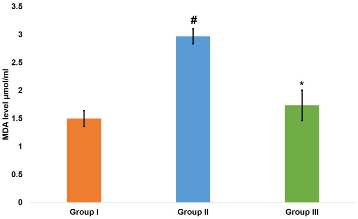
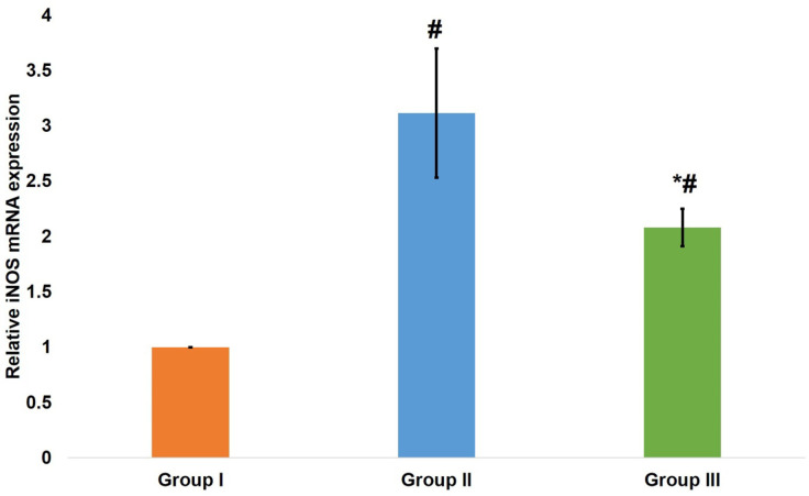
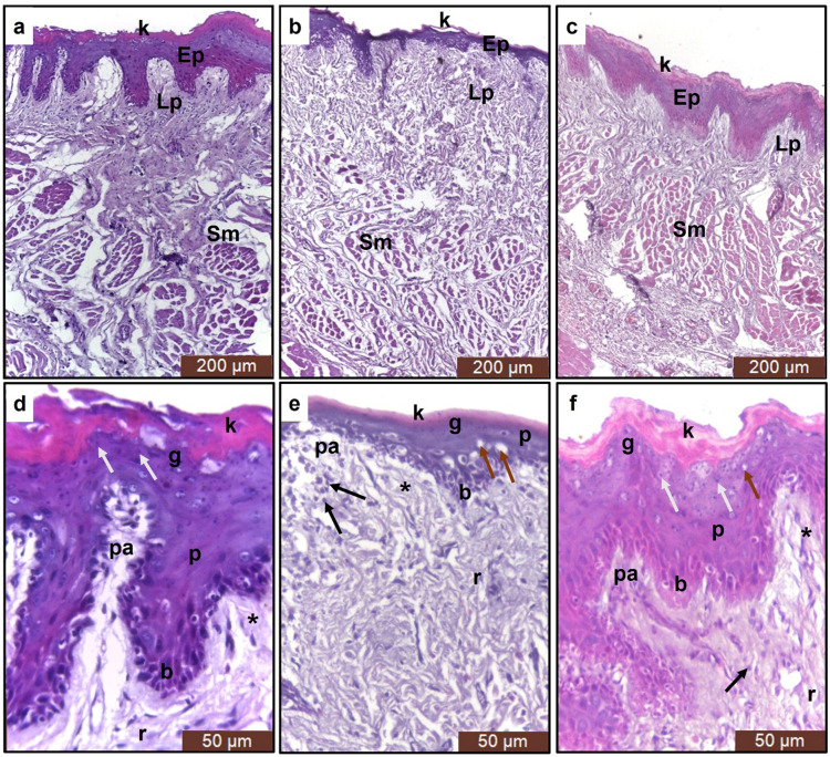
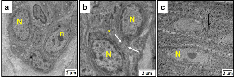
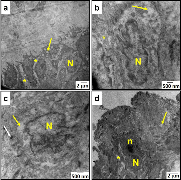
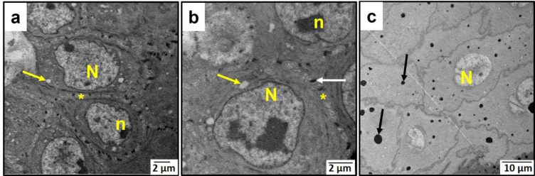
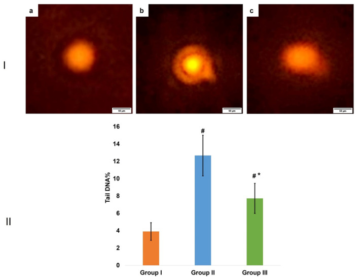
Most living organisms experience time-dependent functional deterioration as they age. To combat aging, aspirin was proposed as an already well-studied drug. However, its antiaging effect is neither well studied nor understood. So, this study intended to assess the proposed antiaging effect of aspirin. Three groups of seven adult male albino rats were established. The control group received saline, the aging model group got a daily single D-galactose subcutaneous injection (300 mg/kg), and the aspirin group consisted of D-galactose-induced aged rats that received a daily aspirin oral dose (60 mg/kg). Drugs were given for 8 weeks. Then, malondialdehyde (MDA) blood level was evaluated, and rats were euthanized. Buccal mucosa samples were obtained for inducible nitric oxide synthase (iNOS) gene expression, histopathological, ultrastructural, and comet analyses. MDA blood level, iNOS gene expression and DNA damage examined by comet assay displayed a significant reduction in the aspirin group when compared to the aging model group. Histopathological and ultrastructural results showed that aspirin ameliorated most of the degenerative signs caused by D-galactose. Thus, it was deduced that aspirin had promising results as an antiaging pharmaceutical agent. However, more studies are needed regarding its translation to human trials.
Keywords: Antiaging; Aspirin; Buccal mucosa; Comet assay; iNOS.
© 2025. The Author(s).
Conflict of interest statement
Declarations. Competing interests: The authors declare no competing interests.
Figures
Similar articles
-
The Black Book of Psychotropic Dosing and Monitoring.Psychopharmacol Bull. 2024 Jul 8;54(3):8-59. Psychopharmacol Bull. 2024. PMID: 38993656 Free PMC article. Review.
-
Oral aspirin for treating venous leg ulcers.Cochrane Database Syst Rev. 2016 Feb 18;2(2):CD009432. doi: 10.1002/14651858.CD009432.pub2. Cochrane Database Syst Rev. 2016. PMID: 26889740 Free PMC article.
-
Drugs for preventing postoperative nausea and vomiting in adults after general anaesthesia: a network meta-analysis.Cochrane Database Syst Rev. 2020 Oct 19;10(10):CD012859. doi: 10.1002/14651858.CD012859.pub2. Cochrane Database Syst Rev. 2020. PMID: 33075160 Free PMC article.
-
Sertindole for schizophrenia.Cochrane Database Syst Rev. 2005 Jul 20;2005(3):CD001715. doi: 10.1002/14651858.CD001715.pub2. Cochrane Database Syst Rev. 2005. PMID: 16034864 Free PMC article.
-
Signs and symptoms to determine if a patient presenting in primary care or hospital outpatient settings has COVID-19.Cochrane Database Syst Rev. 2022 May 20;5(5):CD013665. doi: 10.1002/14651858.CD013665.pub3. Cochrane Database Syst Rev. 2022. PMID: 35593186 Free PMC article.
References
-
- Harman, D. Aging: overview. Ann. N Y Acad. Sci.928 (1), 1–21. 10.1111/j.1749-6632.2001.tb05631.x (2001). - PubMed
-
- Troen, B. R. The biology of aging. Mt. Sinai J. Med.70 (1), 3–22 (2003). - PubMed
-
- Piskovatska, V., Strilbytska, O., Koliada, A., Vaiserman, A. & Lushchak, O. Health Benefits of Anti-Aging Drugs. In: Harris, J., Korolchuk, V. editors. Biochemistry and Cell Biology of Ageing: Part II Clinical Science. Subcellular Biochemistry, Springer, Singapore; pp. 339–392. (2019). 10.1007/978-981-13-3681-2_13 - PubMed
-
- Desborough, M. J. R. & Keeling, D. M. The aspirin story–from Willow to Wonder drug. Br. J. Haematol.177 (5), 674–683. 10.1111/bjh.14520 (2017). - PubMed
MeSH terms
Substances
LinkOut - more resources
Full Text Sources
Medical
