. 2025 Jun;642(8069):1090-1096.
doi: 10.1038/s41586-025-08896-1. Epub 2025 Apr 16.

Chromosome end protection by RAP1-mediated inhibition of DNA-PK

Affiliations

Chromosome end protection by RAP1-mediated inhibition of DNA-PK

Patrik Eickhoff et al. Nature. 2025 Jun.

Erratum in

Abstract

During classical non-homologous end joining (cNHEJ), DNA-dependent protein kinase (DNA-PK) encapsulates free DNA ends, forming a recruitment platform for downstream end-joining factors including ligase 4 (LIG4)1. DNA-PK can also bind telomeres and regulate their resection2-4, but does not initiate cNHEJ at this position. How the end-joining process is regulated in this context-specific manner is currently unclear. Here we show that the shelterin components TRF2 and RAP1 form a complex with DNA-PK that directly represses its end-joining function at telomeres. Biochemical experiments and cryo-electron microscopy reveal that when bound to TRF2, RAP1 establishes a network of interactions with KU and DNA that prevents DNA-PK from recruiting LIG4. In mouse and human cells, RAP1 is redundant with the Apollo nuclease in repressing cNHEJ at chromosome ends, demonstrating that the inhibition of DNA-PK prevents telomere fusions in parallel with overhang-dependent mechanisms. Our experiments show that the end-joining function of DNA-PK is directly and specifically repressed at telomeres, establishing a molecular mechanism for how individual linear chromosomes are maintained in mammalian cells.


Conflict of interest statement

Competing interests: The authors declare no competing interests.

Figures

Fig. 1
Fig. 1. RAP1 and Apollo redundantly prevent cNHEJ at telomeres in mouse and human cells.
a, Representative FISH of metaphase spreads of Apollofl/fl Lig4+/+ MEFs 108 h after transduction with single guide RNA (sgRNA) targeting Rap1 (sgRap1) and/or Hit&Run Cre. Telomeres were detected with Cy3-(TTAGGG)3 (green) and DNA was stained with DAPI (magenta). White and green arrows highlight chromatid-type and chromosome-type fusions, respectively. See also Extended Data Fig. 1a. Scale bars 10 µm. b,c, Percentage of telomeres involved in chromatid-type (b) or chromosome-type (c) fusions per metaphase after removal of Apollo and/or RAP1 as indicated, in the presence or absence of LIG4. Data from 3 independent experiments, 10 metaphases per experiment (n = 30 total), with median. d, Representative FISH of metaphase spreads from TP53−/− RAP1+/+ (RAP1 is also known as TERF2IP) or TP53−/− RAP1−/− RPE-1 cells 120 h after transduction with Cas9 and control sgRNA (sgControl) or sgAPOLLO as indicated. Scale bars, 10 µm. e, Quantification of the percentage of telomeres fused per metaphase after removal of Apollo as described in d. Data from 3 independent experiments, 10 metaphases per experiment (n = 30 total), with median. See also Extended Data Fig. 1e–g. Ordinary one-way analysis of variance (ANOVA). *P ≤ 0.05, **P ≤ 0.01, ***P ≤ 0.001, ****P ≤ 0.0001; NS, not significant.
Fig. 2
Fig. 2. TRF2, RAP1 and DNA-PK form a terminal complex at telomeric DNA ends.
a, Outline of DNase I footprinting experiment. The 32P-labelled 5′ end is highlighted with a red asterisk. Radiolabelled template is incubated with KU and DNA-PKcs prior to the addition of shelterin, comprising TRF1, TRF2, RAP1, TIN2, POT1 and TPP1. DNase I-digested products are then analysed by denaturing urea polyacrylamide gel electrophoresis (urea-PAGE). bf, DNase I footprinting of telomere end-binding complexes formed in the presence of DNA-PK and shelterin (b), TRF2 and RAP1 (c), TRF2 alone (d), RAP1 alone (e) TRF2 and RAP1 with telomeric or non-telomeric DNA (f). Nucleotides from the 5′ telomeric end indicated. For gel source data see Supplementary Fig. 1.
Fig. 3
Fig. 3. Three distinct interfaces are required for the complex with DNA-PK.
a, Domain organization of RAP1 and TRF2. TRFH, TRF homology domain. bd, DNase I footprinting of telomere end-binding complexes, testing the requirement for RAP1 RCT or TRF2 RBM (b) TRF2 Myb domain, basic domain or both Myb and basic domains (ΔMΔB) (c), or testing the requirement for TRF2 in the presence of Teb1, RAP1 or Teb1–RAP1 (d). Nucleotides from the 5′ telomeric end indicated. See Extended Data Fig. 2 for details. WT, wild type. e, Protein crosslinking analysis of RAP1 and KU in the presence of DNA. Proteins were mixed with crosslinker and reaction products were separated on a denaturing tris-acetate polyacrylamide gel and analysed by silver staining or immunoblotting as indicated. Arrowheads mark the position of crosslinked species containing only KU, or KU and RAP1 as indicated. Bottom and top bands observed with KU alone are presumed to represent KU dimers and tetramers, respectively. f,g, DNase I footprinting of telomere end-binding complexes, testing the requirement for BRCT or Myb domains of RAP1 (f) and rescue of RAP1(ΔBRCT) by fusion to the LIG4 BRCT domain (g). Nucleotides from the 5′ telomeric end indicated. For gel source data see Supplementary Fig. 1.
Fig. 4
Fig. 4. Cryo-EM structure of the RAP1–DNA-PK complex.
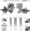
a, Bottom, composite electron density map with protein domains coloured as indicated. Top, schematic of proteins used for structure determination. Uncoloured domains were not visualized. CTD, C-terminal domain; FAT, FRAP–ATM–TRRAP domain; M-HEAT, middle Huntington–EF3–PP2A–TOR1 repeat; N-HEAT, N-terminal Huntington–EF3–PP2A–TOR1 repeat; vWA, von Willebrand A domain. b, Subsection of the structure in a, showing KU70 SAP and RAP1 Myb domains bound to DNA. c,d, The BRCT domain of RAP1 bound to the KU70 vWA domain (c), highlighting RAP1 and KU residues that mediate the interaction (d). eg, DNase I footprinting analysis of telomere end-binding complexes with RAP1 variants R133E (e) and ΔBRCT and KR/DE (f) and KU variant DE/KR (g). Nucleotides from the 5′ telomeric end indicated. For gel source data see Supplementary Fig. 1. h, Cryo-EM model showing binding of RAP1 BRCT to KU70 and KU80 with (bottom) and without (top) the LIG4 BRCT domain from Protein Data Bank (PDB) structure 7LT3 overlaid.
Fig. 5
Fig. 5. TRF2 and RAP1 prevent cNHEJ by directly blocking recruitment of XRCC4–LIG4 to DNA-PK.
a, Outline of the KU pulldown assay. Details in Methods. bd, KU-bound proteins from reactions containing KU70–KU80 (KU70/80), DNA-PKcs, XRCC4–LIG4, TRF2, RAP1 and template DNA together with wild-type RAP1 (b), RAP1(ΔBRCT) or RAP1(KR/DE) (c), or RAP1(ΔMyb) or RAP1(R133E) (d) were separated by SDS–PAGE and immunoblotted as indicated. TRF2, RAP1 and LIG4 were detected with anti-strep tag antibody, KU70 was detected with anti-Flag antibody. Association of TRF2 with KU is mediated by template DNA. For gel source data see Supplementary Fig. 1. e, Percentage of telomeres per metaphase involved in chromosome fusions upon over-expression of mouse RAP1, RAP1(KR/DE) and RAP1(R130E) (equivalent to human RAP1(R133E)) after CRISPR- and Cre-mediated deletion of Rap1 and Apollo, respectively in Apollofl/fl Lig4+/+ MEFs. Data from 3 independent experiments, 10 metaphases per experiment (n = 30 total), with median. Ordinary one-way ANOVA. See Extended Data Fig. 8 for further details. f, Percentage of telomeres fused per metaphase upon CRISPR-mediated deletion of APOLLO in TP53−/− RPE-1 cells with wild-type RAP1 or RAP1(KR/DE). Data from 3 independent experiments, 10 metaphases per experiment (n = 30 total), with median. Statistics as in e. See Extended Data Fig. 8 for further details. g, Model for the direct inhibition of DNA-PK by TRF2 and RAP1 at mammalian telomeres. When assembled on telomeric DNA, DNA-PK and its associated DNA is bound by the Myb and BRCT domains of RAP1. The BRCT domain acts as a circuit breaker, preventing DNA-PK from engaging LIG4.
Extended Data Fig. 1
Extended Data Fig. 1. RAP1 and APOLLO redundantly prevent cNHEJ at telomeres in mouse and human cells.
a. Immunoblot showing effective removal of Rap1 and persistence of TRF2 as indicated after Crispr-mediated Rap1 deletion and/or Cre-mediated deletion of Apollo in ApolloF/F Lig4+/+ or ApolloF/F Lig4−/− MEFs. Beta-actin as loading control. b. Immunoblot showing expression of TRF2 in SV40LT-immortalized TRF2F/F RsCre-ERT1 MEFs transduced with an empty vector control (-), mouse TRF2 WT or TRF2 alleles habouring a F120A mutation and/or deleted of the RAP1 binding motif (∆RBM) and/or deleted of the DNA-damage response motif (∆iDDR) 120 h after deletion of endogenous TRF2 with 4-OHT. Beta-actin as loading control. c. Representative FISH of metaphase spreads from cells as described in b. Telomeres detected with Cy3-(TTAGGG)3 (green). DNA stained with DAPI (magenta). White and green arrows highlight chromatid- and chromosome type fusions respectively. Scale bar = 10 µm d. Quantification of percentage telomeres involved in chromatid and chromosome fusions per metaphase after deletion of endogenous TRF2 in MEFs expressing the indicated TRF2 mutants, as described in b. Data from 3 independent experiments, 10 metaphases per experiment (n = 30 total), with median. Statistics by ordinary One-way ANOVA. ‘ns’ not significant, ****P ≤ 0.0001. e. Sequencing of the RAP1 locus in p53−/− RAP1+/+ and p53−/− RAP1−/− RPE-1 cells. 19 bp deletion in knockout clones generates an altered reading frame terminating in a stop codon after a total of 135 amino acids. f. Immunoblot showing Apollo protein levels 120 h after transduction of p53−/− RAP1+/+ and p53−/− RAP1−/− RPE−1 cells with Cas9 and sgApollo or sgControl. Alpha-tubulin as loading control. g. Chromatid- and chromosome-type telomere fusions quantified after Apollo deletion as in (f). From three independent experiments, 10 metaphases from each, with median. Statistics as in (d). h. Upper panel as in (g) but cells treated with 2 µM DNA-PK inhibitor NU7741 for 48 h prior to cell collection. From two independent experiments, 10 metaphases each, with median. Statistics as in (d). Lower panel as in (f). i. Representative coFISH images of RAP1 KO clone 2 120 h after transduction with Cas9 and sgApollo. Telomeres detected with Cy3-(TTAGGG)3 (red = leading) and FITC-(CCCTTA)3 (green = lagging). Arrows highlight leading:leading telomere fusions. For gel source data see Supplementary Fig. 1.
Extended Data Fig. 2
Extended Data Fig. 2. Deletion constructs, purified proteins and data relating to Figs. 2 and 3.
a. Purified proteins as indicated, separated on a denaturing tris-glycine polyacrylamide gel and stained with Instant Blue. b. DNase I footprinting performed as in Fig. 2a with shelterin or TRF2/RAP1 at increasing concentrations (left panel) or with KU70/80, DNA-PKcs and TRF2/RAP1 each at 250 nM (right panel). Template concentation increased to 20 nM for the experiment on the right. Nucleotides from the 5′ telomeric end indicated. c. DNase I footprinting performed as in Fig. 2a, except shelterin was added coincident with KU70/80 and DNA-PKcs. Lanes 9-22 contained 8 nM template, 100 nM KU/DNA-PKcs and 40 nM shelterin or TRF2:RAP1 as indicated. Cleavage within the 10 bp footprint derives from a change in template sequence register compared with the standard template in Figs. 2–4. Nucleotides from the 5′ telomeric end indicated. d. Purified proteins as indicated, separated on a denaturing tris-glycine polyacrylamide gel and stained with Instant Blue. e. Domain organisation of RAP1 and TRF2 mutants. TRF2 L330R (equivalent to L288R in the shorter TRF2 isoform) prevents binding to RAP1 (Chen et al, 2011). PreScission cleavage site highlighted by 3 C. Also see Supplementary Information Table 2. f. Electrophoretic mobility shift assay of telomeric DNA bound by TRF2. g. DNaseI footprinting performed as in Fig. 3d with proteins omitted as indicated. Nucleotides from the 5′ telomeric end indicated. h. Cleavage of Teb1 DBD from RAP1 using PreScission protease. Proteins were separated on a denaturing tris-glycine polyacrylamide gel and stained with Instant Blue. i. Protein cross-linking analysis of KU, RAP1 and TRF2 containing only RBM and myb domains, in the presence of DNA. Arrowheads mark the position of cross-linked species containing only KU, or KU, RAP and TRF2. For gel source data see Supplementary Fig. 1.
Extended Data Fig. 3
Extended Data Fig. 3. Cryo-EM data processing pipeline for the RAP1:DNA-PK complex on DNA.
Schematic showing the cryoSPARC classification and refinement steps used to obtain the RAP1:DNA-PK structure. Also see Extended Data Table 1.
Extended Data Fig. 4
Extended Data Fig. 4. DNA-PKcs conformation in the RAP1:DNA-PK complex.
DNA-PKcs models from the structures indicated, showing the M-HEAT and N-HEAT domains adopting an ‘inactive’ conformation in the telomere end binding complex.
Extended Data Fig. 5
Extended Data Fig. 5. Binding of the KU70 SAP and RAP1 myb domains to DNA.
a. Density map and model of the KU70 SAP:DNA interaction extracted from the complete structure, showing K575, K595 and K596 coordinating the phosphate backbone b. Density map and model of the RAP1 myb:DNA interaction extracted from the complete structure, showing the recognition helix sitting in the major groove, and R133 acting as an N-terminal arm inserted into the neighbouring minor groove. Region protected from DNase I indicated c. Sequence alignment of the human RAP1 myb domain. Alignment adopts Clustal X colouring d. Comparison of the DNA-bound human RAP1 myb domain (blue) with homeotic protein antennapedia (purple - 1ahd) and HOX-B1 (pink - 1b72). Structures were aligned via the DNA. e. Table of intermolecular chemical crosslinks detected by XLMS. Data were thresholded with an XlinkX score ≥90 and crosslinks between KU and DNA-PKcs were excluded. f. Intermolecular chemical crosslinks detected by XLMS. Data were thresholded with an XlinkX score ≥90. g. Density map and model of the RAP1 loop C-terminal to the myb domain anchored to KU80.
Extended Data Fig. 6
Extended Data Fig. 6. Binding of the RAP1 BRCT domain to KU70 vWA.
a. Sequence alignment of the RAP1 (left), KU70 (middle) and KU80 (right) regions that engage in the BRCT:KU interaction. Asterisks mark K39, R40 and R55 in RAP1, which are changed to aspartate or glutamate in the KR/DE mutant. Also, D496 and E499 in KU70 and D326 in KU80, which are changed to lysine or arginine in the KU DE/KR mutant. Alignment adopts Clustal X colouring b. Nano-scale differential scanning fluorimetry analysis of the RAP1 BRCT or KU mutants as indicated, showing no significant effect of the point mutations on protein folding. c. Protein cross-linking analysis of RAP1 and KU in the presence of DNA. Proteins were mixed with crosslinker and reaction products were separated on a denaturing tris-acetate polyacrylamide gel and analysed by silver staining or immunoblotting as indicated. ΔB and ΔM mark Rap1 BRCT and myb domain deletion mutants respectively. RAP1 KR/DE (KR) contains K39D, R40E and R55E mutations. KU DE/KR (DE) contains KU70 D496K, E499R and KU80 D327K mutations. For gel source data see Supplementary Fig. 1.
Extended Data Fig. 7
Extended Data Fig. 7. Supplementary DNA-PK binding assays related to Fig. 5.
a. KU pulldown experiment without TRF2 or RAP1, executed as described in Fig. 5a. KU DE/KR contains the mutations KU70 D496K, E499R and KU80 D327K b. As in (a), but with reactions containing 5 nM TRF2/RAP1 complex and increasing concentrations of XRCC4/LIG4 as indicated. c. As in (b). ‘X/L pre-bind’ indicates reactions where XRCC4/LIG4 was incubated with KU70/80, DNA-PKcs and DNA template for 5 min prior to the addition of TRF2/RAP1. d. As in (b), but with 30 nM XRCC4/LIG4, and the RAP1 proteins indicated. For gel source data see Supplementary Fig. 1.
Extended Data Fig. 8
Extended Data Fig. 8. Mutation analysis of RAP1 in mouse and human cells.
a. Immunoblot showing over-expression of mouse RAP1, RAP1 KR/DE and RAP1 R130E after Crispr- and Cre-mediated deletion of Rap1 and Apollo respectively in ApolloF/F Lig4+/+ MEFs. Beta-actin as loading control. b. Representative FISH of metaphase spreads of ApolloF/F Lig4+/+ MEFs expressing the indicated RAP1 mutants or a control empty vector (EV) 96-120 h after deletion of endogenous Rap1 with sgRNA and 120 h after deletion of APOLLO with Hit & Run Cre. Telomeres detected with Cy3-(TTAGGG)3 (green). DNA stained with DAPI (magenta). White and green arrows highlight chromatid- and chromosome type fusions respectively. Scale bar = 10 µm c. Quantification of percentage telomeres involved in chromatid fusions per metaphase after expression of Rap1 or the empty vector control (-) and removal of endogenous Rap1 and Apollo as described in b. Data from 3 independent experiments, 10 metaphases per experiment (n = 30 total), with median. Statistics by ordinary One-way ANOVA. ‘ns’ not significant. d. Sequence analysis of the human RAP1 locus in RAP1 WT and RAP1 KR/DE p53−/− RPE-1 cells. Targeted mutations in KR/DE are marked with an asterisk, with altered amino acids highlighted in red e. Immunoblot showing Apollo, RAP1 and TRF2 levels after Crispr-mediated deletion in p53−/− RAP1 WT and p53−/− RAP1 KR/DE RPE-1 cells. Asterisk marks a non-specific band detected by the anti-Apollo antibody. Alpha-tubulin as loading control.f. Representative FISH of metaphase spreads of RAP1 WT and RAP1 KR/DE p53−/− RPE-1 cells 120 h after transduction with Cas9 and sgApollo or sgControl as indicated. Telomeres detected with Cy3-(TTAGGG)3 (green). DNA stained with DAPI (magenta). White and green arrows highlight chromatid- and chromosome type fusions respectively. g. Quantification of the percentage of telomeres involved in chromatid and chromosome fusions per metaphase after removal of Apollo in RAP1 WT and RAP1 KR/DE p53−/− RPE-1 cells as indicated. Data from 3 independent experiments, 10 metaphases per experiment (n = 30 total), with median. Statistics by ordinary One-way ANOVA. ‘ns’ not significant, ****P ≤ 0.0001. For gel source data see Supplementary Fig. 1.
Extended Data Fig. 9
Extended Data Fig. 9. Conservation analysis of the RAP1 BRCT:KU interaction.
Amino acid sequences of RAP1 from the metazoan species indicated were aligned using the Muscle algorithm. The basic patch composed of K39, R40, R41 and R55 in the BRCT domain is shown with basic residues displayed in red. Corresponding structure predictions for the RAP1:KU complex made using AlphaFold 3 are shown for the species indicated, revealing that the BRCT:KU interaction is likely to occur widely across metazoa.

Similar articles

Cited by

  • Restriction of Ku translocation protects telomere ends.
    Mattarocci S, Baconnais S, Roisné-Hamelin F, Pobiega S, Alibert O, Morin V, Deshayes A, Veaute X, Ropars V, Chevreuil M, Mehringer J, Busso D, Mazon G, Fernandez Varela P, Le Cam É, Charbonnier JB, Cuniasse P, Marcand S. Mattarocci S, et al. Nat Commun. 2025 Jul 24;16(1):6824. doi: 10.1038/s41467-025-61864-1. Nat Commun. 2025. PMID: 40707444 Free PMC article.
  • Telomere Crisis Shapes Cancer Evolution.
    Nassour J, Karlseder J. Nassour J, et al. Cold Spring Harb Perspect Biol. 2025 Aug 11:a041688. doi: 10.1101/cshperspect.a041688. Online ahead of print. Cold Spring Harb Perspect Biol. 2025. PMID: 40789644 Free PMC article.

References

    1. Chang, H. H. Y., Pannunzio, N. R., Adachi, N. & Lieber, M. R. Non-homologous DNA end joining and alternative pathways to double-strand break repair. Nat. Rev. Mol. Cell Biol.18, 495–506 (2017). - PMC - PubMed
    1. de Lange, T. Shelterin-mediated telomere protection. Annu. Rev. Genet.52, 223–247 (2018). - PubMed
    1. Hsu, H. L., Gilley, D., Blackburn, E. H. & Chen, D. J. Ku is associated with the telomere in mammals. Proc. Natl Acad. Sci. USA96, 12454–12458 (1999). - PMC - PubMed
    1. d’Adda di Fagagna, F. et al. Effects of DNA nonhomologous end-joining factors on telomere length and chromosomal stability in mammalian cells. Curr. Biol.11, 1192–1196 (2001). - PubMed
    1. Denchi, E. L. & de Lange, T. Protection of telomeres through independent control of ATM and ATR by TRF2 and POT1. Nature448, 1068–1071 (2007). - PubMed

MeSH terms