. 2025 Apr 16;24(1):167.
doi: 10.1186/s12933-025-02725-5.

Impaired cardiac branched-chain amino acid metabolism in a novel model of diabetic cardiomyopathy

Affiliations

Impaired cardiac branched-chain amino acid metabolism in a novel model of diabetic cardiomyopathy

Junko Asakura et al. Cardiovasc Diabetol. .

Abstract

Background: Systemic insulin resistance plays an important role in the pathogenesis of type 2 diabetes and its complications. Although impaired branched-chain amino acid (BCAA) metabolism has been reported to be involved in the development of diabetes, the relationship between cardiac BCAA metabolism and the pathogenesis of diabetic cardiomyopathy (DbCM) remains unclear.

Objectives: The aim of this study was to investigate BCAA metabolism in insulin-resistant hearts by using a novel mouse model of DbCM.

Methods: The cardiac phenotypes of adipocyte-specific 3'-phosphoinositide-dependent kinase 1 (PDK1)-deficient (A-PDK1KO) mice were assessed by histological analysis and echocardiography. The metabolic characteristics and cardiac gene expression were determined by mass spectrometry or RNA sequencing, respectively. Cardiac protein expression was evaluated by Western blot analysis.

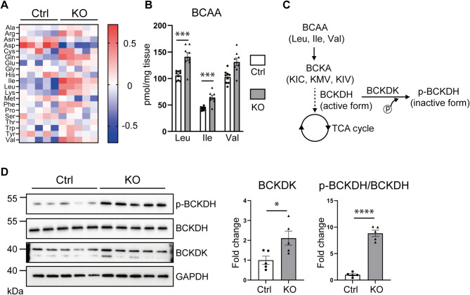
Results: A-PDK1KO mouse hearts exhibited hypertrophy with prominent insulin resistance, consistent with cardiac phenotypes and metabolic disturbances previously reported as DbCM characteristics. RNA sequencing revealed the activation of BCAA uptake in diabetic hearts. In addition, the key enzymes involved in cardiac BCAA catabolism were downregulated at the protein level in A-PDK1KO mice, leading to the accumulation of BCAAs in the heart. Mechanistically, the accumulation of the BCAA leucine caused cardiac hypertrophy via the activation of mammalian target of rapamycin complex 1 (mTORC1).

Conclusions: A-PDK1KO mice closely mimic the cardiac phenotypes and metabolic alterations observed in human DbCM and exhibit impaired BCAA metabolism in the heart. This model may contribute to a better understanding of DbCM pathophysiology and to the development of novel therapies for this disease.

Keywords: Branched-chain amino acid; Cardiac metabolism; Diabetes mellitus; Diabetic cardiomyopathy; Heart failure.


Conflict of interest statement

Declarations. Ethics approval and consent to participate: All animal studies were conducted in accordance with institutional guidelines and the Guide for the Care and Use of Laboratory Animals after receiving the approval of Kobe University. Consent for publication: Not applicable. Competing interests: The Division of Evidence-based Laboratory Medicine, Kobe University Graduate School of Medicine, was established by an endowment fund from Sysmex Corporation, Japan.

Figures

Fig. 1
Fig. 1
A-PDK1KO mice exhibited impaired glucose utilization in the heart. A Plasma glucose concentrations of control mice and A-PDK1KO mice (n = 10, each group). B–D Body weights B, lean mass C, and fat mass D of 14-week-old mice were measured (n = 7, each group). E Representative images of Western blots of heart tissue and quantification of the band intensity in mice intraperitoneally injected with 0.9% saline or 5 U/kg insulin after a 4-hour fast (n = 8, each group). The data are shown as means ± SEs. Significance was calculated by the unpaired Student’s t test. *p value < 0.05, **p value < 0.01, ***p value < 0.001, and ****p value < 0.0001. BW, body weight; Ctrl, control mice; KO, A-PDK1KO mice
Fig. 2
Fig. 2
A-PDK1KO mice exhibited cardiac hypertrophy. A Mouse heart weight (HW), heart weight-to-body weight ratio (HW/BW), and heart weight-to-tibia length ratio (HW/TL) (n = 8, each group). B Representative images of the whole heart. Scale bar: 10 mm. C Representative images of HE-stained heart sections (scale bar: 50 μm) and the cross-sectional area (CSA) of the myocardium quantified by ImageJ software and normalized to the average of the control group (n = 7 per group). D Representative images of M-mode echocardiography. E Interventricular septum thickness at diastole (IVSd), posterior wall thickness at diastole (PWd), relative wall thickness (RWT), and fractional shortening (%FS) were determined by echocardiography (n = 10 in each group). F Gene expression of Nppa, Nppb, and Myh7 quantified by qPCR and normalized to that of GAPDH (n = 6 per group). The data are shown as means ± SEs. Significance was calculated by the unpaired Student’s t test. *p value < 0.05, **p value < 0.01, ***p value < 0.001, and ****p value < 0.0001
Fig. 3
Fig. 3
RNA sequencing of heart tissue. A Principal component analysis (PCA) of RNA sequencing data for heart tissue from mice (n = 5 per group). B A heatmap of gene expression related to the indicated metabolic pathways in heart tissue was created from fragments per kilobase of transcript per million (FPKM) values and is illustrated as the log2-fold change (n = 5, each group)
Fig. 4
Fig. 4
Blood BCAA levels were increased in A-PDK1KO mice. A Heatmap of amino acid concentrations in mouse blood, shown as log2-fold changes. The values are normalized to the median of each amino acid concentration (n = 5, each group). B Blood concentrations of BCAAs (leucine (Leu), isoleucine (Ile), and valine (Val)) and BCKAs (α-keto-isocaproate (KIC), α-keto-β-methylvalerate (KMV), and α-keto-isovalerate (KIV)) were determined by LC‒MS (n = 6 per group). The data are shown as means ± SEs. Significance was calculated by the unpaired Student’s t test or the Mann–Whitney U test, as appropriate. *p value < 0.05, and **p value < 0.01
Fig. 5
Fig. 5
Cardiac BCAA metabolism was impaired in A-PDK1KO mice. A Heatmap of amino acid content in mouse heart tissue, shown as the log2-fold change. The values are normalized to the median content of each amino acid (n = 5, each group). B BCAA levels in mouse heart tissue were analyzed by LC‒MS (n = 8–9 per group). C Schematic diagram of the BCAA oxidation pathway. D Western blot analysis of BCKDK and phospho-BCKDH in the heart tissue of mice; the expression levels of BCKDK and phospho-BCKDH were normalized to those for GAPDH and BCKDH, respectively. (n = 5, each group). The data are shown as means ± SEs. Significance was calculated by the unpaired Student’s t test. *p value < 0.05, ***p value < 0.001, and ****p value < 0.0001. Leu, leucine; Ile, isoleucine; Val, valine; KIC, α-keto-isocaproate; KMV, α-keto-β-methylvalerate; and KIV, α-keto-isovalerate
Fig. 6
Fig. 6
Association between leucine-mTOR signaling and cardiac hypertrophy in A-PDK1KO mice. A Correlation between blood leucine levels and the heart weight-to-body weight ratio (HW/BW) in mice (Ctrl; n = 8, KO; n = 8). B Western blot analysis of phospho-S6K (p-p70-S6K) and S6K (p70-S6K) in the heart tissue of mice (n = 10, each group). The data are shown as means ± SEs. Significance was calculated by the unpaired Student’s t test. *p value < 0.05
Fig. 7
Fig. 7
BT2 treatment mitigated cardiac hypertrophy in A-PDK1KO mice. A Scheme of the experimental approach. 4–5-week-old A-PDK1KO mice were orally administered 40 mg/kg/day of BT2 or vehicle for 4 weeks. B Mouse heart weight (HW), heart weight-to-body weight ratio (HW/BW), and heart weight-to-tibia length ratio (HW/TL) (n = 8, each group). C Representative images of M-mode echocardiography. D Interventricular septum thickness at diastole (IVSd), posterior wall thickness at diastole (PWd), relative wall thickness (RWT), and fractional shortening (%FS) were measured by echocardiography (n = 8, each group). E BCAA content in the cardiac tissue or plasma of A-PDK1KO mice treated with BT2 or vehicle (n = 10, each group). F Western blot analysis of BCKDH and phospho-BCKDH in the heart tissue of the mice; the expression levels of phospho-BCKDH were normalized to those for BCKDH. (n = 6, each group). G Western blot analysis of phospho-S6K (p-p70-S6K) and S6K (p70-S6K) in the heart tissue of mice (n = 6, each group). The data are shown as means ± SEs. Significance was calculated by the unpaired Student’s t test or the Mann–Whitney U test, as appropriate. *p value < 0.05, **p value < 0.01, ***p value < 0.001, and ****p value < 0.0001
Fig. 8
Fig. 8
A schematic model of the underlying mechanism of cardiac hypertrophy in A-PDK1KO mice as a model of diabetic cardiomyopathy. BCAA, branched-chain amino acid; FFAs, free fatty acids; BCKDK, branched chain ketoacid dehydrogenase kinase; and mTORC1, mammalian target of rapamycin complex 1

References

    1. Pan KL, Hsu YC, Chang ST, Chung CM, Lin CL. The role of cardiac fibrosis in diabetic cardiomyopathy: from pathophysiology to clinical diagnostic tools. Int J Mol Sci 2023, 24(10). - PMC - PubMed
    1. Collaborators GBDD. Global, regional, and National burden of diabetes from 1990 to 2021, with projections of prevalence to 2050: a systematic analysis for the global burden of disease study 2021. Lancet. 2023;402(10397):203–34. - PMC - PubMed
    1. Tinajero MG, Malik VS. An update on the epidemiology of type 2 diabetes: a global perspective. Endocrinol Metab Clin North Am. 2021;50(3):337–55. - PubMed
    1. Kim AH, Jang JE, Han J. Current status on the therapeutic strategies for heart failure and diabetic cardiomyopathy. Biomed Pharmacother. 2022;145:112463. - PubMed
    1. Nichols GA, Hillier TA, Erbey JR, Brown JB. Congestive heart failure in type 2 diabetes: prevalence, incidence, and risk factors. Diabetes Care. 2001;24(9):1614–9. - PubMed

MeSH terms

Substances

LinkOut - more resources