. 2025 Apr;14(4):e70074.
doi: 10.1002/jev2.70074.

Yersinia pestis Actively Inhibits the Production of Extracellular Vesicles by Human Neutrophils

Affiliations

Yersinia pestis Actively Inhibits the Production of Extracellular Vesicles by Human Neutrophils

Katelyn R Sheneman et al. J Extracell Vesicles. 2025 Apr.

Abstract

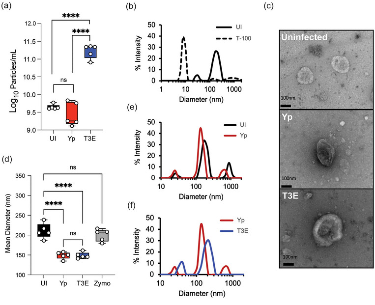
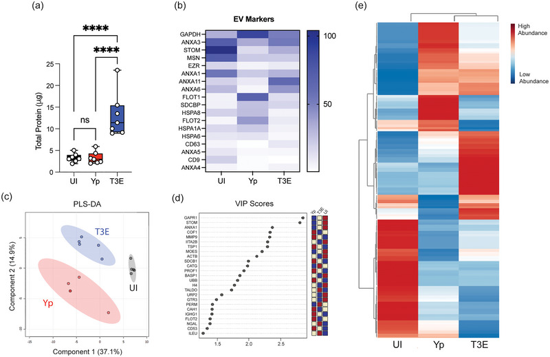
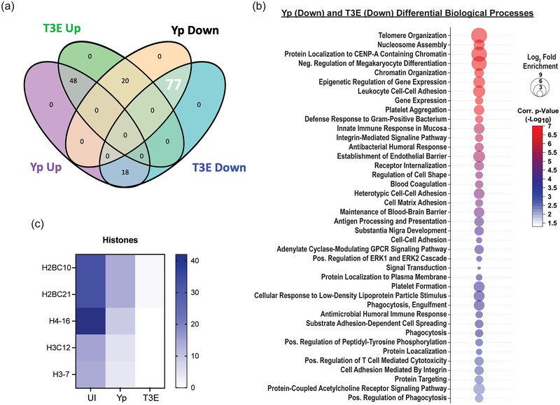
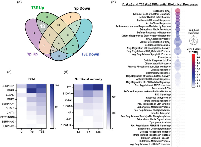
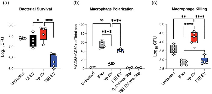
Yersinia pestis is the etiologic agent of the plague. A hallmark of plague is subversion of the host immune response by disrupting host signalling pathways required for inflammation. This non-inflammatory environment permits bacterial colonization and has been shown to be essential for disease manifestation. Previous work has shown that Y. pestis inhibits phagocytosis and degranulation by neutrophils. Manipulation of these key vesicular trafficking pathways suggests that Y. pestis influences extracellular vesicle (EV) secretion, cargo selection, trafficking and/or maturation. Our goals were to define the EV population produced by neutrophils in response to Y. pestis and determine how these vesicles might influence inflammation. Towards these goals, EVs were isolated from human neutrophils infected with Y. pestis or a mutant lacking bacterial effector proteins known to manipulate host cell signalling. Mass spectrometry data revealed that cargoes packaged in EVs isolated from mutant infected cells were enriched with antimicrobial and cytotoxic proteins, contents which differed from uninfected and Y. pestis infected cells. Further, EVs produced in response to Y. pestis lacked inflammatory properties observed in those isolated from neutrophils responding to the mutant. Together, these data demonstrate that Y. pestis actively inhibits the production of antimicrobial EVs produced by neutrophils, likely contributing to immune evasion.

Keywords: Yersinia pestis; Yop effectors; human neutrophils (hPMNs); plague; type 3 secretion system (T3SS).


Conflict of interest statement

The authors declare no conflicts of interest.

Figures

FIGURE 1
FIGURE 1
Characterization of EVs released by human neutrophils during Y. pestis interactions. hPMNs were infected with Y. pestis (Yp; red) or Y. pestis lacking the Yop effectors (T3E; blue) for 1 h prior to EV isolation via ultracentrifugation. (a) Total EV particle quantification via NTA. Each point represents EVs from an individual human donor. (b) Representative DLS analysis of EVs from uninfected hPMNs without (UI; solid black line) or with Triton‐X100 treatment (T‐100; dashed line). (c) Representative TEM images of EVs isolated from uninfected (UI), Yp, or T3E‐infected hPMNs. Original magnification ∼40,000x. (d) Average diameter of EVs isolated from uninfected, zymosan‐treated (Zymo), Yp‐infected, or T3E‐infected hPMNs. Each point represents EVs from an individual human donor. (e‐f) Representative DLS analysis of EVs isolated from UI, Yp, or T3E‐infected hPMNs. (a,d) One‐way ANOVA with Tukey; **** = p ≤ 0.0001. (b,e,f) Representative results of five independent donors. ns = not significant.
FIGURE 2
FIGURE 2
Protein content of EVs is altered by Y. pestis. hPMNs were infected with Y. pestis (Yp; red) or Y. pestis lacking the Yop effectors (T3E; blue) for 1 h prior to EV isolation via ultracentrifugation. (a) Protein quantification of indicated EVs. Each point represents EVs from an individual human donor. One‐way ANOVA with Tukey; **** = p ≤ 0.0001. (b) Average enrichment of recognized EV markers as measured by MS (n = 5). (c) Partial Least Squares Discriminant Analysis (PLS‐DA) plot depicting discriminant analysis of EVs isolated from uninfected (UI), Yp, or T3E‐infected hPMNs. (d) VIP scores contributing to the variance in (c). (e) Heat map depicting distribution of 307 proteins identified as associated with EVs isolated from uninfected (UI), Yp, or T3E‐infected hPMNs (average from five individual donors for each group). Proteins clustered according to Ward's Hierarchical Agglomerative Clustering Method (Alvarez‐Jiménez et al. 2018). ns = not significant.
FIGURE 3
FIGURE 3
EV proteins reduced in response to Y. pestis. (a) Venn diagram highlighting the 77 proteins (white) that were enriched in EVs from UI hPMNs (p < 0.05). (b) Biological process analysis predicted by DAVID () using the 77 enriched proteins (pathway enrichment displayed −Log10 p > 2.5; 1‐sided Student's t test). (c) Prevalence of histone proteins from EVs isolated from UI, Yp‐infected, or T3E‐infected EVs. ns indicates not significant.
FIGURE 4
FIGURE 4
EV proteins enriched in response to Y. pestis. (a) Venn diagram highlighting the 48 proteins (white) that were enriched in EVs from infected hPMNs (p < 0.05) compared to the UI EV population. (b) Biological process analysis predicted by DAVID (Ding et al. 2020) using the 48 enriched proteins (pathway enrichment displayed −Log10 p > 1.5; one‐sided Student's t test). Prevalence of ECM proteins (c) and nutritional immunity proteins (d) from EVs isolated from UI, Yp‐infected, or T3E‐infected EVs.
FIGURE 5
FIGURE 5
Y. pestis T3SS limits antimicrobial and proinflammatory protein packaging within EVs. (a) Venn diagram highlighting the 20 proteins (white) that were enriched in T3E EVs and subsequently reduced in Yp EVs (p < 0.05) compared to the UI EV population. (b) Biological process analysis predicted by DAVID (Ding et al. 2020) using the 20 dysregulated proteins (pathway enrichment displayed −Log10 p > 1.5; one‐sided Student's t test). (c) Fold change of the 20 identified proteins relative to UI. Prevalence of Annexins (d) and antimicrobial proteins (e) from EVs isolated from UI, Yp‐infected, or T3E‐infected EVs.
FIGURE 6
FIGURE 6
Yp‐elicited EVs have limited antimicrobial capacity. (a) Y. pestis was treated for 40 min with EVs isolated from UI, Yp‐infected, or T3E‐infected hPMNs and bacterial viability was determined by serial dilution and conventional colony forming unit (CFU) formation on agar plates. (b) hMDMs were treated with EVs isolated from Yp‐infected or T3E‐infected hPMNs or IFNγ for 24 h and M1 polarization was determined by flow cytometry. Yp EV‐free Sup and T3E EV‐free Sup represent cells treated with supernatants in which EVs were removed by filtration with 100 kDa filters. (c) hMDMs were treated with EVs isolated from Yp‐infected or T3E‐infected hPMNs or IFNγ for 24 h and subsequently infected with Yp at an MOI of 10. Intracellular bacterial survival was assessed with conventional gentamicin protection assay followed by CFU enumeration on agar plates. One‐way ANOVA with Tukey's post test; ns indicates not significant; * = p ≤ 0.05; ** = p ≤ 0.01; *** = p ≤ 0.001; **** = p ≤ 0.0001.
FIGURE 7
FIGURE 7
Yop effector proteins work cooperatively to manipulate EV protein packaging. hPMNs were infected with WT Y. pestis (Yp) or with a Y. pestis mutant expressing only YopE (E+), YopH (H+), or YopK (K+), or mixed at a 1:1 ratio for YopE and YopH (E+H), YopH and YopK (H+K), YopE and YopK (E+K). (a) Total protein in each EV sample was quantified and normalized to the T3E control to account for donor variability. One‐way ANOVA with Dunnett's multiple comparisons test to Yp with Geisser‐Greenhouse correction; ** = p ≤ 0.01; **** = p ≤ 0.0001 (n = 5–10). (b) PLS‐DA plot depicting discriminant analysis of EVs from one‐dimensional reverse phase liquid chromatography tandem mass spectrometry (n = 5).

Update of

Similar articles

Cited by

References

    1. Al Halawani, A. , Mithieux S. M., Yeo G. C., Hosseini‐Beheshti E., and Weiss A. S.. 2022. “Extracellular Vesicles: Interplay With the Extracellular Matrix and Modulated Cell Responses.” International Journal of Molecular Sciences 23, no. 6: 3389. - PMC - PubMed
    1. Alvarez‐Jiménez, V. D. , Leyva‐Paredes K., García‐Martínez M., et al. 2018. “Extracellular Vesicles Released From Mycobacterium Tuberculosis‐Infected Neutrophils Promote Macrophage Autophagy and Decrease Intracellular Mycobacterial Survival.” Frontiers in Immunology 9: 272. - PMC - PubMed
    1. Amjadi, M. F. , Avner B. S., Greenlee‐Wacker M. C., Horswill A. R., and Nauseef W. M.. 2021. “Neutrophil‐derived Extracellular Vesicles Modulate the Phenotype of Naïve human Neutrophils.” Journal of Leukocyte Biology 110: 917–925. - PMC - PubMed
    1. Andersson, K. , Magnusson K.‐E., Majeed M., Stendahl O., and FäLlman M.. 1999. “Yersinia Pseudotuberculosis‐induced Calcium Signaling in Neutrophils Is Blocked by the Virulence Effector YopH.” Infection and Immunity 67, no. 5: 2567–2574. - PMC - PubMed
    1. Andor, A. , Trulzsch K., Essler M., et al. 2001. “YopE of Yersinia, a GAP for Rho GTPases, Selectively Modulates Rac‐dependent Actin Structures in Endothelial Cells.” Cellular Microbiology 3, no. 5: 301–310. - PubMed

LinkOut - more resources