. 2025 Apr 16;22(1):101.
doi: 10.1186/s12985-025-02725-7.

Cytomegalovirus infection initiates inflammatory bowel disease by activating a positive MyD88/NF-κB feedback loop in allogeneic skin transplantation mice

Affiliations

Cytomegalovirus infection initiates inflammatory bowel disease by activating a positive MyD88/NF-κB feedback loop in allogeneic skin transplantation mice

Ming-Xian Chen et al. Virol J. .

Abstract

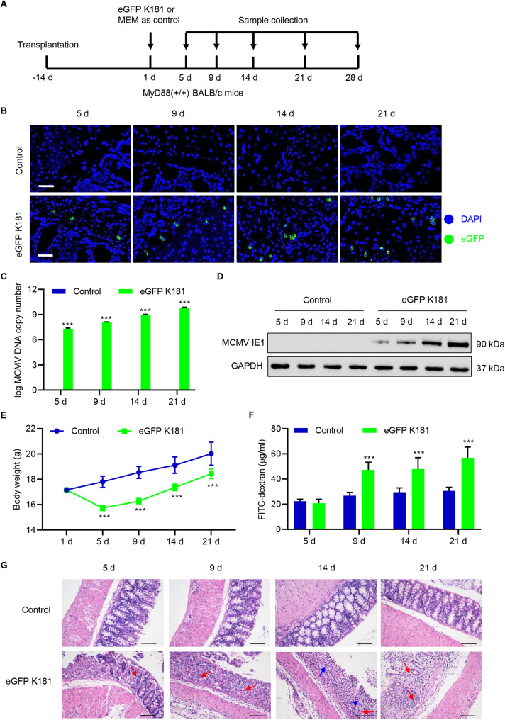
Infection with the cytomegalovirus (CMV) is common. Inflammatory bowel disease (IBD) is characterized by chronic inflammation in the gastrointestinal tract. CMV infection is involved in IBD pathogenesis. The abnormal activation of myeloid differentiation factor 88 (MyD88)/nuclear factor- kappa B (NF-κB) signaling, which results in inflammatory cytokine overexpression, is an important factor in IBD pathogenesis. The present study aimed to examine the effect of CMV infection on NF-κB activation and its role in IBD pathogenesis. Since BALB/c rather than C57BL/6 mice belong to the murine CMV (MCMV) susceptible strain, allogeneic skin transplantation was conducted between MyD88 (+/+) or MyD88-knockout Myd88 (-/-) BALB/c (recipient) mice and C57BL/6 (donor) mice. Thereafter, the immune function of the recipient mice was reduced by immunosuppressant cyclosporine, which is meaningful in the pathogenesis of IBD caused by MCMV in immunocompromised mice. MCMV strain K181-eGFP (eGFP K181) or hMIEP-eGFP K181 (knockout MCMV IE1-3 promoter) was used to infect MyD88 (+/+) BALB/c mice while eGFP K181 was also used to infect MyD88 (-/-) BALB/c mice on day 14 post allogeneic skin transplantation. MCMV DNA was detected via nested polymerase chain reaction. Hematoxylin-Eosin staining was used to assess colon necrosis and inflammatory cell infiltration. The serum levels of tumor necrosis factor-alpha, interleukin (IL)-1β, IL-6, IL-8, IL-12, flagellin, lipopolysaccharide, and myeloperoxidase were detected by ELISA and immune reaction. Immunoblots were applied to measure protein levels. eGFP K181 infection significantly induced colon permeability, necrosis, inflammatory cell infiltration, and inflammation in allogeneic skin transplantation mice. hMIEP-eGFP K181 infection significantly inhibited colon permeability, necrosis, inflammatory cell infiltration, and inflammation compared with eGFP K181 infection in allogeneic skin transplantation mice. Moreover, the MyD88-dependent NF-κB signaling pathway was involved in the pathogenesis of eGFP K181-induced colon permeability and inflammation in allogeneic skin transplantation mice. Our findings highlight the importance of CMV infection and the Myd88/NF-κB signaling pathway in IBD and might provide a new direction for the development of drugs for IBD.

Keywords: Cytomegalovirus; Immediate-early protein; Inflammatory bowel disease; Inflammatory cytokines; Myd88/NF-κB.


Conflict of interest statement

Declarations. Ethics approval and consent to participate: All the experimental protocols were approved by the Laboratory Animal Ethics Committee of the Zhejiang Acedemy of Traditional Chinese Medicine [no. (2021) 016]. Consent for publication: Not applicable. Competing interests: The authors declare no competing interests.

Figures

Fig. 1
Fig. 1
Effect of eGFP K181 infection on colon barrier permeability in allogeneic skin transplantation mice. 1 × 105 PFU eGFP K181 were inoculated intranasally into allogeneic skin transplantation BALB/c mice on day 14 posttransplantation. (A) A schematic representation of the assay indicating time points of transplantation and eGFP K181 infection. (B) MCMV infection in colon tissues was confirmed by immunofluorescence with positive eGFP (Scale bar: 50 μm); (C) MCMV DNA and (D) IE1 levels in colon tissues were measured by PCR and Western blot, respectively; (E) Body weight and (F) colon barrier permeability were measured; (G) Necrosis (blue arrow) and inflammatory cell infiltration (red arrow) of colon tissues at 5, 9, 14, or 21 d postinfection (Scale bar: 100 μm). Data are shown as the mean ± SD of six mice per group. *** P < 0.001 vs. control
Fig. 2
Fig. 2
Effect of eGFP K181 infection on the NF-κB signaling pathway in allogeneic skin transplantation mice. 1 × 105 PFU eGFP K181 were inoculated intranasally into allogeneic skin transplantation BALB/c mice on day 14 posttransplantation. The serum levels of (A) TNF-α, (B) IL-1β, (C) IL-6, (D) IL-8, and (E) IL-12 were measured by ELISA; (F) The expression levels of IKKα, IKKβ, p65, p52, TRIF, MyD88, TRAF6, and TAK1 in colon tissues were measured by Western blot at 5, 9, 14, or 21 d postinfection. (G) The levels of anti-flagellin IgG and anti-LPS IgG in serum and MPO in colon tissues were measured at 28 d postinfection. Data are shown as the mean ± SD of six mice per group. * P < 0.05, ** P < 0.01, *** P < 0.001 vs. control
Fig. 3
Fig. 3
Effect of hMIEP-eGFP K181 infection on colon barrier permeability in allogeneic skin transplantation mice. 1 × 105 PFU hMIEP-eGFP K181 or eGFP K181 were inoculated intranasally into allogeneic skin transplantation BALB/c mice on day 14 posttransplantation. (A) A schematic representation of the assay indicating time points of transplantation and MCMV infections. (B) MCMV infection in colon tissues was confirmed by immunofluorescence with positive eGFP (Scale bar: 50 μm); (C) MCMV DNA and (D) IE1 levels in colon tissues were measured by PCR and Western blot, respectively; (E) Body weight and (F) colon barrier permeability were measured; (G) Necrosis (blue arrow) and inflammatory cell infiltration (red arrow) of colon tissues at 5, 9, 14, or 21 d postinfection (Scale bar: 100 μm). Data are presented as the mean ± SD of six mice per group. *** P < 0.001 vs. eGFP K181
Fig. 4
Fig. 4
Effect of hMIEP-eGFP K181 infection on the NF-κB signaling pathway in allogeneic skin transplantation mice. 1 × 105 PFU hMIEP-eGFP K181 or eGFP K181 were inoculated intranasally into allogeneic skin transplantation BALB/c mice on day 14 posttransplantation. The serum levels of (A) TNF-α, (B) IL-1β, (C) IL-6, (D) IL-8, and (E) IL-12 were measured by ELISA; (F) The expression levels of IKKα, IKKβ, p65, p52, TRIF, MyD88, TRAF6, and TAK1 in colon tissues were measured by Western blot at 5, 9, 14, or 21 d postinfection. Data are shown as the mean ± SD of six mice per group. * P < 0.05, *** P < 0.001 vs. eGFP K181
Fig. 5
Fig. 5
Effect of eGFP K181 infection on colon barrier permeability in allogeneic skin transplantation mice. 1 × 105 PFU eGFP K181 were inoculated intranasally into allogeneic skin transplantation MyD88(−/−) and MyD88(+/+) BALB/c mice on day 14 posttransplantation. (A) A schematic representation of the assay indicating time points of transplantation and eGFP K181 infection. (B) MCMV infection in colon tissues was confirmed by immunofluorescence with positive eGFP (Scale bar: 50 μm); (C) MCMV DNA and (D) IE1 levels in colon tissues were measured by PCR and Western blot, respectively; (E) Body weight and (F) colon barrier permeability were measured; (G) Necrosis (blue arrow) and inflammatory cell infiltration (red arrow) of colon tissues at 5, 9, 14, or 21 d postinfection (Scale bar: 100 μm). Data are shown as the mean ± SD of six mice per group. * P < 0.05, *** P < 0.001 vs. Myd88(+/+)
Fig. 6
Fig. 6
Effect of eGFP K181 infection on colon inflammation and the NF-κB signaling pathway in allogeneic skin transplantation mice. 1 × 105 PFU eGFP K181 were inoculated intranasally into allogeneic skin transplantation MyD88(−/−) and MyD88(+/+) BALB/c mice on day 14 posttransplantation. The serum levels of (A) TNF-α, (B) IL-1β, (C) IL-6, (D) IL-8, and (E) IL-12 were measured by ELISA; (F) The expression levels of IKKα, IKKβ, p65, p52, TRIF, MyD88, TRAF6, and TAK1 in colon tissues were measured by Western blot at 5, 9, 14, or 21 d postinfection. (G) The levels of anti-flagellin IgG and anti-LPS IgG in serum and MPO in colon tissues were measured at 28 d postinfection. Data are shown as the mean ± SD of six mice per group. ** P < 0.01, *** P < 0.001 vs. Myd88(+/+)
Fig. 7
Fig. 7
A graphical summary of the study. MCMV infection initiates IBD by activating a positive MyD88/NF-κB feedback loop in allogeneic skin transplantation mice

References

    1. Lv YL, Han FF, Jia YJ, Wan ZR, Gong LL, Liu H, et al. Is cytomegalovirus infection related to inflammatory bowel disease, especially steroid-resistant inflammatory bowel disease? A meta-analysis. Infect Drug Resist. 2017;10:511–9. 10.2147/IDR.S149784. PubMed PMID: 29276397; PubMed Central PMCID: PMC5733908. - PMC - PubMed
    1. Seyedian SS, Nokhostin F, Malamir MD. A review of the diagnosis, prevention, and treatment methods of inflammatory bowel disease. J Med Life. 2019;12(2):113–22. PubMed PMID: 31406511; PubMed Central PMCID: PMC6685307. doi: 10.25122/jml-2018-0075. - PMC - PubMed
    1. Shapiro JM, Subedi S, LeLeiko NS. Inflammatory Bowel Disease. Pediatrics in review. 2016;37(8):337–47. 10.1542/pir.2015-0110. PubMed PMID: 27482063. - PubMed
    1. Collaborators GBDIBD, Global Burden of Disease Study 2017. The global, regional, and National burden of inflammatory bowel disease in 195 countries and territories, 1990–2017: a systematic analysis for the. Lancet Gastroenterol Hepatol. 2020;5(1):17–30. 10.1016/S2468-1253(19)30333-4. PubMed PMID: 31648971; PubMed Central PMCID: PMC7026709. - PMC - PubMed
    1. Gatherer D, Depledge DP, Hartley CA, Szpara ML, Vaz PK, Benkő M, et al. ICTV virus taxonomy profile: herpesviridae 2021. J Gen Virol. 2021;102(10). 10.1099/jgv.0.001673. PubMed PMID: 34704922; PubMed Central PMCID: PMCPMC8604186. Epub 2021/10/28. - PMC - PubMed

Publication types

MeSH terms