Multicenter Study
. 2025 Apr;32(4):e70128.
doi: 10.1111/ene.70128.

An Investigation of Corticospinal Tract Microstructural Integrity in ARSACS Using a Profilometry MRI Analysis: Results From the PROSPAX Study

Collaborators, Affiliations
Multicenter Study

An Investigation of Corticospinal Tract Microstructural Integrity in ARSACS Using a Profilometry MRI Analysis: Results From the PROSPAX Study

Alessandra Scaravilli et al. Eur J Neurol. 2025 Apr.

Abstract

Background: Spasticity represents a core clinical feature of Autosomal Recessive Spastic Ataxia of Charlevoix-Saguenay (ARSACS) patients. Nonetheless, its pathophysiological substrate is poorly investigated. We assessed the microstructural integrity of the corticospinal tract (CST) using diffusion MRI (dMRI) via profilometry analysis to understand its possible role in the development of spasticity in ARSACS.

Materials and methods: In this multi-center prospective study, data of 37 ARSACS (M/F = 21/16; 33.4 ± 12.4 years) and 29 controls (M/F = 13/16; 42.1 ± 17.2 years) acquired within the PROSPAX consortium were collected from January 2021 to October 2022 and analyzed. Differences in terms of global CST microstructural integrity were probed, as well as a possible spatial distribution of the damage along the tract via profilometry analysis. Possible correlations between clinical severity, including the Spastic Paraplegia Rating Scale (SPRS), were also tested.

Results: A significant global involvement of the CST was found in ARSACS compared to controls (all tests with p < 0.001), with a spatially defined pattern of more pronounced microstructural integrity loss occurring right below and above the pons, a structure that was also confirmed to be thickened in these patients (p < 0.001). A bilateral negative correlation emerged between the microstructural integrity of the CST and clinical indices of spasticity expressed via SPRS (p = 0.02 for both CSTs).

Conclusion: A clinically meaningful microstructural involvement of CST is present in ARSACS patients, with a spatially defined pattern of damage occurring right below and above a thickened pons. An evaluation of the microstructure of this bundle might serve as a possible biomarker in this condition.

Keywords: ARSACS; ataxia; corticospinal tract; diffusion tensor imaging; magnetic resonance imaging.


Conflict of interest statement

S.C. have received fees for Advisory Board by Amicus. All other co‐authors declare no conflicts of interest.

Figures

FIGURE 1
FIGURE 1
Study flowchart. Flowchart showing how the final number of subjects analyzed in this study was reached after the application of inclusion and exclusion criteria. ARSACS = Autosomal Recessive Spastic Ataxia of Charlevoix‐Saguenay, HC = healthy controls.
FIGURE 2
FIGURE 2
Differences between groups in terms of mean microstructure of the CST. Violin plots showing the differences between ARSACS (orange) and HC (green) in terms of CST mean microstructural metrics. ARSACS = Autosomal Recessive Spastic Ataxia of Charlevoix‐Saguenay, CST = corticospinal tract, FA = fractional anisotropy, HC = healthy controls, MD = mean diffusivity, RD = radial diffusivity, NDIw = neurite density index.
FIGURE 3
FIGURE 3
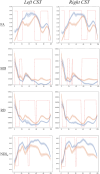
Results of the profilometry analysis. Results of the profilometry analysis showing the spatial distribution of microstructural damage along the CST (x‐axis, ranging from 0‐pyramids‐ to 100‐cortex) in ARSACS (orange lines) and HC (blue lines). Red lines indicate where, along the CST, significant differences between groups occurs. ARSACS = Autosomal Recessive Spastic Ataxia of Charlevoix‐Saguenay, CST = corticospinal tract, FA = fractional anisotropy, HC = healthy controls, MD = mean diffusivity, RD = radial diffusivity, NDIw = neurite density index.
FIGURE 4
FIGURE 4
Localization and the degree of CST microstructural damage. Representation of the spatial profile and magnitude of microstructural damage affecting the CST using a color‐coded scale weighted by Cohen‐d values. CST = corticospinal tract.
FIGURE 5
FIGURE 5
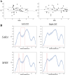
Results of the correlation analyses between microstructural changes and clinical scores in ARSACS. In (A), scatterplots of the correlation analyses between mean FA value along the entire CST (y‐axes) and SARA (left) and SPRS (right) scores (x‐axes) showing the absence of a significant correlation when this approach was used. In (B) results of the correlation analyses using a profilometry approach between FA values along the CST of ARSACS subjects and SARA (upper row) and SPRS scores (lower row) showing the occurrence of a significant and spatially distributed correlation between CST microstructure and clinical scores. Red lines indicate significant correlations between the microstructural damage and the corresponding clinical variable along the CST (x‐axis, ranging from 0‐pyramids‐ to 100‐cortex). ARSACS = Autosomal Recessive Spastic Ataxia of Charlevoix‐Saguenay, CST = corticospinal tract, FA = fractional anisotropy, SARA = Scale for the Assessment and Rating of Ataxia, SPRS = Spastic Paraplegia Rating Scale.

Similar articles

References

    1. Engert J. C., Bérubé P., Mercier J., et al., “ARSACS, a Spastic Ataxia Common in Northeastern Québec, Is Caused by Mutations in a New Gene Encoding an 11.5‐Kb ORF,” Nature Genetics 24, no. 2 (2000): 120–125, 10.1038/72769. - DOI - PubMed
    1. Baets J., Deconinck T., Smets K., et al., “Mutations in SACS Cause Atypical and Late‐Onset Forms of ARSACS,” Neurology 75, no. 13 (2010): 1181–1188, 10.1212/WNL.0b013e3181f4d86c. - DOI - PubMed
    1. Fogel B. L. and Perlman S., “Clinical Features and Molecular Genetics of Autosomal Recessive Cerebellar Ataxias,” Lancet Neurology 6, no. 3 (2007): 245–257, 10.1016/S1474-4422(07)70054-6. - DOI - PubMed
    1. Bouhlal Y., Amouri R., El Euch‐Fayeche G., and Hentati F., “Autosomal Recessive Spastic Ataxia of Charlevoix–Saguenay: An Overview,” Parkinsonism & Related Disorders 17, no. 6 (2011): 418–422, 10.1016/j.parkreldis.2011.03.005. - DOI - PubMed
    1. Vermeer S., Meijer R. P. P., Pijl B. J., et al., “ARSACS in the Dutch Population: A Frequent Cause of Early‐Onset Cerebellar Ataxia,” Neurogenetics 9, no. 3 (2008): 207–214, 10.1007/s10048-008-0131-7. - DOI - PMC - PubMed

Publication types

Supplementary concepts

LinkOut - more resources