Review
. 2025 Apr 17;6(6):1040-1049.
doi: 10.34067/KID.0000000820.

The Pathologic Actions of Phosphate in CKD

Affiliations
Review

The Pathologic Actions of Phosphate in CKD

Abul Fajol et al. Kidney360. .

Abstract

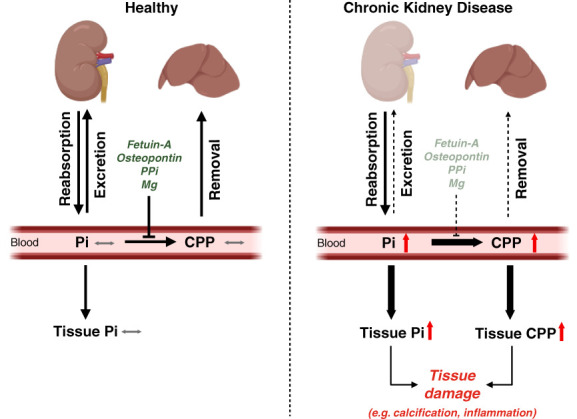
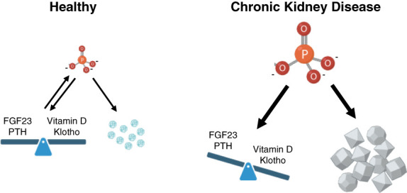
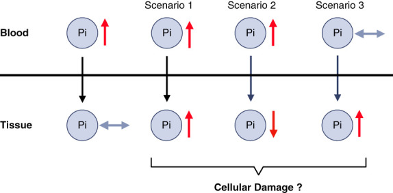
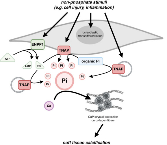
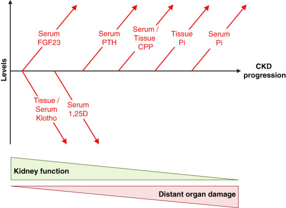
CKD is associated with high serum levels of phosphate (also called hyperphosphatemia), which is a main driver of soft tissue calcifications and potentially other pathologic changes that are associated with CKD. However, it remains unclear in what form and through which mechanisms and targets elevated phosphate can damage cells and tissues. Rises in serum phosphate levels are accompanied by changes in the endocrine regulators of phosphate metabolism and result in the formation of calcium-phosphate crystals, and all three events can have pathologic actions on various tissues. Furthermore, tissues can accumulate phosphate from the circulation, and cells can generate free phosphate in their environment independently from circulating phosphate, which both result in local elevations of phosphate that could also contribute to tissue damage. It is important to better understand the various scenarios underlying the pathologic actions of hyperphosphatemia, as some of them suggest that measuring extracellular serum phosphate, which is the gold standard to estimate overall phosphate status of the body, is not sufficient to do so. Understanding the pathologic actions of phosphate on a conceptual level should not only help to design more efficient detection tools for phosphate but also to identify phosphate-induced pathomechanisms which could provide us with novel drug targets to tackle phosphate-driven pathologies in CKD. Here, we discuss the different concepts and scenarios that could underlie the widespread pathologic actions of hyperphosphatemia in CKD.

Trial registration: ClinicalTrials.gov NCT04095039 NCT03573089.

Keywords: CKD; hyperphosphatemia; vascular calcification.


Conflict of interest statement

Disclosure forms, as provided by each author, are available with the online version of the article at http://links.lww.com/KN9/B41.

Figures

Figure 1
Figure 1
The regulation and effects of Pi and CPPs in CKD. In heathy conditions, renal excretion and reabsorption of inorganic Pi maintain blood Pi at normal levels. Thereby, tissues receive sufficient Pi to maintain cellular functions and structures, while tissue Pi elevations with potential cytotoxic effects are avoided. Fetuin-A, osteopontin, PPi, and Mg prevent the formation of calcium-Pi crystals in the blood and in soft tissues and thereby protect from ectopic calcifications in scenarios of systemic Pi elevations, for example after food intake. Fetuin-A binds calcium and Pi to form CPP, which are quickly removed from the circulation by the liver and are not deposited in tissues. In CKD, reduced renal excretion causes an elevation in inorganic Pi levels in the blood, which might result in increased Pi levels in tissues. CKD is also accompanied by reduced levels of fetuin-A, osteopontin, PPi, and Mg, which together with high Pi levels and reduced hepatic clearance capacity might cause the appearance of CPPs in the blood and eventually in tissues. The accumulation of inorganic Pi and CPPs in tissues and cells induces various pathologic changes, such as local calcification and inflammation. CPP, calciprotein particle; Mg, magnesium; Pi, phosphate; PPi, pyrophosphate.
Figure 2
Figure 2
The potential culprits in CKD-associated hyperphosphatemia. Pi homeostasis is regulated by the hormones, FGF23, PTH, and vitamin D and by the FGF23 coreceptor klotho. Together, these factors maintain blood levels of inorganic Pi (PO43−) in a physiologic range at which Pi cannot crystalize. Instead, Pi and calcium form amorphous particles which can be removed from the circulation. In CKD, reduced renal excretion causes an elevation in inorganic Pi levels in the blood, which results in changes in the endocrine regulators with the goal to maintain Pi homeostasis. With disease progression, the endocrine system eventually fails, leading to highly elevated blood levels of inorganic Pi which together with calcium forms crystals in the blood and in soft tissues. One of the key questions in the field is whether elevated inorganic Pi, the unbalanced endocrine regulators of Pi homeostasis and/or the appearance of calcium-Pi crystals cause tissue damage, such as ectopic calcifications, that occur in CKD. Defining the precise culprit(s) will be important to better understand the underlying pathomechanisms and to identify novel drug targets. FGF23, fibroblast growth factor 23; PTH, parathyroid hormone.
Figure 3
Figure 3
The relationship between Pi levels in blood versus tissue. It is important that tissues have the capacity to maintain normal levels of inorganic Pi, even if Pi levels in the blood rise, for example, after food intake. However, very high and prolonged elevations of circulating Pi, as in CKD, seem to affect Pi tissue content in different ways. High blood Pi might result in high tissue Pi (scenario 1), as the case in the calcified vasculature, and most likely in all calcifying tissues, as well as in the lung. High blood Pi can also result in low tissue Pi (scenario 2), as described for skeletal muscle. Finally, some tissues might increase Pi content before a rise in blood Pi (scenario 3), which seems to be the case for the liver. Overall, different tissues seem to show different responses to elevations in blood Pi, which might also depend on the pathologic context. Furthermore, rises in tissue Pi, as the case in scenarios 1 and 3 might not have pathologic effects, at least not initially, but aim to temporarily store Pi or to remove Pi from the system. It is important to note that in scenarios 2 and 3, Pi levels in the blood do not reflect changes in tissue Pi content.
Figure 4
Figure 4
The regulation of local Pi levels. Cells seem to be able to regulate the concentration of inorganic Pi in their extracellular environment by expressing enzymes on their surface that can generate Pi. This includes ENPP1 which generates PPi and AMP from ATP, and TNAP, which then hydrolyzes PPi to generate free Pi. TNAP can remove Pi from various substrates, including several forms of organic Pi. Cells can also release EV that contain TNAP which generates free Pi within the extracellular matrix. The relevance of this mechanism under physiologic conditions is not understood. If uncontrolled and accompanied by the activation of osteogenic gene programs, elevated Pi and calcium form crystals that are deposited on collagen fibers and cause tissue calcifications. Importantly, this cellular process to increase local Pi content is not initiated by elevations in blood Pi levels, but by other pathologic stimuli. Ca, calcium; CaPi, calcium-phosphate; ENPP1, ectonucleotide pyrophosphatase/phosphodiesterase 1; EV, extracellular vesicle; Pi, phosphate; TNAP, tissue-nonspecific alkaline phosphatase.
Figure 5
Figure 5
The changes in Pi metabolism in the course of CKD progression. With declining kidney function, the endocrine regulators of Pi metabolism undergo significant changes in their serum levels to compensate for the reduced capacity of renal Pi excretion with the goal to keep systemic Pi concentrations in a normal range. The levels of FGF23 and klotho seem to be altered first, followed by changes in active vitamin D (1,25D) and PTH. The serum levels of inorganic Pi only increase in very late stages of CKD, raising the question whether Pi can contribute to distant organ damage that is initiated early in CKD. While the described alterations in Pi metabolism are well established, more recent research has shown that Pi-containing CPP appear early in the blood. Furthermore, CPPs and inorganic Pi might accumulate in tissues before a rise in serum Pi levels. It is possible that these earlier elevations in CPPs and in tissue Pi content contribute to CKD-associated tissue damage.

Similar articles

Cited by

References

    1. Razzaque MS. Phosphate toxicity: new insights into an old problem. Clin Sci (Lond). 2011;120(3):91–97. doi: 10.1042/CS20100377 - DOI - PMC - PubMed
    1. Vervloet MG Sezer S Massy ZA, et al. The role of phosphate in kidney disease. Nat Rev Nephrol. 2017;13(1):27–38. doi: 10.1038/nrneph.2016.164 - DOI - PubMed
    1. Virkki LV, Biber J, Murer H, Forster IC. Phosphate transporters: a tale of two solute carrier families. Am J Physiol Renal Physiol. 2007;293(3):F643–F654. doi: 10.1152/ajprenal.00228.2007 - DOI - PubMed
    1. Hruska KA, Mathew S, Lund R, Qiu P, Pratt R. Hyperphosphatemia of chronic kidney disease. Kidney Int. 2008;74(2):148–157. doi: 10.1038/ki.2008.130 - DOI - PMC - PubMed
    1. Chande S, Bergwitz C. Role of phosphate sensing in bone and mineral metabolism. Nat Rev Endocrinol. 2018;14(11):637–655. doi: 10.1038/s41574-018-0076-3 - DOI - PMC - PubMed

MeSH terms

Associated data