. 2025 May 6;482(9):413-432.
doi: 10.1042/BCJ20240473.

Single-molecule observations of human small heat shock proteins in complex with aggregation-prone client proteins

Affiliations

Single-molecule observations of human small heat shock proteins in complex with aggregation-prone client proteins

Lauren Rice et al. Biochem J. .

Abstract

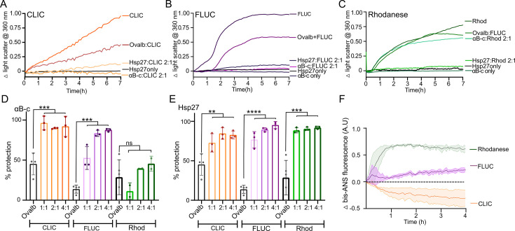
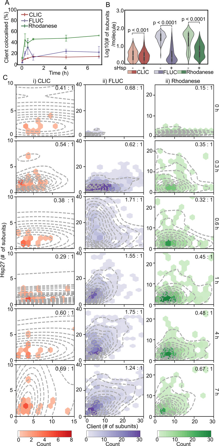
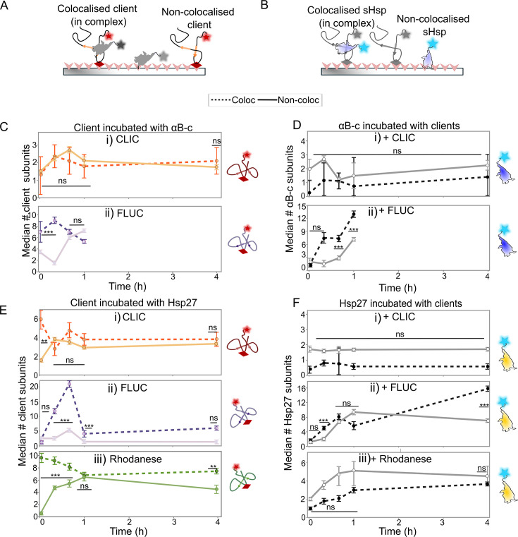
Small heat shock proteins (sHsps) are molecular chaperones that act to prevent the aberrant aggregation of misfolded proteins. Whilst it is suggested that sHsps prevent aggregation by binding to misfolded client proteins, the dynamic and heterogeneous nature of sHsps has hindered attempts to establish the mechanistic details of how sHsp-client protein complexes form. Single-molecule approaches have emerged as a powerful tool to investigate dynamic and heterogeneous interactions such as those that can occur between sHsps and their client proteins. Here, we use total internal reflection fluorescence microscopy to observe and characterise the complexes formed between model aggregation-prone client proteins (firefly luciferase, rhodanese and chloride intracellular channel 1 protein), and the human sHsps αB-crystallin (αB-c; HSPB5) and Hsp27 (HSPB1). We show that small (monomeric or dimeric) forms of both αB-c and Hsp27 bind to misfolded or oligomeric forms of the client proteins at early stages of aggregation, resulting in the formation of soluble sHsp-client complexes. Stoichiometric analysis of these complexes revealed that additional αB-c subunits accumulate onto pre-existing sHsp-client complexes to form larger species - this does not occur to the same extent for Hsp27. Instead, Hsp27-client interactions tend to be more transient than those of αB-c. Elucidating these mechanisms of sHsp function is crucial to our understanding of how these molecular chaperones act to inhibit protein aggregation and maintain cellular proteostasis.

Keywords: Hsp27; molecular chaperones; photobleaching; proteostasis; single-molecule fluorescence; αB-crystallin.


Conflict of interest statement

The authors declare that there are no conflicts associated with the manuscript.

Figures

Figure 1
Figure 1. Differences in the capacity of the human sHsps, αB-c and Hsp27 to inhibit the heat-induced amorphous aggregation of client proteins.
(A-C) Example of light scatter traces from the heat-induced aggregation of the client proteins. Recombinant client protein (A-CLIC at 30 µM, B-FLUC at 4 µM or C-rhodanese at 4 µM) was incubated at 42°C for 7 hours in the presence or absence of a 2:1 molar ratio (sHsp:client) of either αB-c or Hsp27, or the control protein ovalbumin (ovalb), and the change in light scatter at 360 nm over time monitored. Data shown are the normalised change in light scatter. (D-E) The percent reduction in aggregation (light scatter) of CLIC, FLUC and rhodanese afforded by varying molar ratios of αB-c (D) and Hsp27 (E), and the control protein ovalbumin at the highest molar ratio tested (4:1, ovalb:client). Data reported are the average ± S.D. of three independent experiments. Data were analysed by one-way ANOVA and Tukey’s post-hoc test, where ****, *** and ** indicates P<0.0001, 0.001 and 0.01, respectively; ns indicates P>0.05. (F) Exposed hydrophobicity of client proteins was monitored by bis-ANS fluorescence. CLIC, FLUC and rhodanese (200 nM) were incubated at 42°C for 4 hours in the presence of 20 µM bis-ANS; the change in fluorescence emission from bis-ANS at 480 nm was monitored over time. Data are reported as the mean ± S.E. of three independent experiments. CLIC, chloride intracellular channel 1; FLUC, firefly luciferase; sHsps, small heat shock proteins.
Figure 2
Figure 2. The number of αB-c subunits within sHsp–client protein complexes increases over time in a client-dependent manner.
αB-c-AF488 and CLIC- or FLUC- AF647 were incubated (42°C for up to 7 hour) in the absence or presence of αB-c (2:1 molar ratio; αB-c:client); aliquots were taken at time points throughout the incubation. Following incubation, samples were immediately cross-linked, diluted and incubated in flow cells for 15 min before imaging using TIRF microscopy. (A) Schematic depicting the workflow used to identify sHsp–client complexes formed during incubation. Representative TIRF microscopy image shows the fluorescence emission from FLUC-AF647 and αB-c-AF488 when incubated together, observed in separate emission channels. Overlaying these channels (‘Merge’) allows identification of colocalised (white arrows) and non-colocalised molecules. (B) The percentage (%) of CLIC-AF647 (red) or FLUC-AF647 (purple) molecules colocalised with αB-c-AF488 at each time point, reported as the mean ± S.D. of three independent replicates. (C) Violin plots show the distributions of CLIC-AF647 (red) and FLUC-AF647 (purple) molecule sizes (log10 number of subunits/molecule) after 1 hour (for FLUC) or 4 hours (for CLIC) incubation with (‘+’) or without (‘-’) αB-c-AF488. Molecule size reported includes all molecules (i.e. both colocalised and non-colocalised with αB-c). Results include measurements from three independent experiments and, where marked, statistical comparisons between distributions was performed via Kruskal-Wallis test for multiple comparisons with Dunn’s procedure (p values indicated). (D) Hexbin plots show the relative abundance of protein subunits and median molar ratio (sHsp:client) (inset) of αB-c-AF488 (y-axis) and CLIC-AF647 (i) or FLUC-AF647 (ii) (x-axis) within complexes at each time point during incubation. Each hexbin plot is overlaid with the kernel probability density (dashed line) of complexes at each time point. The depth of colour in each hexbin indicates the number of molecules in that bin, the scale for each plot is indicated by the respective colour bar. Data shown are from all molecules in complexes measured across the three independent experiments. CLIC, chloride intracellular channel 1; FLUC, firefly luciferase; sHsps, small heat shock proteins.
Figure 3
Figure 3. The number of subunits of Hsp27 within complexes increases in a client-dependent manner.
Hsp27-AF488 and the client proteins (CLIC-, FLUC- or rhodanese-AF647) were incubated (42°C for up to 7 hour) in the absence or presence of Hsp27 (2:1 molar ratio, Hsp27:client); aliquots were taken at time points throughout the incubation. Following incubation, samples were immediately cross-linked, diluted and incubated in flow cells for 15 min before imaging using TIRF microscopy. (A) The percentage (%) of CLIC-AF647 (red), FLUC-AF647 (purple) or rhodanese-AF647 (green) molecules colocalised with Hsp27-AF488 at each time point, reported as the mean ± S.D. of three independent replicates . (B) Violin plots show the distributions of CLIC-AF647 (red), FLUC-AF647 (purple) and rhodanese-AF647 (green) molecule sizes (log10 number of subunits/molecule) after 7-hour incubation with (‘+’) or without (‘-’) Hsp27-AF488. Molecule size reported includes all molecules (i.e. both colocalised and non-colocalised with Hsp27). Results include measurements from three independent experiments and, where marked, statistical comparisons between distributions was performed via Kruskal-Wallis test for multiple comparisons with Dunn’s procedure (p values indicated). (C) Hexbin plots show the relative abundance of protein subunits and median molar ratio (sHsp:client) (inset) of Hsp27-AF488 (y-axis) and each of the clients (CLIC-AF647 (i), FLUC-AF647 (ii) and rhodanese-AF647 (iii)) (x-axis) within complexes at each time point during incubation. Each hexbin plot is overlaid with the kernel probability density (dashed line) of complexes at each time point. The depth of colour in each hexbin indicates the number of molecules in that bin, the scale for each plot is indicated by the respective colour bar. Data shown are from all molecules in complexes measured across the three independent experiments. CLIC, chloride intracellular channel 1; FLUC, firefly luciferase; sHsps, small heat shock proteins.
Figure 4
Figure 4. sHsps maintain client proteins in small oligomeric states.
AF488-labelled sHsps (αB-c or Hsp27) were incubated with the AF647-labelled client proteins (CLIC, FLUC or rhodanese) (42°C for 4 hour, 2:1 molar ratio) and aliquots were taken throughout the incubation. Samples were immediately cross-linked, diluted and incubated in flow cells for 15 min before imaging using TIRF microscopy. The molecule size (# of subunits/molecule) was calculated for all molecules, and the data were filtered for the (A) client and (B) sHsp molecules that were colocalised with one another (i.e. in complexes) and those that were not colocalised (i.e. not in complexes). (C-E) The median (±S.E.) number of subunits per molecule of both colocalised (‘Coloc’) and non-colocalised (‘Non-coloc’) molecules was plotted at each time point for the clients CLIC (i), FLUC (ii) or rhodanese (E-iii) incubated with (C) αB-c or (E) Hsp27. The size of the sHsps in these treatments are shown in the corresponding plots for (D) αB-c and (F) Hsp27. The median is calculated at each time point from all molecules from three independent experiments. Comparisons were made via a two-way ANOVA with Tukey’s HSD post-hoc testing performed to compare the number of subunits between colocalisation states (i.e. ‘Coloc’ versus ‘Non-coloc’) at each time point; ns = not significant (P>0.05); *** P<0.0005; **P<0.005. CLIC, chloride intracellular channel 1; FLUC, firefly luciferase; sHsps, small heat shock proteins.

Similar articles

Cited by

References

    1. Ecroyd H., Bartelt-Kirbach B., Ben-Zvi A., Bonavita R., Bushman Y., Casarotto E., et al. The beauty and complexity of the small heat shock proteins: a report on the proceedings of the fourth workshop on small heat shock proteins. Cell Stress Chaperones. 2023;28:621–629. doi: 10.1007/s12192-023-01360-x. - DOI - PMC - PubMed
    1. Haslbeck M., Weinkauf S., Buchner J. Small heat shock proteins: simplicity meets complexity. J. Biol. Chem. 2019;294:2121–2132. doi: 10.1074/jbc.REV118.002809. - DOI - PMC - PubMed
    1. Bartelt-Kirbach B., Golenhofen N. Reaction of small heat-shock proteins to different kinds of cellular stress in cultured rat hippocampal neurons. Cell Stress Chaperones. 2014;19:145–153. doi: 10.1007/s12192-013-0452-9. - DOI - PMC - PubMed
    1. Kato H., Araki T., Itoyama Y., Kogure K., Kato K. An immunohistochemical study of heat shock protein-27 in the hippocampus in a gerbil model of cerebral ischemia and ischemic tolerance. Neuroscience. 1995;68:65–71. doi: 10.1016/0306-4522(95)00141-5. - DOI - PubMed
    1. Regini J.W., Ecroyd H., Meehan S., Bremmell K., Clarke M.J., Lammie D., et al. The interaction of unfolding α-lactalbumin and malate dehydrogenase with the molecular chaperone αB-crystallin: a light and X-ray scattering investigation. Mol. Vis. 2010;16:2446–2456. - PMC - PubMed

Publication types

MeSH terms