. 2025 Jun 1;328(6):F830-F849.
doi: 10.1152/ajprenal.00226.2024. Epub 2025 Apr 17.

Proximal tubule pannexin 1 contributes to mitochondrial dysfunction and cell death during acute kidney injury

Affiliations

Proximal tubule pannexin 1 contributes to mitochondrial dysfunction and cell death during acute kidney injury

Nabin Poudel et al. Am J Physiol Renal Physiol. .

Abstract

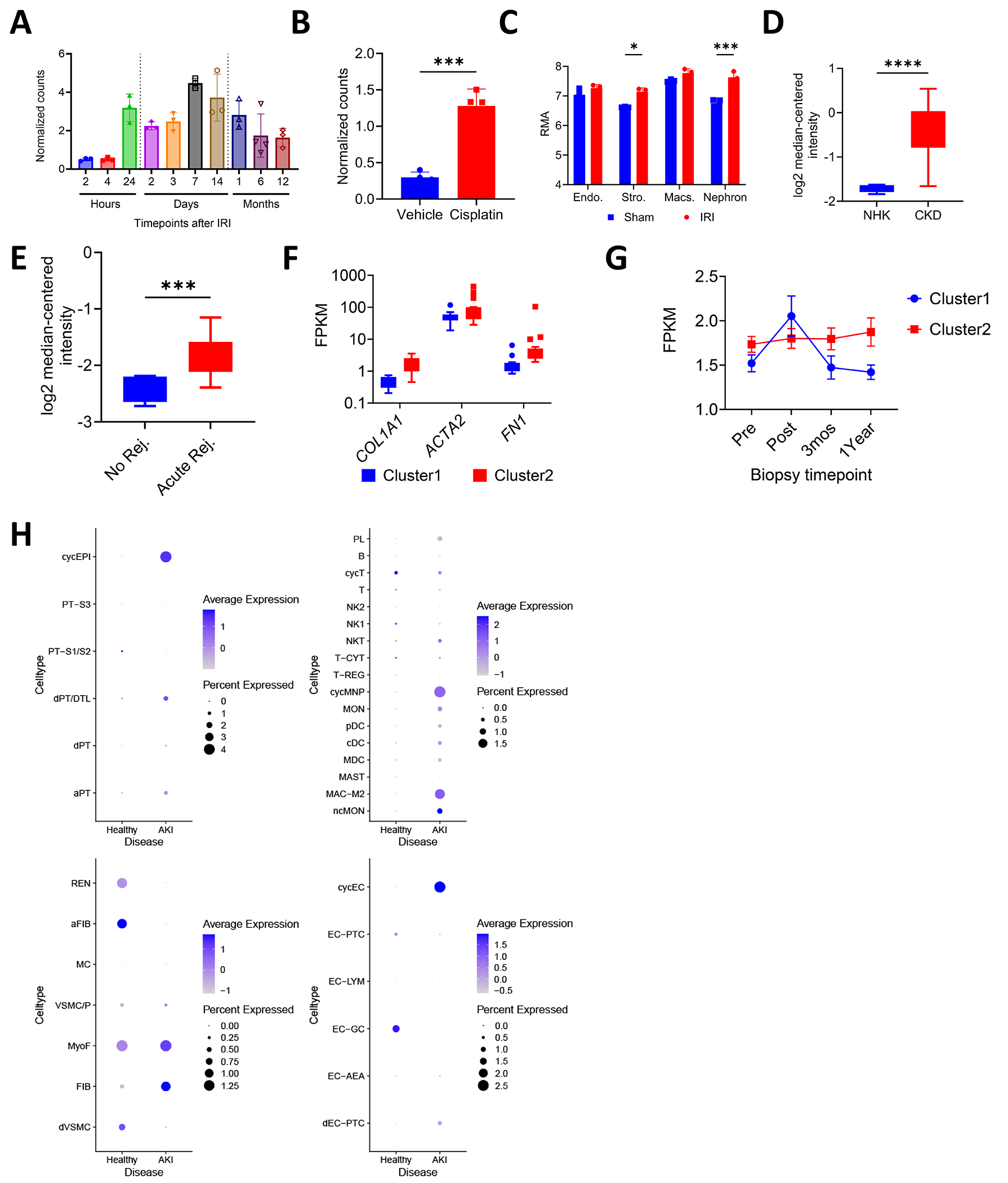
Pannexin 1 (Panx1) is a membrane-associated channel that, when activated, facilitates the release of small metabolites into the extracellular environment. These metabolites signal as damage-associated molecular patterns (DAMP) and initiate inflammation. Upregulation and activation of Panx1 is one of the early events during inflammatory injury. Animal models show that a lack of Panx1 is protective against acute kidney injury (AKI). How Panx1 modulates AKI is poorly understood. We utilized both in vivo and in vitro models of PANX1 overexpression to study mitochondrial function, cell death, and inflammation to evaluate how Panx1 contributes to AKI. We used two models of AKI, ischemia-reperfusion injury (IRI) and cisplatin-induced AKI (cis-AKI), in animals that overexpress PANX1 globally or specifically in the proximal tubule or in the endothelium. Cisplatin-induced injury was investigated in vitro in PANX1-overexpressing proximal tubule cells in culture. Both global and proximal tubule-specific overexpression of PANX1 exacerbated AKI, whereas endothelium-specific overexpression had no effect. Panx1-dependent metabolite release and alterations in the intracellular compartment in proximal tubules independently contributed to cell death in vitro. PANX1 overexpression impaired mitochondrial function and increased mitochondrial reactive oxygen species (ROS) production. PANX1 overexpression resulted in increased inflammation in the kidneys during cis-AKI. We showed that PANX1 overexpression resulted in overt renal injury during AKI that is in part mediated by reduced mitochondrial function, increased cell death, and inflammation. Selective strategies to inhibit Panx1 could help prevent or treat AKI.NEW & NOTEWORTHY Despite the huge medical, economical, and quality of life burden that AKI poses to patients, there are no Food and Drug Administration (FDA)-approved therapeutic or pharmaceutical interventions for AKI. Pannexin 1 (Panx1), which is upregulated in patients with AKI as well as in animals that develop experimental AKI, plays a crucial role in mediating both inflammation and cell death during AKI. Our findings suggest clinical interventions with molecules that inhibit Panx1 channel activity could improve outcomes in AKI patients.

Keywords: ATP; acute kidney injury; cell death; mitochondria; pannexins.


Conflict of interest statement

DISCLOSURES

All the authors declared no competing interests.

Figures

Fig. 1:
Fig. 1:. Panx1 is upregulated during renal injury.
Public datasets on various renal diseases were analyzed in silico for expression of Panx1 during disease progression. The datasets, their GEO accession, data source, and original publication are provided in Table 1. A) Expression of Panx1 mRNA in mouse kidney is higher as early as 24 hours after IRI-AKI and stays at a higher level during kidney fibrosis progression. B) Expression of Panx1 in mouse kidneys is higher at 72 hours after cisplatin injection (Normalized counts, raw counts normalized to counts per million (cpm)). C) Tissue compartment-specific microarray analysis reveals increased Panx1 mRNA expression in mouse renal stromal cells (Stro.) and nephron compartments, but not in macrophages (Macs) or endothelial cells (Endo.), 24 hours after IRI. RMA, Robust Multichip Average. D) Patients with chronic kidney disease (CKD) have higher renal PANX1 expression compared to normal healthy kidneys (NHK) (n=5 (NHK), 48 (CKD)). E) Patients with acute renal graft rejection have elevated PANX1 levels in the kidneys (n=10 (No Rejection, Rej.), 7 (Acute Rej.)). F) Higher expression of fibrotic markers alpha-smooth muscle actin (ACTA2), alpha-1 type I collagen (COL1A1), and fibronectin (FN1) in 1 year protocol kidney biopsies from patients in Cluster 2 compared to kidney biopsies from patients in Cluster 1; FPKM: fragments per kilobase of exon per million mapped fragments. G) Pseudo-time analysis of PANX1 mRNA expression in transplanted kidneys showing an early increase and later decline in PANX1 in Cluster 1 and a sustained higher level of PANX1 in Cluster 2. Pre, biopsy taken before transplant; Post, biopsy taken after transplant; 3Mos, biopsy taken 3 months after transplant; 1Year, biopsy taken 1 year after transplant (n=13 (Cluster1), 19 (Cluster2) H) Dot plot showing upregulated expression of PANX1 in various cell subpopulation Clusters, including proximal tubules, immune cells, interstitial cells, and endothelial cells, from kidneys of AKI patients compared to healthy living donors. (*p<0.05, ***p<0.001, ****p<0.0001; Data are represented as mean ± S.D.)
Fig. 2:
Fig. 2:. Panx1 overexpression exacerbates AKI.
(A) mRNA expression relative to Actb (β-actin) and (B) protein levels of human PANX1 and (C) mRNA expression of mouse Panx1 in kidneys from PANX1Tg animals compared to wildtype littermate controls (WT). (D-F) Exacerbation of IRI-induced AKI in PANX1Tg animals (D, experimental model) showing elevated (E) plasma creatinine levels and (F) mRNA expression levels of renal injury marker Lcn2 (also known as Ngal) in kidneys of PANX1Tg mice compared to their littermate controls. (G-K) Exacerbation of cisplatin-induced AKI (cis-AKI) in PANX1Tg animals (G, experimental model) showing (H) elevated plasma creatinine levels, (I) elevated mRNA expression levels of renal injury marker Lcn2 in kidneys, (J) representative H&E staining in the kidneys (scale bar = 100 μM. Insets show kidney outer medulla at higher magnification, scale bar = 400 μM), and (K) quantification of acute tubular necrosis (%ATN, % of total kidney section surface area occupied by tubule injury) from H&E-stained kidney sections of PANX1Tg mice compared to their littermate controls (I-K, day 3). Data are represented as mean ± S.D. (*p<0.05, **p<0.01, ***p<0.001; each symbol represents an experimental animal).
Fig. 3:
Fig. 3:. Proximal tubule but not endothelial Panx1 overexpression exacerbates AKI.
Bilateral IRI or cis-AKI was induced in a transgenic mouse that overexpresses the human isoform of PANX1 specifically in kidney PT epithelial cells (PTPTg) or in endothelial cells (ECs; ECPTg), and the extent of AKI was evaluated. (A-C) Exacerbation of IRI-AKI in PTPTg animals (A, experimental model) showing elevated (B) plasma creatinine levels and (C) mRNA expression levels of renal injury marker Lcn2 (also known as Ngal) in kidneys compared to their littermate controls (PTWT). (D-F) Exacerbation of cis-AKI in PTPTg animals (D, experimental model) showing elevated (E) plasma creatinine levels and (F) mRNA expression levels of renal injury marker Lcn2 in kidneys compared to PTWT. (G-H) No difference in the extent of injury in ECPTg and ECWT mice subjected to G) IRI and H) cis-AKI. Data are represented as mean ± S.D. (*p<0.05, **p<0.01, ****p<0.0001; each symbol represents an experimental animal)
Fig. 4:
Fig. 4:. Panx1 overexpression causes overt cell death during cis-AKI.
(A) Representative images of kidneys from WT and PANX1Tg animals subjected to cis-AKI and stained using FITC-conjugated TUNEL labeling kit to identify apoptotic/dead cells. Panels at far right are digital magnification of areas identified by red boxes in middle panels showing both apoptotic nuclei (colocalization with DAPI; white arrows) as well as non-apoptotic dead cells (red arrows). Kidneys from PANX1Tg animals show more areas of non-apoptotic TUNEL staining patterns compared to kidneys from WT littermate controls (scale bar = 100 μm). (B-C) Characterization of stable cell lines of TKPTS cells stably transfected with human PANX1 cDNA (OX) showing (B) mRNA expression of human PANX1, and (C) elevated PANX1 functional activity reflected by higher Panx1-mediated TO-PRO-3 dye uptake in OX cells compared to WT. MFI, mean fluorescence intensity. (D-E) In vitro cisplatin challenge in TKPTS cells stably transfected with human PANX1 cDNA showing (D) gating strategy to identify apoptotic/dead cells using annexin V and 7-AAD, and (E) higher level of cell death in OX cells compared to WT cells after treatment with varying concentrations of cisplatin. (F) In vitro cisplatin challenge in primary proximal tubule epithelial cells isolated from wildtype (WT) or PANX1 transgenic (PTg) mice showing a higher percentage of cell death in PTECS from PTg mice after after treatment with varying concentrations of cisplatin. 0 Cisplatin, saline vehicle control. Data are represented as mean ± S.D. (****p<0.0001; each symbol represents an experimental replicate.)
Fig. 5:
Fig. 5:. Panx1-mediated increase in cell death is due to intracellular and/or extracellular cell death signals.
(A-B) Elevated Panx1 levels make proximal tubule cells more susceptible to cell death. Cisplatin challenge in a 1:1 mixed WT and hPANX1 overexpressing (OX) cell culture model (A) caused more cell death in OX cells than in WT cells (B). (C-E) Conditioned media from PANX1 overexpressing cells induced greater cell death in an experimental model of transfer of conditioned media collected 18 hours after cisplatin challenge (C). Conditioned media from cisplatin-challenged OX cells induced greater cell death in WT cells (OX→WT) compared to that from WT cells (WT→WT)(D), and conditioned media from cisplatin-challenged OX cells induced greater cell death in OX cells (OX→OX) compared to that from WT cells (WT→OX)(E). 0 Cisplatin, saline vehicle control. Data are represented as mean ± S.D. (*p<0.05, **p<0.01, ****p<0.0001; each symbol represents an experimental replicate.)
Fig. 6:
Fig. 6:. PANX1 overexpression results in reduced mitochondrial function.
Assessment of mitochondrial changes in wildtype (WT) and PANX1 overexpressing (OX) TKPTS cells after cisplatin challenge. (A) Mitochondrial stress test showing reduced mitochondrial function in OX cells compared to WT cells at baseline and after cisplatin challenge. OX cells had (B) reduced basal mitochondrial respiration, (C) reduced ATP production, (D) reduced maximum respiratory capacity, (E) increased mitochondrial ROS production, and (F) reduced mitochondrial membrane potential at baseline and after cisplatin challenge compared to WT cells. TMRE; tetramethylrhodamine ethyl ester. Data are represented as mean ± S.D. (*p<0.05, **p<0.01, ****p<0.0001; each symbol represents an experimental replicate.)
Fig. 7:
Fig. 7:. Panx1 overexpression results in higher immune cell recruitment in the kidneys during cis-AKI.
A-D) Quantification of leukocyte infiltration in the kidneys of WT and PTg mice at day 0, day 2 and day 3 after initial cisplatin injection shows higher infiltration of (A) total leukocytes, (B) neutrophils (CD45+/CD2/Ly6G+/CD11b+), (C) CD8+ T cells (CD45+/CD2+/CD3+/CD8+/CD4), and (D) CD11b+ dendritic cells (CD45+/CD2/Ly6G/MHCII+/CD11c+/CD11b+) in the kidneys of PTPTg animals compared to WT animals at day 3 post-cis-AKI. E-I) in vitro stimulation of RAW267 cells by the conditioned media from WT or OX cells challenged with various concentrations of cisplatin showing a significant upregulation of mRNA expression of E) Il1b and a trend of upregulation of F) Il6, and G) Cxcl1 and H) Cxcl2, and no change in expression of I) Il10. Data are represented as mean ± S.D. (*p<0.05, **p<0.01; each symbol represents an experimental animal.)
Fig. 8:
Fig. 8:. Panx1 transgene expression is specific to basolateral surface of kidney proximal tubules in PTPTg mice and does not co-localize with mitochondria.
Mouse kidneys were fixed in PLP and 5 μm sections prepared. Directly fluorochrome-conjugated Ab were used to stain the sections and images visualized by Zeiss LSM980 as described in Materials and Methods. (A) Sections were stained with A488-anti-GFP, A555-anti-Flag, and PB-anti-collectrin and image were captured with a 20X (a-e) or a 63X objective and Merged Z-section images in pseudocolors are shown. Arrows indicate Panx1 and GFP transgene expressing tubules coinciding with collectrin staining. Panels c and e, bars equal 50 μm; Panels g and h, bars equal 10 μm. (B) Kidney sections were stained with A488-anti-GFP, A555-anti-Flag, A647-anti-Epcam, and BV421-anti-CD13 and merged Z-stack images are shown in pseudocolors. Arrows indicate Panx1 and GFP transgene-expressing proximal tubule segments. Arrowheads show Epcam-staining tubules distal to the thin descending limb tubules with no transgene expression. Bars in d and e equal 50 μm. (C) Kidney sections were stained with A488-Anti-GFP, A555-anti-Flag, A647-anti-Tom20, and BV421-anti-CD13. Mitochondrial staining occurred in all cells (panels c and e) and CD13 stains the brush border of proximal tubular cells (panels d and e). Anti-Flag staining of Panx1-Flag was perinuclear and overlaps with Tom20 but not CD13 staining (panels a, c, d, and e). GFP staining was evenly distributed within the cells including the nuclear region (panels b and e). Some epithelial cells within the proximal tubules failed to express the Panx1 and GFP transgenes. (D) In sections stained with Ab against Flag, GFP, CD13 and the proximal tubule S2 epithelial cell marker Oat1, colocalization of Panx1 with Oat1 but not CD13 was found (panels a, c, d, e, arrows), supporting the basolateral expression of the Panx1 transgene. In panels A-d, A-e, B-d, B-e, bars equal 10 μm.

Similar articles

References

    1. Baranova A, Ivanov D, Petrash N, Pestova A, Skoblov M, Kelmanson I, Shagin D, Nazarenko S, Geraymovych E, Litvin O, Tiunova A, Born TL, Usman N, Staroverov D, Lukyanov S, Panchin Y. The mammalian pannexin family is homologous to the invertebrate innexin gap junction proteins. Genomics 83: 706–716, 2004. doi: 10.1016/j.ygeno.2003.09.025. - DOI - PubMed
    1. Bao L, Locovei S, Dahl G. Pannexin membrane channels are mechanosensitive conduits for ATP. FEBS Letters 572: 65–68, 2004. doi: 10.1016/j.febslet.2004.07.009. - DOI - PubMed
    1. Chekeni FB, Elliott MR, Sandilos JK, Walk SF, Kinchen JM, Lazarowski ER, Armstrong AJ, Penuela S, Laird DW, Salvesen GS, Isakson BE, Bayliss DA, Ravichandran KS. Pannexin 1 channels mediate ‘find-me’ signal release and membrane permeability during apoptosis. Nature 467: 863–867, 2010. doi: 10.1038/nature09413. - DOI - PMC - PubMed
    1. Hanner F, Lam L, Nguyen MTX, Yu A, Peti-Peterdi J. Intrarenal localization of the plasma membrane ATP channel pannexin1. American Journal of Physiology-Renal Physiology 303: F1454–F1459, 2012. doi: 10.1152/ajprenal.00206.2011. - DOI - PMC - PubMed
    1. Good ME, Eucker SA, Li J, Bacon HM, Lang SM, Butcher JT, Johnson TJ, Gaykema RP, Patel MK, Zuo Z, Isakson BE. Endothelial cell Pannexin1 modulates severity of ischemic stroke by regulating cerebral inflammation and myogenic tone. JCI Insight 3, 2018. doi: 10.1172/jci.insight.96272. - DOI - PMC - PubMed

MeSH terms

Grants and funding

LinkOut - more resources