. 2025 Jun 1;328(6):G663-G676.
doi: 10.1152/ajpgi.00027.2025. Epub 2025 Apr 17.

Saturated phosphatidic acids induce mTORC1-driven integrated stress response contributing to glucolipotoxicity in hepatocytes

Affiliations

Saturated phosphatidic acids induce mTORC1-driven integrated stress response contributing to glucolipotoxicity in hepatocytes

Rui Guo et al. Am J Physiol Gastrointest Liver Physiol. .

Abstract

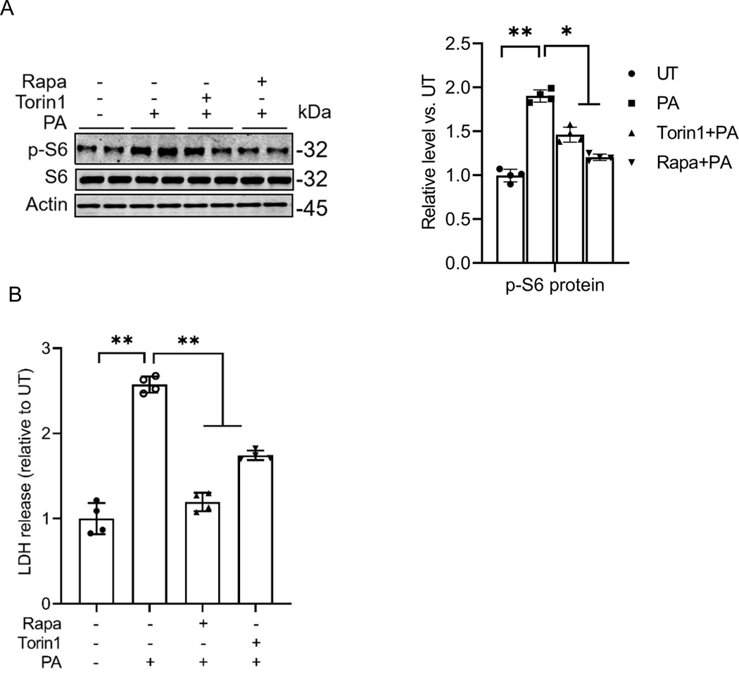
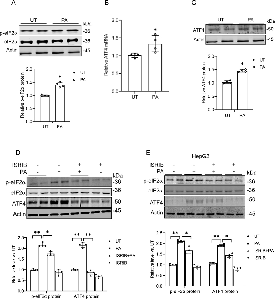
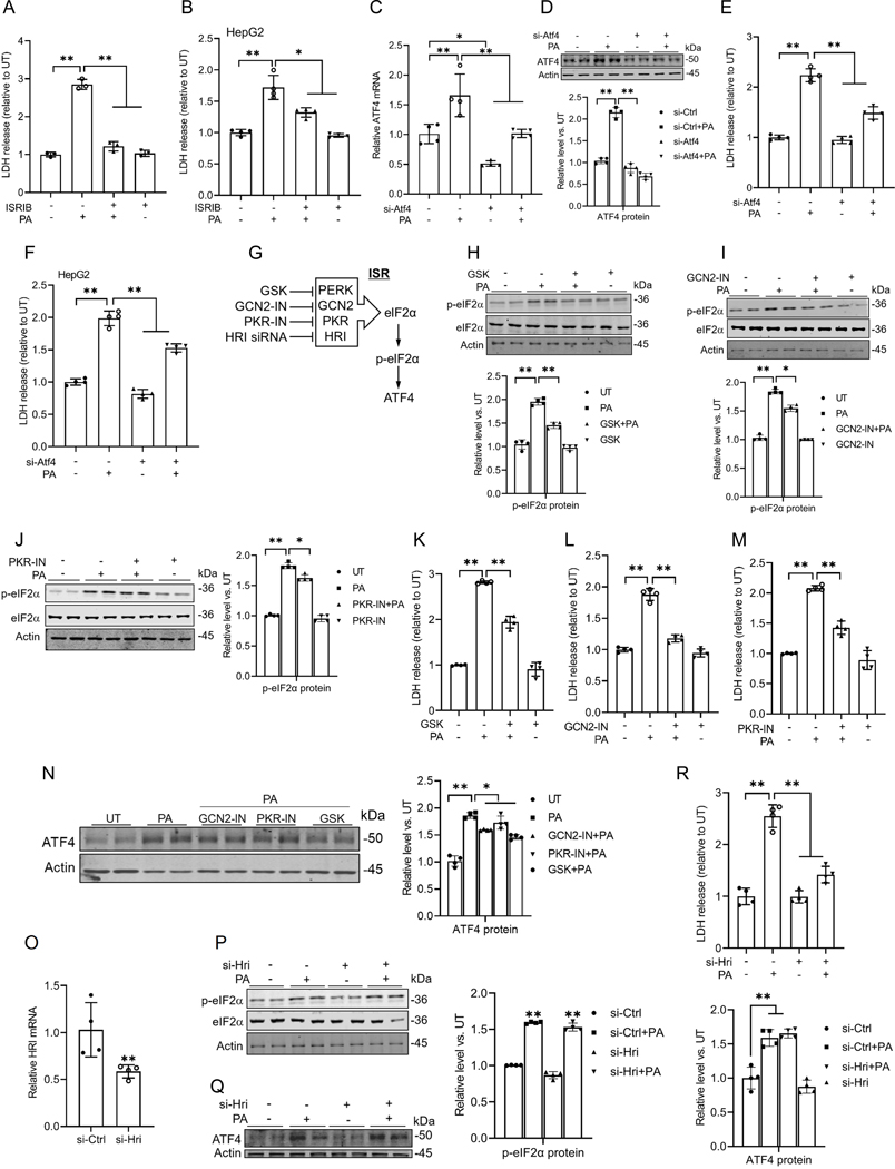
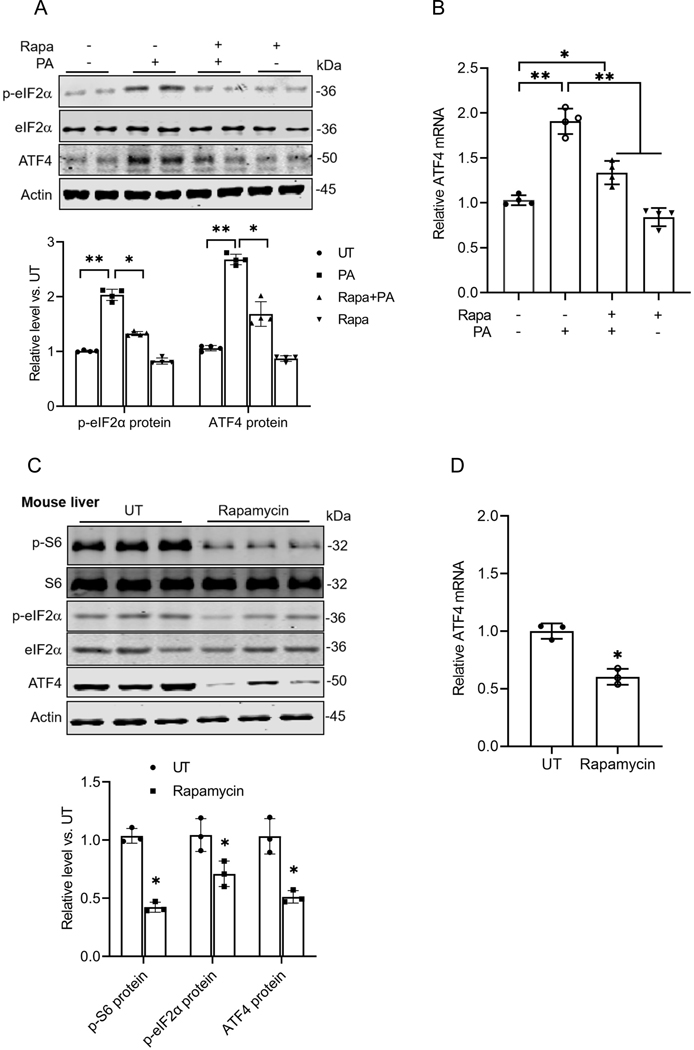
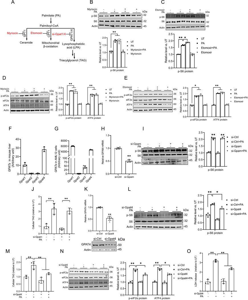
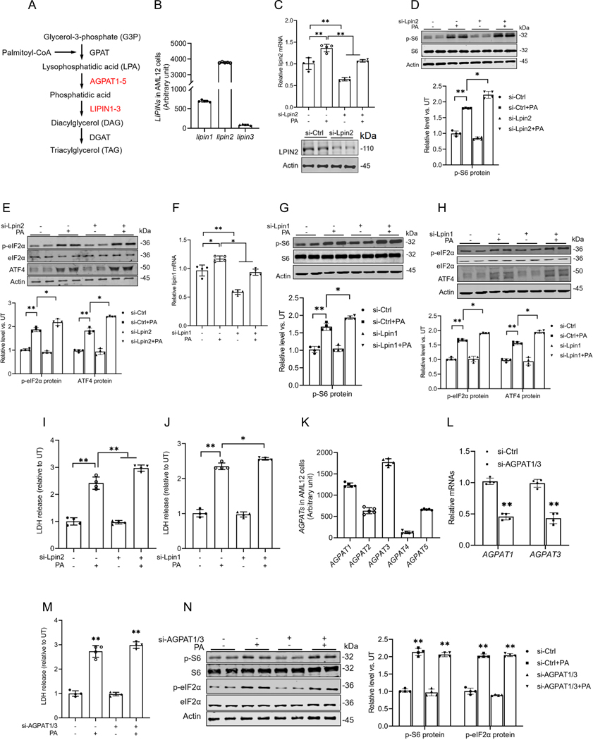
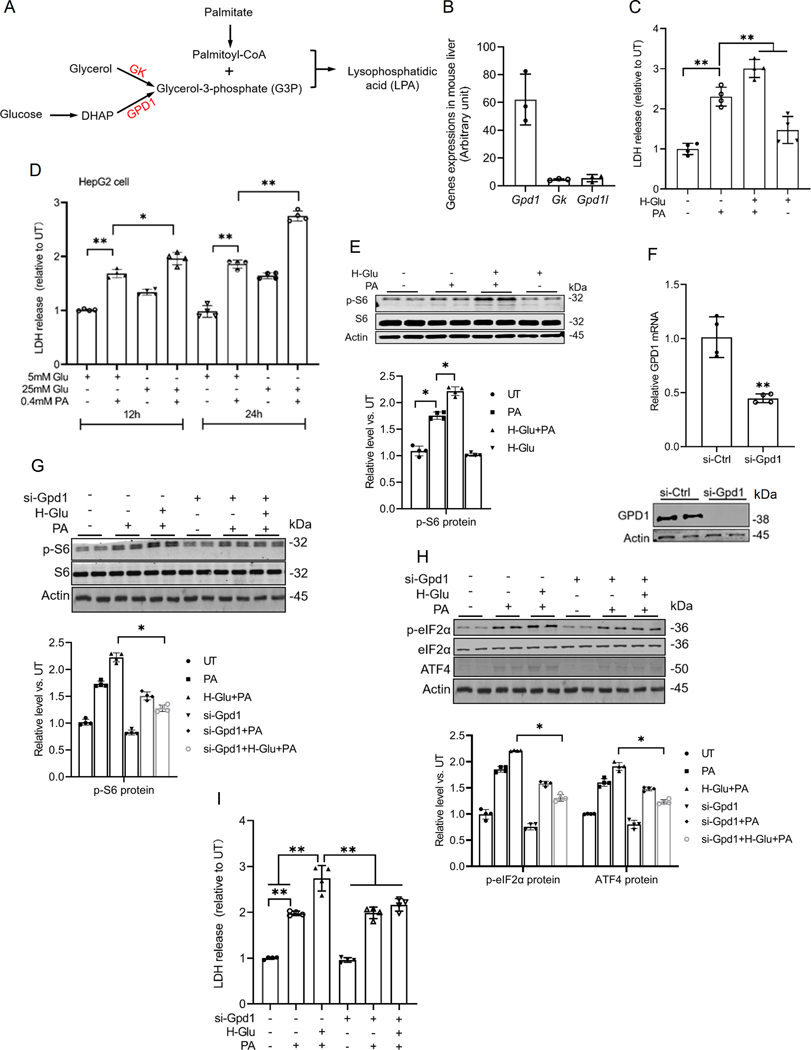
Hepatic glucolipotoxicity, characterized by the synergistic detrimental effects of elevated glucose levels combined with excessive lipid accumulation in hepatocytes, plays a central role in the pathogenesis of various metabolic liver diseases. Despite recent advancements, the precise mechanisms underlying this process remain unclear. Using cultured AML12 and HepG2 cells exposed to excess palmitate, with and without high glucose, as an in vitro model, we aimed to elucidate the cellular and molecular mechanisms underlying hepatic glucolipotoxicity. Our data showed that palmitate exposure induced the integrated stress response (ISR) in hepatocytes, evidenced by increased eukaryotic translation initiation factor 2 alpha (eIF2α) phosphorylation (serine 51) and upregulated activating transcription factor 4 (ATF4) expression. Moreover, we identified mammalian target of rapamycin complex 1 (mTORC1) as a novel upstream kinase responsible for palmitate-triggered ISR induction. Furthermore, we showed that either mTORC1 inhibitors, ISRIB (an ISR inhibitor), or ATF4 knockdown abolished palmitate-induced cell death, indicating that the mTORC1-eIF2α-ATF4 pathway activation plays a mechanistic role in mediating palmitate-induced hepatocyte cell death. Our continuous investigations revealed that glycerol-3-phosphate acyltransferase (GPAT4)-mediated metabolic flux of palmitate into the glycerolipid synthesis pathway is required for palmitate-induced mTORC1 activation and subsequent ISR induction. Specifically, we uncovered that saturated phosphatidic acid production contributes to palmitate-triggered mTORC1 activation. Our study provides the first evidence that high glucose enhances palmitate-induced activation of the mTORC1-eIF2α-ATF4 pathway, thereby exacerbating palmitate-induced hepatotoxicity. This effect is mediated by the increased availability of glycerol-3-phosphate, a substrate essential for phosphatidic acid synthesis. In conclusion, our study highlights that the activation of the mTORC1-eIF2α-ATF4 pathway, driven by saturated phosphatidic acid overproduction, plays a mechanistic role in hepatic glucolipotoxicity.NEW & NOTEWORTHY Integrated stress response (ISR) activation contributes to palmitate-induced lipotoxicity in hepatocytes. mTORC1 acts as an upstream kinase essential for palmitate-mediated ISR activation and hepatocyte death. The formation of saturated phosphatidic acid mechanistically regulates hepatic mTORC1 activation induced by palmitate. Glucose-enhanced generation of saturated phosphatidic acid amplifies palmitate-induced hepatotoxicity, contributing to glucolipotoxicity.

Keywords: ISR; glucolipotoxicity; mTORC1; palmitate; phosphatidic acid.


Conflict of interest statement

Competing interests

The authors declare no competing interests.

Figures

Fig. 1
Fig. 1. mTORC1 activation contributes to palmitate-induced lipotoxicity in hepatocytes.
(A) AML12 cells were pretreated with mTORC1 inhibitors (rapamycin, 0.05 mM and torin1, 0.25 μM) for 2 h before palmitate addition (0.4 mM). Protein abundance of phosphorylated and total S6 was detected by Western blot. (B) LDH release. *p < 0.05 and **p < 0.01 indicate statistically significant differences.
Fig. 2
Fig. 2. Palmitate activates the ISR (integrated stress response) in hepatocytes.
(A) Western blot analysis of phosphorylated and total eIF2α expression with/without palmitate (0.4mM) exposure in AML12 cells. (B) qRT-PCR assay of ATF4 gene expression with/without palmitate exposure in AML12 cells. (C) Western blot detection of ATF4 protein abundance with/without palmitate exposure in AML12 cells. (D, E) AML12 and HepG2 cells were pretreated with ISRIB (10 μM), an inhibitor of the integrated stress response (ISR), for 2 h before palmitate exposure (0.4 mM). The hepatic eIF2α phosphorylation and ATF4 expressions were detected by Western blot. *p < 0.05 and **p < 0.01 indicate statistically significant differences. Note: The same internal control (actin) band was utilized for Figures A & C since all protein bands originated from the same membrane.
Fig. 3
Fig. 3. The ISR induction contributes to palmitate-induced hepatotoxicity.
(A, B) The cell death of AML12 and HepG2 cells was determined 16 h later by LDH release measurement. ISRIB treatment rescued the hepatocyte death induced by palmitate. (C, D) AML12 cells were transfected with either scramble siRNA or Atf4 siRNA (si-Atf4) for 24 h before palmitate exposure (0.4 mM) for 16 h. qRT-PCR and Western blot analysis of ATF4 expression. (E, F) ATF4 gene knockdown alleviated palmitate-induced cell death in AML12 and HepG2 cells, determined by LDH release. (G) The schematic illustration of four canonical upstream kinases. (H-M) AML12 cells were pretreated with GSK-2606414 (PERK inhibitor, 10 μM), GCN2 inhibitor (GCN2-IN, 10 μM), or PKR inhibitor (PKR-IN, 10 μM) for 2 h before palmitate addition (0.4 mM). Protein abundances of phosphorylated and total eIF2α were detected by Western blot, and cell death was determined 16 h later by LDH release. (N) Protein abundances of ATF4 were detected by Western blot. (O) The transfection efficiency of HRI siRNA in AML12 cells was assayed by qRT-PCR. (P-R) AML12 cells were transfected with either scramble siRNAor HRI siRNA (si-Hri) for 24 h before palmitate exposure (0.4 mM) for 16 h. The eIF2α phosphorylation and ATF4 expressions were detected by Western blot, and cell death was determined by LDH release. *p < 0.05 and **p < 0.01 indicate statistically significant differences.
Fig. 4
Fig. 4. mTORC1 is an upstream signal contributing to palmitate-triggered ISR activation.
(A, B) AML12 cells were pretreated with rapamycin for 2 h before palmitate addition (0.4 mM). Protein abundances of phosphorylated and total eIF2α, as well as ATF4, were detected using Western blot, while gene expression was assessed through qRT-PCR. (C) Hepatic protein abundances of phosphorylated and total S6, phosphorylated and total eIF2α, and ATF4 were detected by Western blot in mice administrated with/without rapamycin (n=3/group). (D) qRT-PCR analysis of hepatic ATF4 expression in mice administrated with/without rapamycin (n=3/group). Rapa: rapamycin. *p < 0.05 and **p < 0.01 indicate statistically significant differences.
Fig. 5
Fig. 5. Metabolic flow into the glycerolipid synthesis pathway contributes to palmitate-triggered mTORC1 activation.
(A) Schematic illustration of major metabolic pathways of palmitate. (B-E) AML12 cells were pretreated with either myriocin (100 μM), the SPT1 inhibitor, or etomoxir (20 μM), the CPT1 inhibitor, for 2 h before palmitate addition (0.4 mM). The protein abundances of phosphorylated and total S6/eIF2α, and ATF4 were detected by Western blot. (F) The gene expression of 4 isoforms of GPATs in mouse liver. (G) The expression of 4 isoforms of GPATs in AML12 cells. AML12 cells were transfected overnight with Gpam siRNA, prior to palmitate exposure for 16 h. (H) The transfection efficiency of Gpam siRNA in AML12 cells was assayed by qRT-PCR. (I) The protein abundances of phosphorylated and total S6 were detected by Western blot. (J) Intracellular TG content. AML12 cells were transfected overnight with Gpat4 siRNA, prior to palmitate exposure for 16 h. (K) The transfection efficiency of Gpat4 siRNA in AML12 cells was assayed by qRT-PCR and Western blot. (L) The protein abundances of phosphorylated and total S6 were detected by Western blot. (M) Intracellular TG content. (N) The protein abundances of phosphorylated and total eIF2α and ATF4 were detected by Western blot. (O) The cell death was determined by LDH release measurement. *p < 0.05 and **p < 0.01 indicate statistically significant differences.
Fig. 6
Fig. 6. Saturated phosphatidic acid accumulation contributes to palmitate-triggered ISR induction and lipotoxicity in hepatocytes.
(A) Schematic illustration of the metabolic flux of palmitate into the glycerolipid biosynthesis pathway. (B) The gene expression of three isoforms of LIPIN in AML12 cells. (C-E) AML12 cells were transfected overnight with Lpin2 siRNA, prior to palmitate exposure for 16 h. The expression of lipin2 in AML12 cells was tested by qRT-PCR and Western blot. Protein abundances of phosphorylated and total S6, phosphorylated and total eIF2α, and ATF4 were detected by Western blot. (F-H) AML12 cells were transfected overnight with Lpin1 siRNA, prior to palmitate exposure for 16 h. The expression of lipin1 in AML12 cells was tested by qRT-PCR. Protein abundances of phosphorylated and total S6, phosphorylated and total eIF2α, and ATF4 were detected by Western blot. (I, J) AML12 cells were transfected overnight with Lpin1 and Lpin2 siRNAs, prior to palmitate exposure for 16 h. The cell death was determined by LDH, respectively. (K) The gene expression of 5 isoforms of AGPATs in AML12 cells. (L, M) AML12 cells were transfected overnight with AGPAT1/3 siRNAs, prior to palmitate exposure for 16 h exposure. The expressions of AGPAT1 and AGPAT3 in AML12 cells were measured by qRT-PCR and the cell death was determined by LDH release measurement. (N) Protein abundances of phosphorylated and total S6, and phosphorylated and total eIF2α were detected by Western blot. *p < 0.05 and **p < 0.01 indicate statistically significant differences.
Fig. 7
Fig. 7. Glucose enhances palmitate-induced lipotoxicity by promoting the generation of glycerol-3-phosphate, a co-substrate for LPA biosynthesis.
(A) Schematic illustration of the formation of LPA. (B) The expression of Gpd1, Gk, and Gpd1l in mouse liver. AML12 cells were pretreated with high glucose (H-Glu, 25 mM) for 2 h before palmitate addition (0.4 mM). (C) Cell death was determined by LDH release measurement. (D) LDH release in HepG2 cells under different glucose and palmitic acid (PA) conditions. Cells were treated with 5 mM or 25 mM glucose (Glu) in the presence or absence of 0.4 mM PA for 12 h and 24 h. (E) AML12 cells were transfected overnight with Gpd1 siRNA followed by glucose (25 mM) pretreatment for 2 h, prior to a 16 h palmitate exposure. Protein abundance of phosphorylated and total S6 was detected by Western blot. (F) The transfection efficiency of Gpd1 siRNA in AML12 cells was assayed by qRT-PCR and Western blot. (G-I) The protein abundances of phosphorylated and total S6, phosphorylated and total eIF2α, and ATF4 were detected by Western blot, and the cell death was determined by LDH. *p < 0.05 and **p < 0.01 indicate statistically significant differences. Note: The same internal control (actin) band was utilized for Figures G & H since all protein bands originated from the same membrane.

Similar articles

Cited by

References

    1. Lipke K, Kubis-Kubiak A, and Piwowar A. Molecular Mechanism of Lipotoxicity as an Interesting Aspect in the Development of Pathological States-Current View of Knowledge. Cells 11: 2022. - PMC - PubMed
    1. Prentki M, Segall L, Roche E, Thumelin S, Brun T, McGarry JD, Corkey BE, and Assimacopoulos-Jeannet F. [Gluco-lipotoxicity and gene expression in the pancreatic beta cell]. Journ Annu Diabetol Hotel Dieu 17–27, 1998. - PubMed
    1. Unger RH, Clark GO, Scherer PE, and Orci L. Lipid homeostasis, lipotoxicity and the metabolic syndrome. Biochim Biophys Acta 1801: 209–214, 2010. - PubMed
    1. Kuchay MS, Choudhary NS, and Mishra SK. Pathophysiological mechanisms underlying MAFLD. Diabetes Metab Syndr 14: 1875–1887, 2020. - PubMed
    1. Lee JY, Cho HK, and Kwon YH. Palmitate induces insulin resistance without significant intracellular triglyceride accumulation in HepG2 cells. Metabolism 59: 927–934, 2010. - PubMed

Substances

LinkOut - more resources