Feasibility and Safety of a Novel Cable-Transmitted Magnetically Controlled Capsule Endoscopy System for Upper Gastrointestinal Examination
- PMID: 40241979
- PMCID: PMC11997460
- DOI: 10.1002/hcs2.70010
Feasibility and Safety of a Novel Cable-Transmitted Magnetically Controlled Capsule Endoscopy System for Upper Gastrointestinal Examination
Abstract
Background: To evaluate the feasibility and safety of a novel cable-transmitted magnetically controlled capsule endoscopy (CT-MCE) system for upper gastrointestinal examination.
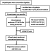
Methods: Twenty-six participants (19 healthy volunteers and seven patients with gastrointestinal symptoms) willing to undergo upper gastrointestinal endoscopy were recruited. Each participant underwent CT-MCE followed by conventional gastroscopy within 24 h. Maneuverability and visibility of the CT-MCE capsule in the upper gastrointestinal tract, adverse events, and discomfort during the procedure were evaluated. The sensitivity and specificity of CT-MCE for diagnosing upper gastrointestinal lesions were evaluated using conventional gastroscopy findings as the standard.
Results: Maneuverability was graded as "good" for all segments of the esophagus. The percentage of participants in which maneuverability was good according to gastric region was as follows: cardia (100.00%), pylorus (96.15%), angulus (92.31%), antrum (88.46%), fundus (84.62%), and body (73.08%). In the duodenal bulb and descending duodenum, it was good in only 20.83% and 16.67% of participants, respectively. Visibility was graded as "excellent" or "good" in the esophagus, Z line, and duodenal bulb in all participants; excellent/good visibility was achieved in the stomach and descending duodenum in 96.15% and 79.17% of participants, respectively. Forty-one lesions were detected overall. The sensitivity and specificity of CT-MCE in diagnosing upper gastrointestinal lesions were 85.00% and 98.15%, respectively. The CT-MCE capsule was successfully removed through the mouth in all participants. No serious adverse events or capsule retention occurred.
Conclusions: CT-MCE showed good feasibility and safety for upper gastrointestinal examination. The system was effective in examining the esophagus and stomach with no risk of capsule retention.
Keywords: capsule endoscopy; diagnostic accuracy; gastrointestinal examination; gastroscopy; safety.
© 2025 The Author(s). Health Care Science published by John Wiley & Sons Ltd on behalf of Tsinghua University Press.
Conflict of interest statement
The authors declare no conflicts of interest.
Figures


Similar articles
-
Accuracy of Magnetically Controlled Capsule Endoscopy, Compared With Conventional Gastroscopy, in Detection of Gastric Diseases.Clin Gastroenterol Hepatol. 2016 Sep;14(9):1266-1273.e1. doi: 10.1016/j.cgh.2016.05.013. Epub 2016 May 20. Clin Gastroenterol Hepatol. 2016. PMID: 27211503
-
Human gastric magnet-controlled capsule endoscopy conducted in a standing position: the phase 1 study.BMC Gastroenterol. 2019 Nov 12;19(1):184. doi: 10.1186/s12876-019-1101-2. BMC Gastroenterol. 2019. PMID: 31718547 Free PMC article.
-
Feasibility and safety of a cable transmission magnetically controlled capsule endoscopy system for examination of the human upper digestive tract.Saudi Med J. 2024 Dec;45(12):1318-1325. doi: 10.15537/smj.2024.45.12.20240645. Saudi Med J. 2024. PMID: 39658110 Free PMC article.
-
Magnetically Controlled Capsule Endoscopy Versus Conventional Gastroscopy: A Systematic Review and Meta-Analysis.J Clin Gastroenterol. 2021 Aug 1;55(7):577-585. doi: 10.1097/MCG.0000000000001540. J Clin Gastroenterol. 2021. PMID: 33883514
-
Current and future role of magnetically assisted gastric capsule endoscopy in the upper gastrointestinal tract.Therap Adv Gastroenterol. 2016 May;9(3):313-21. doi: 10.1177/1756283X16633052. Epub 2016 Feb 21. Therap Adv Gastroenterol. 2016. PMID: 27134661 Free PMC article. Review.
References
-
- Galmiche J.‐P., Sacher‐Huvelin S., Coron E., et al., “Screening for Esophagitis and Barrett's Esophagus With Wireless Esophageal Capsule Endoscopy: A Multicenter Prospective Trial in Patients With Reflux Symptoms,” American Journal of Gastroenterology 103, no. 3 (2008): 538–545, 10.1111/j.1572-0241.2007.01731.x. - DOI - PubMed
LinkOut - more resources
Full Text Sources
