. 2025 Apr 5:50:95-115.
doi: 10.1016/j.bioactmat.2025.03.027. eCollection 2025 Aug.

An engineered M2 macrophage-derived exosomes-loaded electrospun biomimetic periosteum promotes cell recruitment, immunoregulation, and angiogenesis in bone regeneration

Affiliations

An engineered M2 macrophage-derived exosomes-loaded electrospun biomimetic periosteum promotes cell recruitment, immunoregulation, and angiogenesis in bone regeneration

Zhuohao Wen et al. Bioact Mater. .

Abstract

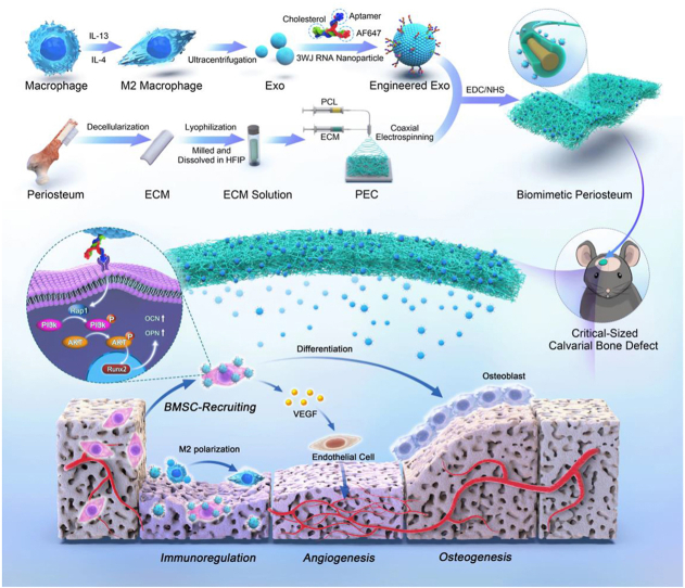
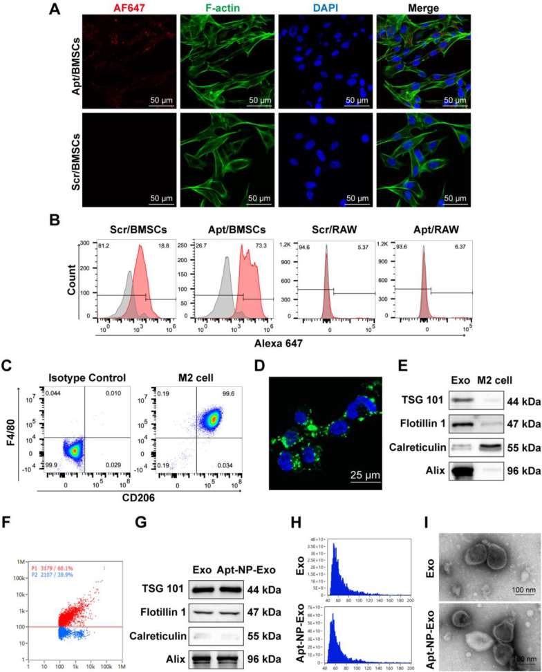
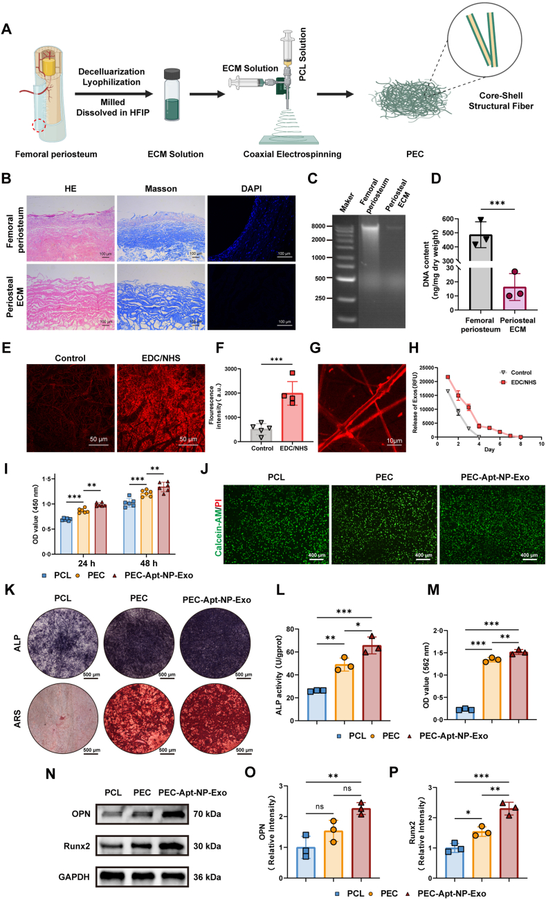
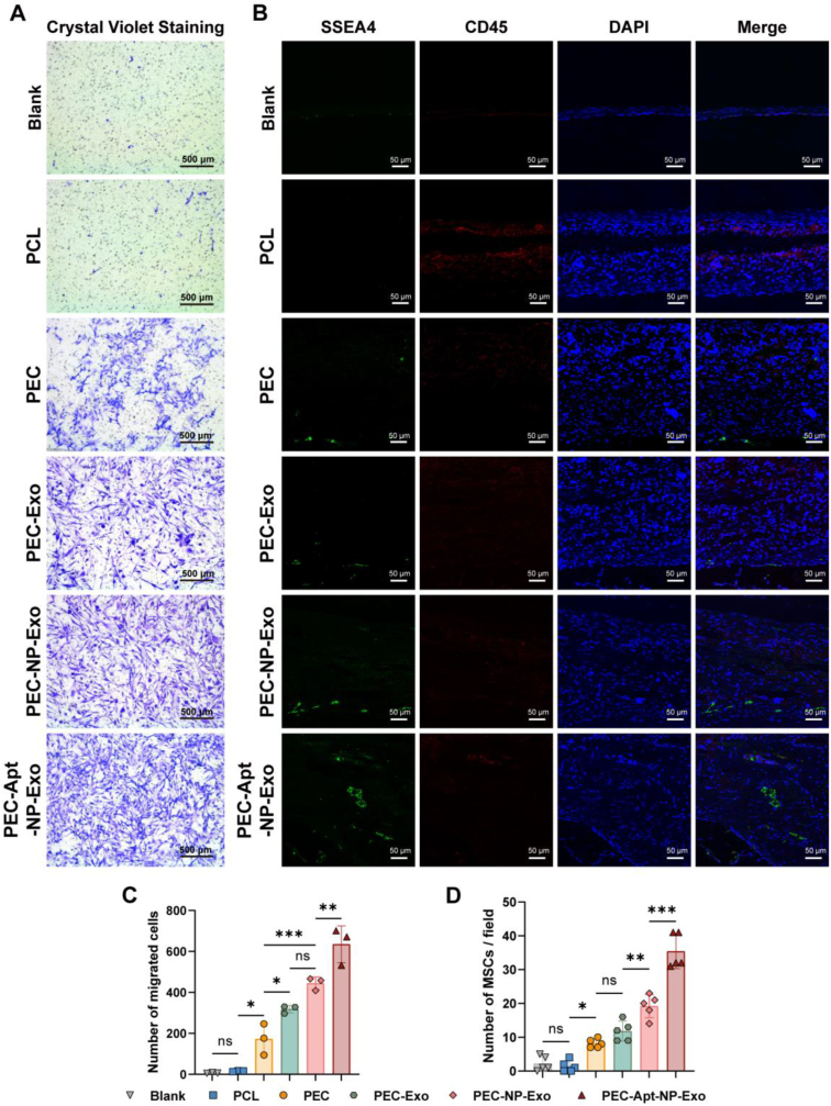
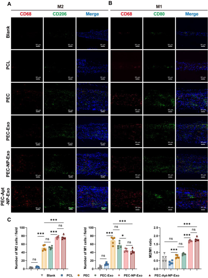
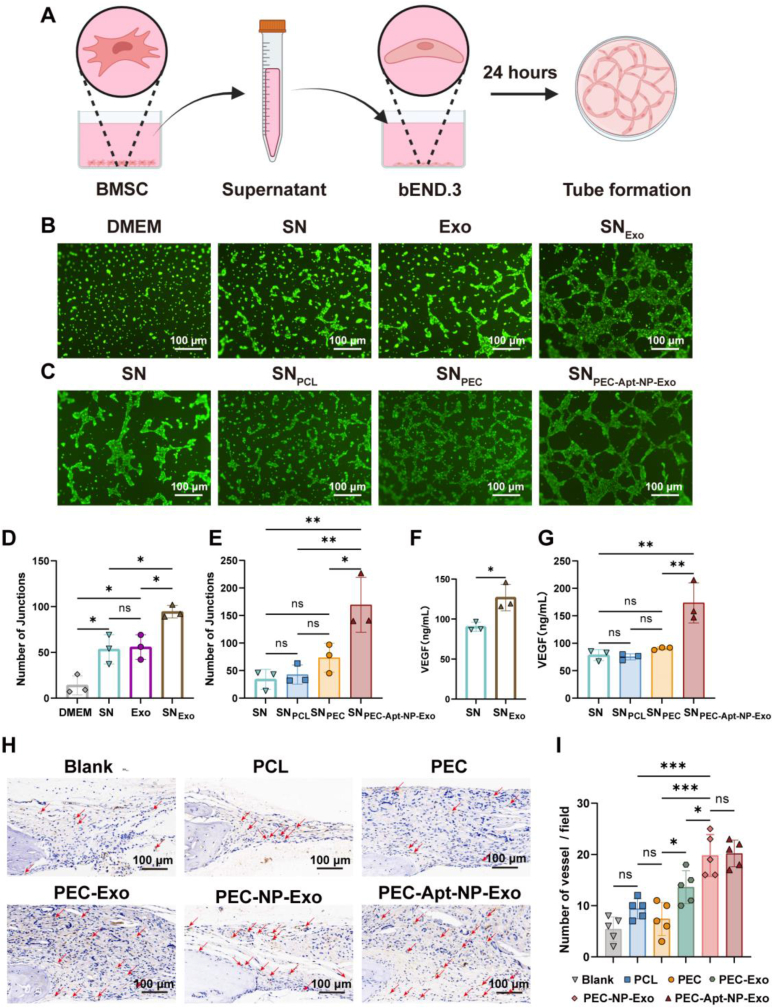
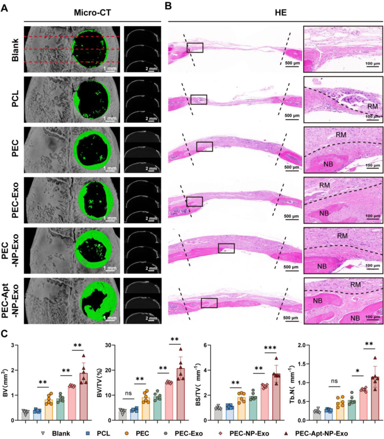
The periosteum, a fibrous tissue membrane covering bone surfaces, is critical to osteogenesis and angiogenesis in bone reconstruction. Artificial periostea have been widely developed for bone defect repair, but most of these are lacking of periosteal bioactivity. Herein, a biomimetic periosteum (termed PEC-Apt-NP-Exo) is prepared based on an electrospun membrane combined with engineered exosomes (Exos). The electrospun membrane is fabricated using poly(ε-caprolactone) (core)-periosteal decellularized extracellular matrix (shell) fibers via coaxial electrospinning, to mimic the fibrous structure, mechanical property, and tissue microenvironment of natural periosteum. The engineered Exos derived from M2 macrophages are functionalized by surface modification of bone marrow mesenchymal stem cell (BMSC)-specific aptamers to further enhance cell recruitment, immunoregulation, and angiogenesis in bone healing. The engineered Exos are covalently bonded to the electrospun membrane, to achieve rich loading and long-term effects of Exos. In vitro experiments demonstrate that the biomimetic periosteum promotes BMSC migration and osteogenic differentiation via Rap1/PI3K/AKT signaling pathway, and enhances vascular endothelial growth factor secretion from BMSCs to facilitate angiogenesis. In vivo studies reveal that the biomimetic periosteum promotes new bone formation in large bone defect repair by inducing M2 macrophage polarization, endogenous BMSC recruitment, osteogenic differentiation, and vascularization. This research provides valuable insights into the development of a multifunctional biomimetic periosteum for bone regeneration.

Keywords: Biomimetic periosteum; Bone regeneration; Engineered exosome; Immune regulation; Osteogenic induction.


Conflict of interest statement

The authors declare no conflict of interest.

Figures

Image 1
Graphical abstract
Scheme 1
Scheme 1
In this study, a bioactive and biomimetic periosteum (i.e., PEC-Apt-NP-Exo) was constructed by combining the electrospun membrane with engineered Exos: (1) the biomimetic electrospun membrane comprised PCL core-ECM shell fibers created via coaxial electrospinning; (2) the bioactive engineered Exos derived from M2 macrophage were modified with BMSC-specific aptamers; and (3) the engineered Exos were covalently bonded to the electrospun membrane. In vitro and in vivo results revealed that PEC-Apt-NP-Exo promoted immune regulation, osteogenesis, and angiogenesis through the Rap1/PI3K/AKT signaling pathway, demonstrating significant efficacy in repairing large bone defects.
Fig. 1
Fig. 1
Construction of Engineered Exos. (A) Representative confocal microscopic images of BMSC Apts after co-culture with BMSCs for 4 h. Red: AF647-labeled Apt; green: F-actin; blue: DAPI-labeled cell nuclei. (B) Flow cytometry analysis was performed to detect the specificity of AF647-labeled BMSC Apts. (C) Flow cytometry analysis of M2 macrophage markers F4/80 and CD206. (D) Confocal microscopic image of DiO-labeled Exos cocultured with BMSCs. (E) WB analysis of exosomal positive and negative markers in Exos and donor cells. (F) Nanoflow cytometry analysis of the binding of Apt nanoparticles to Exos. (G) WB analysis of exosomal positive and negative markers in Exo and Apt-NP-Exo groups. (H) Nanoflow cytometry analysis of size distribution of Exos and Apt-NP-Exos. (I) TEM images of Exos and Apt-NP-Exos.
Fig. 2
Fig. 2
Biological Effects of Engineered Exos on BMSCs. (A) Representative confocal microscopic images and (B) quantitative analysis showed the internalization of Exos, NP-Exos, and Apt-NP-Exos after culture with BMSCs for 4 h. Red: AF647-labeled NP or Apt-NP; green: DiO-labeled Exos; blue: DAPI-labeled cell nuclei. (C) CCK-8 assay was conducted to evaluate the proliferation of BMSCs treated with different concentrations of Apt-NP-Exo at 24 and 48 h. (D) ALP staining of BMSCs on day 7 and ARS staining on day 14. (E) Quantification of ARS staining. (F) ALP activity of BMSCs on day 7. (G) WB assay and (H) quantitative analysis of Runx2 and BMP-2 expression in BMSCs. Data were shown as mean ± SDs. ns, no significance, ∗∗∗P < 0.001, ∗∗P < 0.01, ∗P < 0.05.
Fig. 3
Fig. 3
Preparation of PEC-Apt-NP-Exo and Its Effect on Osteogenic Differentiation of BMSCs. (A) Schematic diagram illustrating the preparation of PEC. (B) Comparative study of femoral periosteum and periosteal ECM through HE, Masson, and DAPI staining analysis. (C) Agarose gel electrophoresis and (D) quantification of DNA content in femoral periosteum and periosteal ECM. (E) Confocal microscopy images and (F) quantification were utilized to evaluate the effectiveness of EDC/NHS in coating engineered Exos onto PECs via catalyzed amide condensation reactions. (G) Confocal microscopy images of core-shell structures. (H) Fluorescence microplate reader assay to evaluate the sustained release of engineered Exos. (I) CCK-8 assay and (J) live-dead staining were conducted to evaluate the effect of various membranes on BMSC growth. Green: calcein-AM, living cells; red: PI, dead cells (K) ALP staining of BMSCs on day 7 and ARS staining on day 14. (L) ALP activity of BMSCs on day 7. (M) Quantification of ARS staining. (N) WB assay and (O, P) quantitative analysis of Runx2 and OPN expression in BMSCs. Data were shown as mean ± SDs. ns, no significance, ∗∗∗P < 0.001, ∗∗P < 0.01, ∗P < 0.05.
Fig. 4
Fig. 4
PEC-Apt-NP-Exo promoting BMSC recruitment in the Early Stages of Bone Defects. (A) In vitro chemotactic behavior and (C) quantification of BMSCs on various membranes. (B) Representative images and (D) quantification of immunofluorescence staining one week after various membranes were implanted into critical-sized calvarial bone defects in mice. BMSCs were defined as SSEA4-positive and CD45-negative cells. Green: SSEA4; red: CD45; blue: DAPI-labeled cell nucleus. Data were shown as mean ± SDs. ns, no significance, ∗∗∗P < 0.001, ∗∗P < 0.01, ∗P < 0.05.
Fig. 5
Fig. 5
PEC-Apt-NP-Exo Regulating the Early Immune Environment of Bone Defects. Immunofluorescence staining of (A) M2 macrophages (red: CD68; green: CD206; blue: nuclei) and (B) M1 macrophages (red: CD68; green: CD80; blue: nuclei) one week after the implantation of various membranes into critical-sized calvarial bone defects in mice. (C) Quantification of the number and ratio of M2 macrophages to M1 macrophages. Data were shown as mean ± SDs. ns, no significance, ∗∗∗P < 0.001, ∗∗P < 0.01, ∗P < 0.05.
Fig. 6
Fig. 6
Biomimetic Periosteum Promoting Angiogenesis by Increasing the Secretion of VEGF in BMSCs. (A) Schematic diagram of the experiment in which BMSCs regulate angiogenesis in bEND.3 cells. (B, C) Representative fluorescence images showing the promotion of tube formation after culturing BMSCs with (B) Exo or (C) various membranes, respectively. (D, E) Quantification of the number of junctions in the tube network. ELISA assay analyzes the VEGF content in the supernatant of BMSCs after co-culture with (F) Exos or (G) various membranes. (H) Representative images and (I) vascular quantification of CD31 immunohistochemical staining 4 weeks after various membranes was implanted into critical-sized calvarial bone defects in mice. Red arrows indicate blood vessels. Data were shown as mean ± SDs. ns, no significance, ∗∗∗P < 0.001, ∗∗P < 0.01, ∗P < 0.05.
Fig. 7
Fig. 7
PEC-Apt-NP-Exo Promoting the Regeneration of Critical-Sized Calvarial Bone Defects in Mice. Representative images of (A) micro-CT and (B) HE staining after 8-week implantation of various membranes on defects. RM: residual materials; NB: new bone; the dashed lines indicate the boundaries of the residual materials. (C) Quantitative analysis of osteogenesis-related parameters based on the 3D reconstruction micro-CT images. BV: bone volume; BV/TV: bone volume fraction; BS/TV: bone surface to tissue volume ratio; Tb.N: trabecular number. Data were shown as mean ± SDs. ns, no significance, ∗∗∗P < 0.001, ∗∗P < 0.01, ∗P < 0.05.
Fig. 8
Fig. 8
PEC-Apt-NP-Exo Promoting New Bone Formation by Enhancing Osteoblast Differentiation in the Late Stage of Bone Defects. (A) Histological staining and (B) quantification of OPN, OCN, POSTN, and Masson after 8 weeks of implantation of various membranes on defects. Data were shown as mean ± SDs. ns, no significance, ∗∗∗P < 0.001, ∗∗P < 0.01, ∗P < 0.05.
Fig. 9
Fig. 9
PEC-Apt-NP-Exo Promoting Vascularization and Osteogenesis via the Rap1/PI3K/AKT Signaling Axis. (A) KEGG enrichment analysis following the sequencing of BMSCs cultured with PEC or PEC-Apt-NP-Exo. (B) GO enrichment analysis. BP: biological process, CC: cellular component, MF: molecular function. (C) Volcano plot showing the distribution of genes related to the Rap1 signaling pathway. (D) Heatmap of significantly differentially expressed genes in the Rap1 signaling pathway. (E) Protein expression levels of Rap1, p-PI3K, PI3K, p-AKT, and AKT were detected by WB in BMSCs. (F) Quantitative expression levels of Rap1, p-PI3K, PI3K, p-AKT, and AKT. (G) Schematic diagram illustrating the action mechanism of the PEC-Apt-NP-Exo membrane. Data were shown as mean ± SDs. ns, no significance, ∗∗∗P < 0.001, ∗∗P < 0.01, ∗P < 0.05.

Similar articles

References

    1. Allen M.R., Hock J.M., Burr D.B. Periosteum: biology, regulation, and response to osteoporosis therapies. Bone. 2004;35(5):1003–1012. - PubMed
    1. Lin Z., Fateh A., Salem D.M., Intini G. Periosteum: biology and applications in craniofacial bone regeneration. J. Dent. Res. 2014;93(2):109–116. - PMC - PubMed
    1. Colnot C. Skeletal cell fate decisions within periosteum and bone marrow during bone regeneration. J. Bone Miner. Res. 2009;24(2):274–282. - PMC - PubMed
    1. Zhang X., Xie C., Lin A.S., Ito H., Awad H., Lieberman J.R., Rubery P.T., Schwarz E.M., O'Keefe R.J., Guldberg R.E. Periosteal progenitor cell fate in segmental cortical bone graft transplantations: implications for functional tissue engineering. J. Bone Miner. Res. 2005;20(12):2124–2137. - PMC - PubMed
    1. Loi F., Cordova L.A., Pajarinen J., Lin T.H., Yao Z., Goodman S.B. Inflammation, fracture and bone repair. Bone. 2016;86:119–130. - PMC - PubMed

LinkOut - more resources