Review
. 2023 Dec 5;5(2):830-850.
doi: 10.1016/j.fmre.2023.09.007. eCollection 2025 Mar.

Plant-derived nanovesicles: Promising therapeutics and drug delivery nanoplatforms for brain disorders

Affiliations
Review

Plant-derived nanovesicles: Promising therapeutics and drug delivery nanoplatforms for brain disorders

Ruoning Wang et al. Fundam Res. .

Abstract

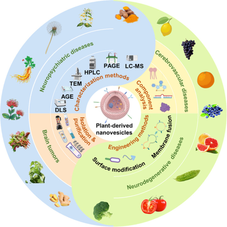
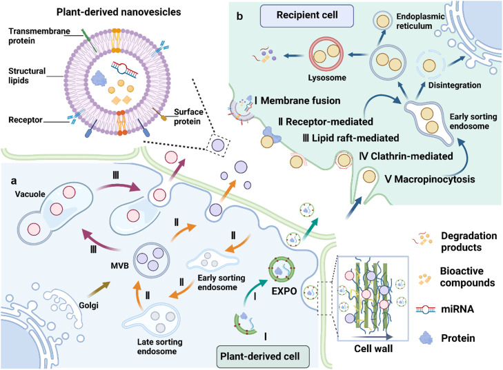
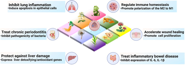
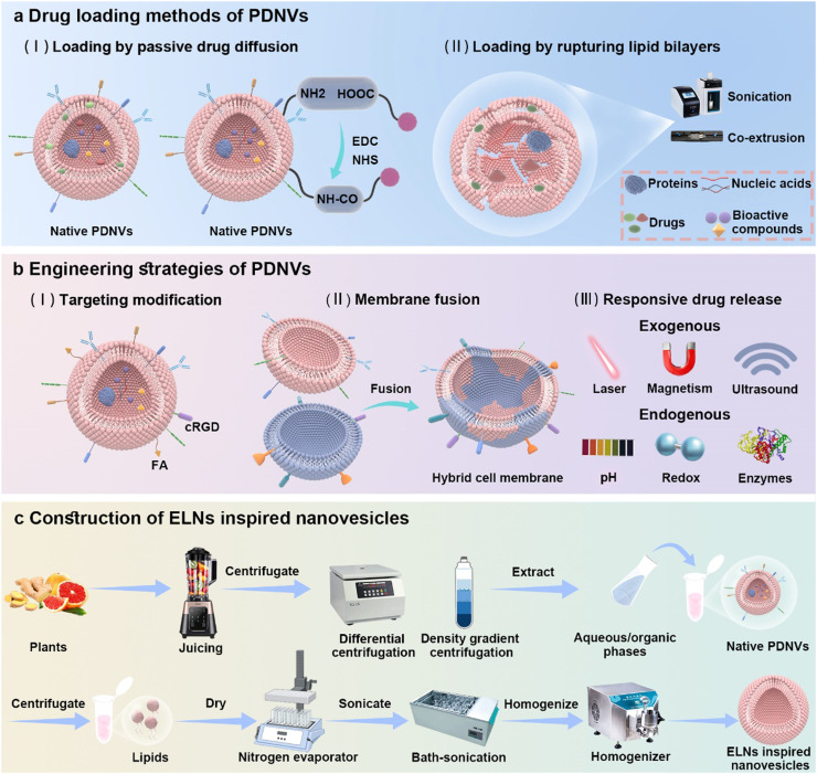
Plant-derived nanovesicles (PDNVs), including plant extracellular vesicles (EVs) and plant exosome-like nanovesicles (ELNs), are natural nano-sized membranous vesicles containing bioactive molecules. PDNVs consist of a bilayer of lipids that can effectively encapsulate hydrophilic and lipophilic drugs, improving drug stability and solubility as well as providing increased bioavailability, reduced systemic toxicity, and enhanced target accumulation. Bioengineering strategies can also be exploited to modify the PDNVs to achieve precise targeting, controlled drug release, and massive production. Meanwhile, they are capable of crossing the blood-brain barrier (BBB) to transport the cargo to the lesion sites without harboring human pathogens, making them excellent therapeutic agents and drug delivery nanoplatform candidates for brain diseases. Herein, this article provides an initial exposition on the fundamental characteristics of PDNVs, including biogenesis, uptake process, isolation, purification, characterization methods, and source. Additionally, it sheds light on the investigation of PDNVs' utilization in brain diseases while also presenting novel perspectives on the obstacles and clinical advancements associated with PDNVs.

Keywords: Brain disorders; Drug delivery platforms; Edible plants; Plant-derived nanovesicles; Traditional Chinese medicine.


Conflict of interest statement

The authors declare that they have no conflicts of interest in this work.

Figures

Image, graphical abstract
Graphical abstract
Fig 1
Fig. 1
Biogenesis and uptake process of PDNVs. (a) Biogenesis process of EVs in plant-derived cells. (I) EXPO pathway. (II) MVBs pathway. (III) Vacuolar pathway. (b) Uptake process of ELNs in recipient cells. (I) Membrane fusion. (II) Receptor-mediated endocytosis. (III) Lipid raft-mediated endocytosis. (IV) Clathrin-mediated endocytosis. (V) Macropinocytosis. This figure is created with biorender.com.
Fig 2
Fig. 2
Applications of PDNVs in the treatment of various disorders, including inhibiting lung inflammation, treating chronic periodontitis, protecting against liver damage, regulating immune homeostasis, accelerating wound healing and treating IBD. This figure is created with biorender.com.
Fig 3
Fig. 3
PDNVs perform as drug delivery nanocarriers. (a) Drug loading methods of PDNVs. (I) Loading by passive drug diffusion. (II) Loading by rupturing lipid bilayers. (b) Engineering strategies of PDNVs. (I) Targeting modification. (II) Membrane fusion. (III) Responsive drug release. (c) Construction of ELNs inspired nanovesicles.
Fig 4
Fig. 4
PDNVs are administered through different routes and cross the BBB into the brain to exert different therapeutic effects. (a) Intravenous administration. (b) Oral administration. (c) Intranasal administration. This figure is created with biorender.com.
Fig 5
Fig. 5
PDNVs are utilized in the treatment of glioma. (a) Grapefruit EVs-DOX loaded nanoparticles for glioma treatment. (b) The glioma-bearing brain tissues' 3D confocal pictures following intravenous injection of EV-DN2. N and T stand for healthy and tumor tissues, respectively. Scale bar: 100 µm. (c) Various treatment groups' glioma tissues were stained with CD34 to identify endothelial vessels and Ki67 for cell proliferation. Scale bar=100 µm. At 96 h after administration of Cy7-EVs, Cy7-DNs, and Cy7-EV-DN2 (d) without and with (e) perfusion, Cy7 and Luc signals were measured in LN229-luc glioma mice in vitro, reproduced from Wang et al. (2021) with permission from American Chemical Society. (f) 6-week-old wild-type B6 mice received intracranial injections of 2 × 10⁴ GL26-luc cells each. Afterward, mice with tumors were given either FA-pGNVs/siRNA-luc or FA-pGNVs/siRNA scramble control intravenously every day. The mice were photographed during the times listed in (f). (g) Imaging data in brain tumor associated photons in DiR+ FA-pGNVs/miR17-DY547 or pGNVs/miR17-DY54-treated mice, reproduced from Zhang et al. (2016) with permission from Elsevier.
Fig 6
Fig. 6
Applications of Carex and MC-ELNs in the treatment of brain diseases. (a) Illustration of the separation of Carex from carrots and how antioxidative properties and molecular changes are studied in cardiomyoblast and neuroblastoma cells. (b) TEM was used to examine the morphology of Carex. (c) The representative size distribution of Carex was analyzed using NTA. (d-f) Analysis of the levels of Nrf-2 (d), HO-1 (e), and NQO-1 (f) mRNA expression in SH-SY5Y cells after 6-OHDA treatment using RT-PCR. All values are expressed as mean ± SD (n = 3); * p < 0.05, ** p < 0.01, *** p < 0.001, reproduced from Rhee et al. (2021) with permission from Multidisciplinary Digital Publishing Institute. (g) MC-ELNs prevent BBB damage from ischemia-reperfusion and prevent neuronal apoptosis, most likely by increasing the activity of the AKT/GSK3β signaling pathway. (h) Representative photos demonstrate the CA1 neurons density in the hippocampal region. The live neurons are denoted by black arrows. (i) The cerebral infarct area in middle cerebral artery occlusion (MCAO) rats treated with MC-ELNs at various concentrations is visible on representative TTC staining images. (j) Images of the ipsilateral striatum stained with TUNEL for each group. (k) Images of the hippocampus DG region stained with TUNEL for each study group, reproduced from Qi et al. (2022) with permission from Frontiers Media SA.
Fig 7
Fig. 7
GaELNs and OatN reduce inflammation in the brain and enhance mouse brain memory. (a) GaELNs (1 × 10¹⁰) that were PKH26 and DiR dye-labeled were orally administered to HFD-fed mice for 24 h, and imaging analysis was used to show the in vivo distribution of the GaELNs in several organs. (b) Using confocal imaging, the uptake of GaELNs by brain microglial and neuronal cells was assessed. IBA-1 and β-tubulin III were specific markers for labeling microglial and neuronal cells, respectively. (c) The brain's inflammatory status is shown through brain histological sections. (d) A NORT analysis of movement traces from lean, HFD, and GaELNs treated HFD mice. Illustrations of movement traces from lean, HFD, and HFD mice treated with GaELNs determined by NORT, reproduced from Zhang et al. (2022) with permission from Ivyspring International. (e) Mice were administered clodronate (CLO) through intracisternal injection before receiving PKH26-labeled OatN via gavage. After brain tissue had been sectioned and stained with anti-Iba-1, confocal imaging assays were performed. Scale bars, 20 mm. (f) A diagram depicts OatN biological effects on microglia cells during alcohol-induced chronic brain inflammation in mice. (g) Mice were gavage-fed OatN three times per week for 28 days while fed either a control liquid diet or a control liquid diet containing 5% ethanol (Eth). EEG data were represented by four spots in the mice's brains. (h) Commercially available BG was used to inhibit BV2 cells before PKH26 labeled OatN was introduced to the cell media, reproduced from Zhang et al. (2022). with permission from Wiley.

Similar articles

Cited by

References

    1. Pinedo M., de la Canal L., de Marcos Lousa C. A call for Rigor and standardization in plant extracellular vesicle research. J. Extracell. Vesicles. 2021;10(6):e12048. - PMC - PubMed
    1. Dad H.A., Gu T.W., Zhu A.Q., et al. Plant exosome-like nanovesicles: Emerging therapeutics and drug delivery nanoplatforms. Mol. Ther. 2021;29(1):13–31. - PMC - PubMed
    1. Halperin W., Jensen W.A. Ultrastructural changes during growth and embryogenesis in carrot cell cultures. J. Ultrastruct. Res. 1967;18(3):428–443. - PubMed
    1. Ju S.W., Mu J.Y., Dokland T., et al. Grape exosome-like nanoparticles induce intestinal stem cells and protect mice from DSS-induced colitis. Mol. Ther. 2013;21(7):1345–1357. - PMC - PubMed
    1. Kim J., Li S., Zhang S., et al. Plant-derived exosome-like nanoparticles and their therapeutic activities. Asian J. Pharmaceut. Sci. 2022;17(1):53–69. - PMC - PubMed

LinkOut - more resources