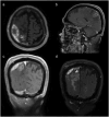
Odontogenic brain abscess caused by Porphyromonas gingivalis and Streptococcus constellatus: a case report and review article
- PMID: 40242593
- PMCID: PMC12001842
- DOI: 10.1080/20002297.2025.2485197
Odontogenic brain abscess caused by Porphyromonas gingivalis and Streptococcus constellatus: a case report and review article
Abstract
Background: Odontogenic brain abscess is a rare, but potentially fatal, central nervous system infection, with insidious onset and unclear etiology.
Methods: This case reports a 70-year-old male patient who developed an odontogenic brain abscess secondary to periodontal infection and underwent neurological surgery. Extract pus during surgery for the metagenomic next-generation sequencing (mNGS).
Results: The mNGS of pus samples obtained from brain abscess aspiration identified the periodontal pathogens Porphyromonas gingivalis and Streptococcus constellatus. Consequently, he was referred to the department of stomatology for further examination and treatment.
Conclusions: Our study found that major periodontal pathogens including P. gingivalis and S. constellatus were essential in the development of odontogenic brain abscesses; thus, timely intervention and preventive measures are important for treatment.
Keywords: Brain abscess; Porphyromonas gingivalis; Streptococcus constellatus; mNGS; periodontitis.
© 2025 The Author(s). Published by Informa UK Limited, trading as Taylor & Francis Group.
Conflict of interest statement
No potential conflict of interest was reported by the author(s).
Figures


Similar articles
-
Multiple Lung Cavity Lesions, Thoracic Wall Abscess and Vertebral Destruction Caused by Streptococcus constellatus Infection: A Case Report.Infect Drug Resist. 2023 Aug 15;16:5329-5333. doi: 10.2147/IDR.S416483. eCollection 2023. Infect Drug Resist. 2023. PMID: 37601557 Free PMC article.
-
Augmented pathogen detection in brain abscess using metagenomic next-generation sequencing: a retrospective cohort study.Microbiol Spectr. 2024 Oct 3;12(10):e0032524. doi: 10.1128/spectrum.00325-24. Epub 2024 Sep 12. Microbiol Spectr. 2024. PMID: 39264158 Free PMC article.
-
Clinical Characteristics of Lung Abscess Caused by Streptococcus Constellatus Infection.Clin Lab. 2024 Jul 1;70(7). doi: 10.7754/Clin.Lab.2024.240329. Clin Lab. 2024. PMID: 38965969
-
Rare brain and pulmonary abscesses caused by oral pathogens started with acute gastroenteritis diagnosed by metagenome next-generation sequencing: A case report and literature review.Front Cell Infect Microbiol. 2022 Sep 30;12:949840. doi: 10.3389/fcimb.2022.949840. eCollection 2022. Front Cell Infect Microbiol. 2022. PMID: 36250052 Free PMC article. Review.
-
Descending necrotizing mediastinitis caused by Streptococcus constellatus: A case report and review of the literature.Medicine (Baltimore). 2023 Apr 7;102(14):e33458. doi: 10.1097/MD.0000000000033458. Medicine (Baltimore). 2023. PMID: 37026905 Free PMC article. Review.
References
Publication types
LinkOut - more resources
Full Text Sources
