. 2025 Apr 4;29(6):111.
doi: 10.3892/etm.2025.12861. eCollection 2025 Jun.

Harmine hydrochloride induces G0/G1 cell cycle arrest and apoptosis in oral squamous carcinoma cells

Affiliations

Harmine hydrochloride induces G0/G1 cell cycle arrest and apoptosis in oral squamous carcinoma cells

Weiting Lin et al. Exp Ther Med. .

Abstract

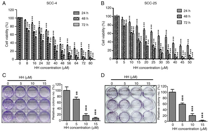
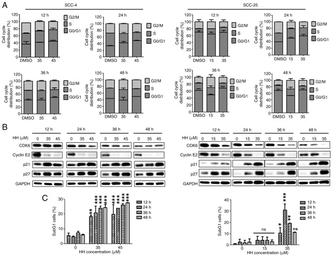
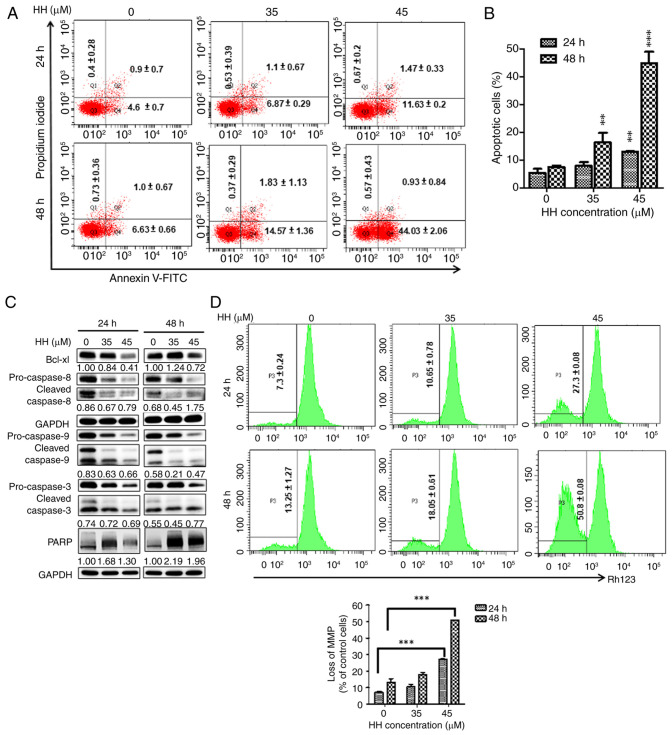
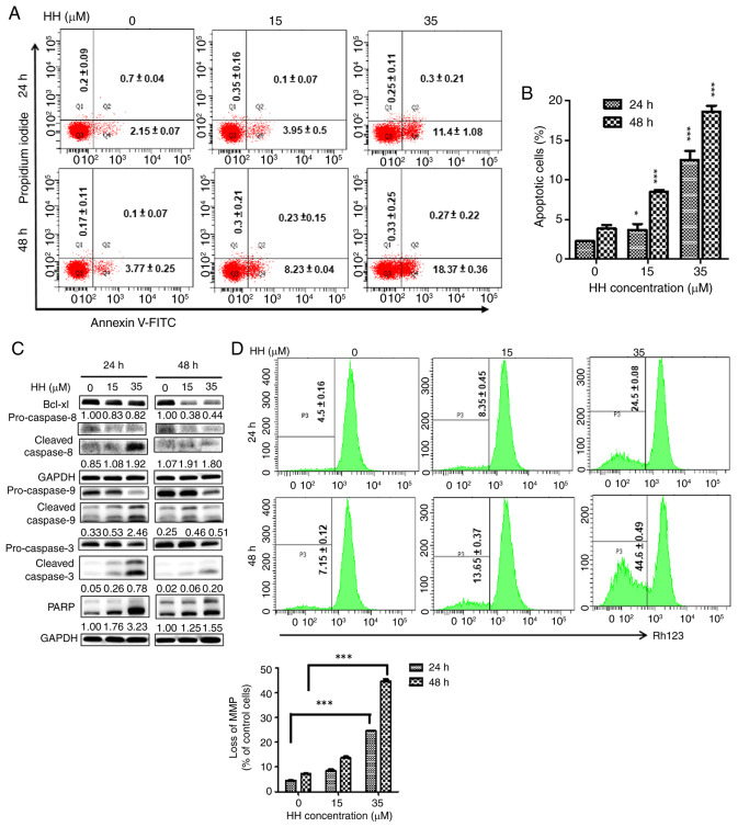
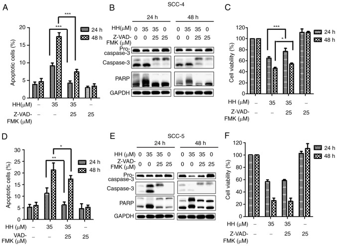
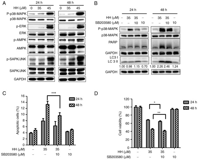
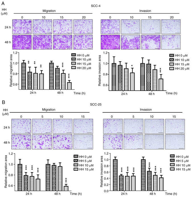
Oral squamous cell carcinoma (OSCC) represents the most frequently occurring form of oral cancer. However, despite the availability of advanced treatment modalities, the global 5-year survival rate for patients with advanced OSCC remains at ~50-60%. Devising alternative therapeutic strategies for oral cancer has therefore become an urgent need. Harmine, a β-carboline alkaloid, has recently been shown to exhibit anticancer activity. Compared with harmine, harmine hydrochloride (HH), a derivative of harmine, has improved water solubility and stability, so can absorb into tissues more readily. Therefore, the present study aimed to investigate the anticancer activity of HH in OSCC cells. A Cell Counting Kit-8 assay was performed to assess the cytotoxic effects of HH on the OSCC cell lines, SCC-4 and SCC-25. Flow cytometric analysis was subsequently employed to examine both the cell cycle profile and the extent of apoptosis. Western blotting was used to assess the expression levels of the regulatory proteins involved in these biological activities, and treatment with a pan-caspase inhibitor (Z-VAD-FMK) confirmed the involvement of the apoptotic pathway. Furthermore, western blotting was used to investigate which signaling pathways were affected in the HH-treated cells. Taken together, the findings of the present study demonstrated that HH was cytotoxic in OSCC cells. HH treatment induced G0/G1 phase cell cycle arrest and apoptosis. Additionally, the MAPK pathway was shown to be involved in HH-induced apoptosis in SCC-4 cells. Therefore, HH exhibited anticancer activity, and may be a putative therapeutic agent for the treatment of OSCC in the future.

Keywords: antitumor activity; apoptosis; harmine hydrochloride; oral squamous cell carcinoma.


Conflict of interest statement

The authors declare that they have no competing interests.

Figures

Figure 1
Figure 1
Cytotoxicity of HH on oral cancer cells. (A) SCC-4 cells were treated with the indicated concentration of HH for 24, 48 and 72 h and cell viability was assessed using the Cell Counting Kit-8 assay. In SCC-4 cells treated with HH, cell viability was significantly reduced compared with the control group (HH 0 µM) at 16 µM following 24 h and at 8 µM following 48 or 72 h. (B) SCC-25 cells were treated with the indicated concentration of HH for 24, 48 and 72 h and cell viability was assessed using the Cell Counting Kit-8 assay. In SCC-25 cells treated with HH, cell viability was significantly reduced compared with the control group (HH 0 µM) at 10 µM following 24 h and at 5 µM following 48 or 72 h. (C) SCC-4 and (D) SCC-25 cells were treated with HH for 7 days and the colony formation capacity of cell was assessed. Colonies were quantified using ImageJ software (version 1.43, National Institutes of Health). HH 0 µM represents DMSO as the solvent for dissolving HH and serves as the control group. Data are presented as mean ± SD (n=3). **P<0.01 and ***P<0.001. HH, harmine hydrochloride.
Figure 2
Figure 2
HH induces G0/G1 cell cycle arrest in oral cancer cells. (A) SCC-4 (left panel) and SCC-25 (right panel) cells were treated with the indicated concentrations of HH for 12, 24, 36 or 48 h, followed by cell cycle distribution analysis using flow cytometric analysis. (B) SCC-4 (left panel) and SCC-25 (right panel) cell cycle regulation protein expression was assessed using western blotting analysis. (C) The proportion of sub-G1 phase cells after HH treatment is shown. HH 0 µM represents DMSO as the solvent for dissolving HH and serves as the control group. Data are presented as mean ± SD (n=3). *P<0.05, **P<0.01, ***P<0.01 vs. control. HH, harmine hydrochloride; ns, not significant.
Figure 3
Figure 3
HH increases apoptosis of SCC-4 cells. The lower right quadrant represents early apoptotic cells, indicated as (FITC+/PI-); the upper right quadrant represents late apoptotic cells, indicated as (FITC+/PI+); the upper left quadrant represents necrotic cells, indicated as (FITC-/PI+); and the lower left quadrant represents live cells, indicated as (FITC-/PI-). The total number of apoptotic cells is the sum of the cells in the lower right and upper right quadrants. (A) SCC-4 cells were treated with HH and apoptotic cells were assessed via flow cytometric analysis. (B) The percentage of apoptotic cells was quantified from three independent experiments. (C) Apoptosis-associated protein expression levels following HH treatment were analyzed using western blotting. (D) MMP was evaluated using flow cytometric analysis in SCC-4 cells following HH treatment. HH 0 µM represents DMSO as the solvent for dissolving HH and serves as the control group. Data are presented as mean ± SD (n=3). **P<0.01, ***P<0.001 vs. control. HH, harmine hydrochloride; MMP, mitochondrial membrane potential; PARP, poly (ADP-ribose) polymerase; Rh123, rhodamine 123.
Figure 4
Figure 4
HH increases apoptosis of SCC-25 cells. The lower right quadrant represents early apoptotic cells, indicated as (FITC+/PI-); the upper right quadrant represents late apoptotic cells, indicated as (FITC+/PI+); the upper left quadrant represents necrotic cells, indicated as (FITC-/PI+); and the lower left quadrant represents live cells, indicated as (FITC-/PI-). The total number of apoptotic cells is the sum of the cells in the lower right and upper right quadrants. (A) SCC-25 cells were treated with HH and apoptotic cells were analyzed using flow cytometry. (B) The percentage of apoptotic cells was determined. (C) Apoptosis-associated protein expression levels following HH treatment were analyzed using western blotting analysis. (D) MMP was analyzed using flow cytometry in SCC-25 cells following HH treatment. HH 0 µM represents DMSO as the solvent for dissolving HH and serves as the control group. Data are presented as mean ± SD (n=3). *P<0.01, ***P<0.001 vs. control. HH, harmine hydrochloride; MMP, mitochondrial membrane potential; PARP, poly (ADP-ribose) polymerase; Rh123, rhodamine 123.
Figure 5
Figure 5
HH-mediated increase in apoptosis HH in oral cancer cells is caspase-dependent. Prior to the application of HH, SCC-4 cells were pretreated with Z-VAD-FMK. (A) The apoptotic cells were subjected to flow cytometric analysis. (B) The expression levels of caspase-3 and PARP proteins were analyzed using western blotting. (C) Cell viability was determined by CCK-8 assay. Before the application of HH, SCC-25 cells were pretreated with Z-VAD-FMK. (D) The apoptotic cells were analyzed using flow cytometric analysis. (E) Western blotting was employed to detect the protein expression levels of caspase-3 and PARP. (F) Cell viability was determined using the CCK-8 assay. HH 0 µM represents DMSO as the solvent for dissolving HH and serves as the control group. Data are presented as mean ± SD (n=3). *P<0.05, **P<0.01, ***P<0.001 vs. control. HH, harmine hydrochloride; CCK-8, Cell Counting Kit-8; PARP, poly (ADP-ribose) polymerase; Z, Z-VAD-FMK; ns, not significant.
Figure 6
Figure 6
HH-mediated apoptosis in SCC-4 cells is mediated via activation of the MAPK pathway. (A) SCC-4 cells were treated with HH for 24 and 48 h. The protein expression levels of p38, ERK, AMPK and JNK proteins was analyzed by western blotting. (B) SCC-4 cells were pre-incubated with SB203580 prior to HH treatment and the protein expression levels of PARP and LC3 were assessed using western blotting. (C) The ratio of apoptotic cells was determined using flow cytometric analysis. *P<0.05, **P<0.01 and ***P<0.001. HH, harmine hydrochloride; p, phosphorylated.
Figure 7
Figure 7
HH inhibits OSCC cells migration and invasion. (A) SCC-4 or (B) SCC-25 cells were seeded onto the upper chamber of the inserts and treated with HH. For the invasion assay, Matrigel was added to the upper chamber. After 24 and 48 h, non-migrated or non-invaded cells were scraped off, and the cells that passed through the insert were stained with crystal violet. The ratio of migration and invasion cells was determined using ImageJ software (version 1.43, National Institutes of Health). Scale bar, 1 mm. HH 0 µM represents DMSO as the solvent for dissolving HH and serves as the control group. *P<0.05, **P<0.01, ***P<0.001 vs. control. HH, harmine hydrochloride.

Similar articles

References

    1. Dong L, Xue L, Cheng W, Tang J, Ran J, Li Y. Comprehensive survival analysis of oral squamous cell carcinoma patients undergoing initial radical surgery. BMC Oral Health. 2024;24(919) doi: 10.1186/s12903-024-04690-z. - DOI - PMC - PubMed
    1. Siegel RL, Giaquinto AN, Jemal A. Cancer statistics, 2024. CA Cancer J Clin. 2024;74:12–49. doi: 10.3322/caac.21820. - DOI - PubMed
    1. Pekarek L, Garrido-Gil MJ, Sánchez-Cendra A, Cassinello J, Pekarek T, Fraile-Martinez O, García-Montero C, Lopez-Gonzalez L, Rios-Parra A, Álvarez-Mon M, et al. Emerging histological and serological biomarkers in oral squamous cell carcinoma: Applications in diagnosis, prognosis evaluation and personalized therapeutics (Review) Oncol Rep. 2023;50(213) doi: 10.3892/or.2023.8650. - DOI - PMC - PubMed
    1. Tan Y, Wang Z, Xu M, Li B, Huang Z, Qin S, Nice EC, Tang J, Huang C. Oral squamous cell carcinomas: State of the field and emerging directions. Int J Oral Sci. 2023;15(44) doi: 10.1038/s41368-023-00249-w. - DOI - PMC - PubMed
    1. Deng H, Sambrook PJ, Logan RM. The treatment of oral cancer: An overview for dental professionals. Aust Dent J. 2011;56:244–252. doi: 10.1111/j.1834-7819.2011.01349.x. - DOI - PubMed

LinkOut - more resources