. 2025 Apr 2:16:1501907.
doi: 10.3389/fimmu.2025.1501907. eCollection 2025.

Nasal delivery of killed Bacillus subtilis spores protects against influenza, RSV and SARS-CoV-2

Affiliations

Nasal delivery of killed Bacillus subtilis spores protects against influenza, RSV and SARS-CoV-2

Rong Xu et al. Front Immunol. .

Abstract

Introduction: Spores of the bacterium Bacillus subtilis (B. subtilis) have been shown to carry a number of properties potentially beneficial for vaccination. Firstly, as vehicles enabling mucosal delivery of heterologous antigens and secondly, as stimulators of innate immunity. Here, we have examined the specificity of protection conferred by the spore-induced innate response, focusing on influenza H1N1, respiratory syncytial virus (RSV), and coronavirus-2 (SARS-CoV-2) infections.

Methods: In vivo viral challenge murine models were used to assess the prophylactic anti-viral effects of B. subtilis spores delivered by intranasal instilling, using an optimised three-dose regimen. Multiple nasal boosting doses following intramuscular priming with SARS-CoV-2 spike protein was also tested for the capability of spores on enhancing the efficacy of parenteral vaccination. To determine the impact of spores on immune cell trafficking to lungs, we used intravascular staining to characterise cellular participants in spore-dosed pulmonary compartments (airway and lung parenchyma) before and after viral challenge.

Results: We found that mice pre-treated with spores developed resistance to all three pathogens and, in each case, exhibited a significant improvement in both survival rate and disease severity. Intranasal spore dosing expanded alveolar macrophages and induced recruitment of leukocyte populations, providing a cellular mechanism for the protection. Most importantly, virus-induced inflammatory leukocyte infiltration was attenuated in spore-treated lungs, which may alleviate the associated collateral tissue damage that leads to the development of severe conditions. Remarkably, spores were able to promote the induction of tissue-resident memory T cells, and, when administered following an intramuscular prime with SARS-CoV-2 spike protein, increased the levels of anti-spike IgA and IgG in the lung and serum.

Conclusions: Taken together, our results show that Bacillus spores are able to regulate both innate and adaptive immunity, providing heterologous protection against a variety of important respiratory viruses of high global disease burden.

Keywords: Bacillus spores; SARS-CoV-2; influenza; mucosal immunity; respiratory syncytial virus (RSV).


Conflict of interest statement

Authors HH and SC were employed by the company SporeGen Ltd. Authors YH and LN were employed by company Huro Biotech. Authors DH and WL were employed by company Destiny Pharma Plc. The remaining authors declare that the research was conducted in the absence of any commercial or financial relationships that could be construed as a potential conflict of interest.

Figures

Figure 1
Figure 1
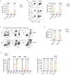
Three weekly doses of heat-killed B. subtilis protect mice from IAV and RSV infections. (A), Schematic graph showing the spore dosing regimens used in this study. (B–E), male C57BL/6 mice (n = 5 per group) were dosed intranasally with 2 or 3 doses of heat-killed spores (1 dose per week) or DPBS control, and challenged with a lethal dose of H1N1 (A/PR/8 strain, 30 PFU/50μl) influenza virus 7-days following the last nasal administration of spores. (B) Survival of infected mice. Mice were weighed and scored daily for 14 days following the infection. The change of body weight (C) was calculated as percentages to the starting weight recorded at the day of infection. (D) Average disease scores for each group, assessed as indicated. (E) Lung viral loads were determined by 50% tissue culture infectious dose (TCID50) at 2, 4, 6 days post the infection, n = 5 per group at all three timepoints. (F, G), Female BALB/c mice were treated with 3 weekly doses of heat-killed spores and challenged with RSV (2.5 x 10⁵ PFU/50μl). (F) Change of body weight following the viral challenge (n = 5 per group). (G) Lung viral titres in infected mice measured by immunoplaque assay at indicated timepoints, n = 5 per group for each timepoint. Data shown as mean ± SEM unless otherwise stated. Ordinary two-way ANOVA with Sidak’s multiple comparisons test was used for statistical analysis of (E, G). Data were from one of two independent experiments. p values that are lower than 0.05 are indicated.
Figure 2
Figure 2
Spore dosing confers protection against SARS-CoV-2 infections. (A–E), male hACE2 transgenic mice were dosed intranasally with 3 weekly doses of heat-killed spores or DPBS as controls and left for a week prior to the viral challenge with SARS-CoV-2 Beta variant. Infected mice were monitored daily for 14 days, and the disease progress was assessed by survival (A), weight loss (B) and severity scores (C). Data shown were pooled from 2 independent experiments, n = 5 for the mock infection group; n = 15 for SARS-CoV-2 infected groups with or without spore pre-treatment. Survival curves were compared using the Log-rank (Mantel-Cox) test. (D) Lung viral RNA copies in infected mice at day 2, 4, 6 post challenge, n = 5 per group for each timepoint. Data were analysed by ordinary two-way ANOVA with Sidak’s multiple comparisons test. (E) IHC staining of lung tissue sections for SARS-CoV-2 nucleocapsid, counterstained with hematoxylin (represents n = 3 mice per group). Scale bars, 50 μm. B, bronchiole; V, vasculature. (F) Male hACE2 transgenic mice were treated with 3 doses of spores and left for 27 days before infected with SARS-CoV-2 Omicron variant. The change of body weight post viral infection is presented as means ± SEM, n = 5 per group.
Figure 3
Figure 3
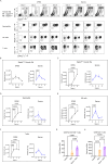
Spore dosing induces immune cell recruitment and expansion of alveolar macrophages. (A–E), male C57BL/6 mice were dosed intranasally with 3 doses of heat-killed spores or control (DPBS). Immune cell recruitment in the lung was analysed 7 days after the last dose of spores. Intravascular staining with CD45.2-FITC was performed to distinguish lung parenchymal and vascular leukocytes. (A) Numbers of neutrophils (CD45+CD11b+Ly6G+), NK cells (CD45+CD3-NKp46+) and T cells (CD45+CD11b-CD3+) harvested from bronchoalveolar lavage (BAL), n = 7 per group pooled from 2 independent experiments. (B) Representative contour plots showing percentage of infiltrated neutrophils (top panel), NK cells (middle panel) and T cells (bottom panel) in the lung parenchyma (gated as CD45.2- populations) following spore dosing. Numbers of these parenchymal immune cells are shown by the scattered dot plot with bars, n = 5 per group. (C) Flow cytometry gating of 2 alveolar macrophages (Mϕ) populations based on the expression levels of SiglecF in spore-treated lungs, and the numbers of SiglecFhigh and SiglecFlow alveolar Mϕ are shown in (D), n = 5 per group. Data were from one of two independent experiments. (E) Levels of cytokines in BAL, n = 8 for the DPBS control group and n = 9 for the spore-dosed group. Data were pooled from two independent experiment. Ordinary two-way ANOVA with Sidak’s multiple comparisons test was used for analysis.
Figure 4
Figure 4
Altered leukocyte trafficking and enhanced TRM induction in spore-dosed lungs following viral infections. (A–F), male C57BL/6 mice were dosed intranasally with 3 doses of heat-killed spores or controls (DPBS) and challenged by H1N1 (A/PR/8 strain) influenza virus 7 days following the last spore dose. Lung tissues were collected on day 0, 2, 4, 6 post viral challenge for analysis, n = 3 per group for each timepoint. Ordinary one-way ANOVA with Dunnett’s multiple comparison tests was used for analysis. Data were from one of two independent experiments. (A), Representative contour plots showing the proportions of SiglecFhigh and SiglecFlow alveolar Mϕ within total CD45+ cells, and percentages of infiltrated neutrophils, NK cells and T cells in the lung parenchyma (gated as CD45.2- populations) following viral challenge. (B, C) Numbers of SiglecFhigh (B) and SiglecFlow (C) alveolar Mϕ over the infection course in spore pre-treated and DPBS pre-treated control mice. (D–F) Numbers of infiltrated (CD45.2- parenchymal cells) neutrophils (D), NK cells (E) and T cells (F) over the infection course. (G, H) Female BALB/c mice were treated with 3 weekly doses of heat-killed spores or control treated with DPBS and challenged by RSV. Numbers of tissue resident memory CD8+ T cells (CD69+CD103+, G) and CD4+ T cells (CD69+, H) in Bronchoalveolar lavage (BAL) harvested at day 4 post viral challenge. n = 3 for the Naïve and n = 5 per group for the RSV infected. Data were from one of two independent experiments and analysed using ordinary two-way ANOVA with Sidak’s multiple comparisons test.
Figure 5
Figure 5
Intranasal spore dosing boosts antibody production against SARS-CoV-2 spike protein. Mice were intranasally administrated with killed spores following a parenteral prime. The dosing regimens for the four study groups (n = 7 per group, pooled from two independent experiments) are shown in (A). Group 1 was a naïve control group with animals receiving no treatment. Groups 2-4 all received an intra-muscular prime of recombinant Spike protein (5 μg). Three weeks later Groups 3 and 4 were dosed intranasally with spores using either 4 doses spread over 2-weeks (Group 3; days 21, 23, 35 & 37) or 9 consecutive doses (Group 4; days 21, 23, 25, 27, 29, 31, 33, 35 & 37). Group 2 received PBS as controls (days 21, 23, 25 & 37). Anti-Spike IgG in serum (B) and anti-Spike SIgA in lungs (C) was measured at day 49 and day 50 respectively. Data were analysed using ordinary two-way ANOVA with Tukey’s multiple comparisons test.

References

    1. Troeger C, Blacker B, Khalil IA, Rao PC, Cao J, Zimsen SRM, et al. . Estimates of the global, regional, and national morbidity, mortality, and aetiologies of lower respiratory infections in 195 countries, 1990–2016: a systematic analysis for the Global Burden of Disease Study 2016. Lancet Infect Diseases. (2018) 18:1191–210. doi: 10.1016/S1473-3099(18)30310-4 - DOI - PMC - PubMed
    1. Nazareth J, Pan D, Martin CA, Barr I, Sullivan SG, Stephenson I, et al. . Is the UK prepared for seasonal influenza in 2022-23 and beyond? Lancet Infect Dis. (2022) 22:1280–1. doi: 10.1016/S1473-3099(22)00503-5 - DOI - PMC - PubMed
    1. Nicholson WL. Roles of Bacillus endospores in the environment. Cell Mol Life Sci. (2002) 59:410–6. doi: 10.1007/s00018-002-8433-7 - DOI - PMC - PubMed
    1. Hong HA, Hitri K, Hosseini S, Kotowicz N, Bryan D, Mawas F, et al. . Mucosal antibodies to the C terminus of toxin A prevent colonization of clostridium difficile . Infect Immun. (2017) 85(4):e01060-16. doi: 10.1128/IAI.01060-16 - DOI - PMC - PubMed
    1. Duc le H, Hong HA, Fairweather N, Ricca E, Cutting SM. Bacterial spores as vaccine vehicles. Infect Immun. (2003) 71:2810–8. doi: 10.1128/IAI.71.5.2810-2818.2003 - DOI - PMC - PubMed

MeSH terms

LinkOut - more resources