

Parvalbumin-positive primary afferent projections to motoneurons increase after complete spinal transection in neonatal and juvenile rats
- PMID: 40242793
- PMCID: PMC12002873
- DOI: 10.1016/j.ibneur.2025.03.011
Parvalbumin-positive primary afferent projections to motoneurons increase after complete spinal transection in neonatal and juvenile rats
Abstract
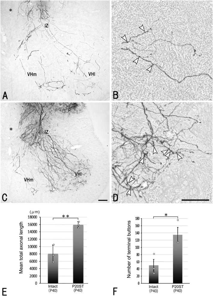
Hindlimb locomotor activity spontaneously recovers after complete spinal cord transection (CST) in neonatal rats, but not in juvenile rats. A previous study in neonatal rats that underwent CST at the thoracic level demonstrated that primary afferent projections increase in the ventral horn and intermediate zone at the lumbar level. It remains unclear whether primary afferent terminals of motoneurons increase and whether primary afferent projections to the spinal cord are altered after CST in juvenile rats. Here, we used biotinylated dextran amine as a tracer to demonstrate that primary afferent projections to the ventral horn and intermediate zone were significantly increased in rats that underwent CST in the juvenile period compared to intact rats of the same age. We then examined Ⅰa afferents using immunohistochemistry for parvalbumin. Our findings revealed an increase in parvalbumin-immunoreactive terminals on motoneurons in both neonatal and juvenile rats after CST compared to intact rats of the same age. These results suggest that proprioceptive afferent terminals on motoneurons are increased after CST in both neonatal and juvenile rats. In neonatal rats, this increase might contribute to the spontaneous recovery of hindlimb motor activity after CST, whereas in juvenile rats, the increase in proprioceptive afferent terminals on motoneurons does not contribute to recovery following CST.
Keywords: Juvenile; Motoneurons; Neonate; Parvalbumin; Primary afferents; Spinal cord injury.
© 2025 The Authors.
Conflict of interest statement
The authors declare that they have no known competing financial interests or personal relationships that could have appeared to influence the work reported in this paper.
Figures


References
-
- Barber R.P., Phelps P.E., Houser C.R., Crawford G.D., Salvaterra P.M., Vaughn J.E. The morphology and distribution of neurons containing choline acetyltransferase in the adult rat spinal cord: an immunocytochemical study. J. Comp. Neurol. 1984;229:329–346. - PubMed
-
- Carr P.A., Yamamoto T., Karmy G., Baimbridge K.G., Nagy J.I. Parvalbumin is highly colocalized with calbindin D28k and rarely with calcitonin gene-related peptide in dorsal root ganglia neurons of rat. Brain Res. 1989;497:163–170. - PubMed
-
- Celio M.R. Calbindin D-28k and parvalbumin in the rat nervous system. Neuroscience. 1990;35:375–475. - PubMed
-
- Krassioukov A.V., Karlsson A.K., Wecht J.W., Wuermser L.A., Mathias C.J., Marino R.J. Assessment of autonomic dysfunction following spinal cord injury: rationale for additions to International Standards for Neurological Assessment. J. Rehabil. Res. Dev. 2007;44:103–112. - PubMed
LinkOut - more resources
Full Text Sources