. 2025 Jun 3;13(6):e0251824.
doi: 10.1128/spectrum.02518-24. Epub 2025 Apr 15.

Inter-kingdom interactions and environmental influences on the oral microbiome in severe early childhood caries

Affiliations

Inter-kingdom interactions and environmental influences on the oral microbiome in severe early childhood caries

Lingjia Weng et al. Microbiol Spectr. .

Abstract

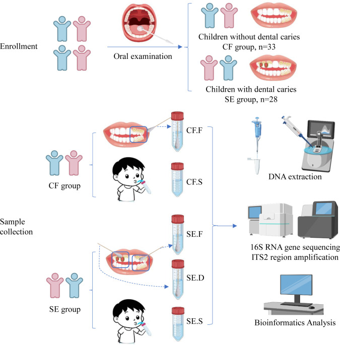
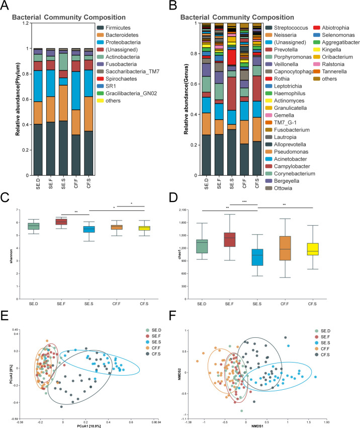
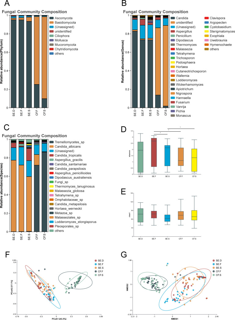
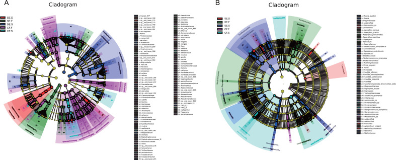
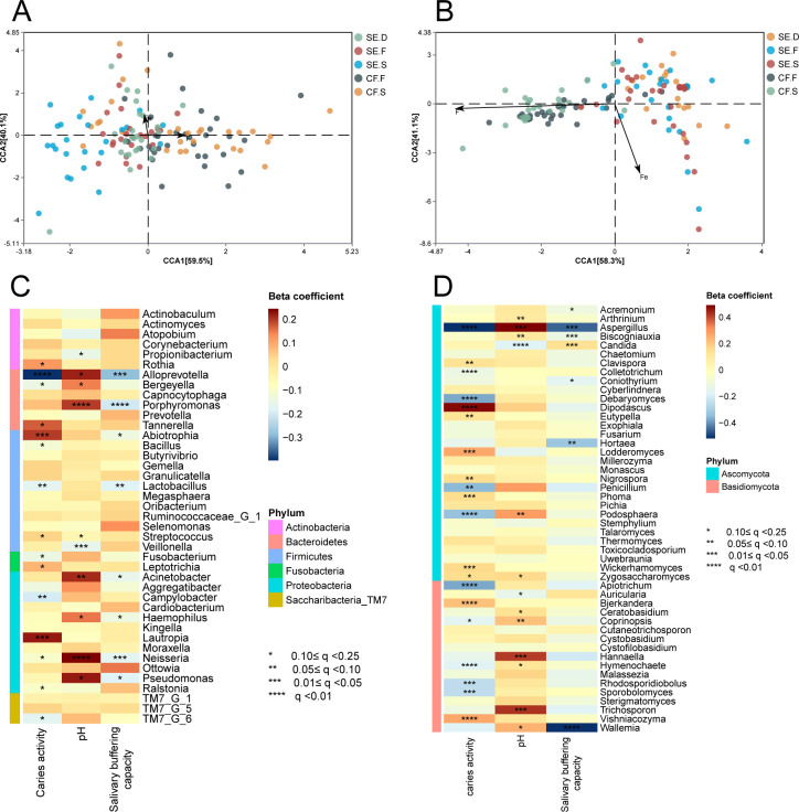
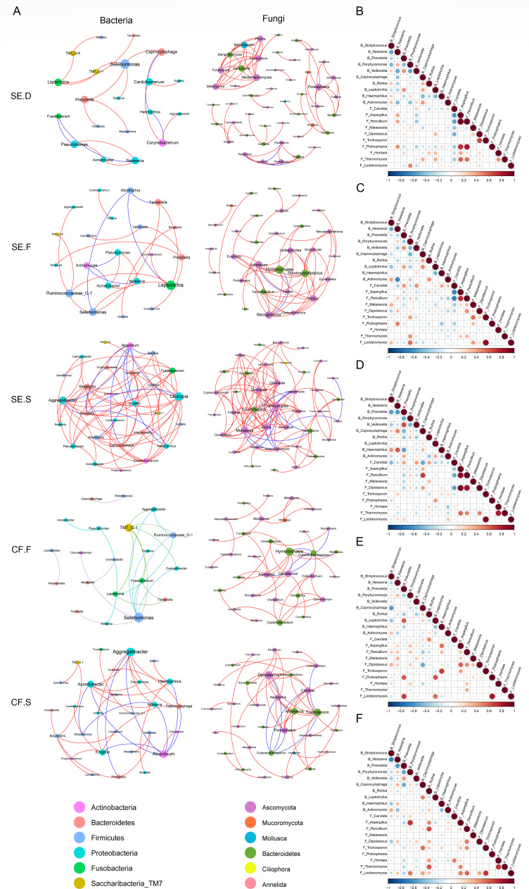
Dental caries arise from intricate interactions among oral microorganisms, impacting ecological stability and disease progression. In this study, we aimed to investigate the microbial diversity and inter-kingdom interactions in severe early childhood caries (S-ECC) and assess the influence of environmental factors such as salivary pH and trace elements. We analyzed 61 children aged 3-4 years with complete deciduous dentition, evaluating salivary pH, buffering capacity, and trace elements (iron, fluoride). We examined the performance of 16S rRNA V1-V9 regions gene and internal transcribed spacer (ITS) primers for bacteria and fungi from plaque and saliva to characterize community compositions and diversity. Findings revealed significant shifts in bacterial diversity in S-ECC saliva samples, marked by decreased diversity and elevated abundance of cariogenic species, particularly Streptococcus mutans. Candida albicans was notably more prevalent in the S-ECC group, implicating its potential role in pathogenesis. Iron and fluoride concentrations showed no significant correlation with microbial community structure. Network analyses uncovered complex intra- and inter-kingdom interactions, underscoring cooperative and competitive dynamics. S-ECC children exhibited higher abundances of bacteria (Streptococcus mutans, Granulicatella, Actinomyces) and fungi (Candida albicans), with specific microbial taxa associated with reduced salivary pH.

Importance: This study illuminates the intricate relationship between bacteria and fungi within the oral microbial community of children, specifically highlighting differences between those with S-ECC and those without caries. Through an extensive analysis of the microbial composition in both saliva and dental plaque, we identified a significant increase in the abundance of specific bacterial taxa (e.g., S. mutans, Granulicatella, Actinomyces) and fungal species (e.g., C. albicans) in the oral cavities of children with S-ECC. This finding underscores the potential role of these microorganisms in the development of caries. Contrary to previous studies that emphasize the importance of iron and fluoride in oral health, our research found no significant correlation between the concentrations of these elements and the composition of oral microbial communities. This result challenges conventional understanding and opens new avenues for future research. Additionally, our findings revealed an association between Veillonella sp., Propionibacterium sp., and Candida sp. and reduced salivary pH. This provides novel insights into the relationship between the oral microenvironment and caries development. The implications of our findings are substantial for the development of prevention and intervention strategies targeting childhood caries. They also underscore the critical need for a deeper exploration of oral microbial interactions and their environmental influences.

Keywords: Candida species; bacteria; bacterial-fungal interactions; dental plaque; early childhood caries; fungi; microbial ecology; saliva.


Conflict of interest statement

The authors declare no conflict of interest.

Figures

Fig 1
Fig 1
Eligible children were enrolled in the study and divided into the severe early (SE) and caries-free (CF) groups after oral examination. In both groups, mixed supragingival plaque and nonirritating saliva were collected from available healthy tooth surfaces. In the SE group, mixed supragingival plaque from caries-damaged tooth surfaces was also collected. The samples from different groups and sections were then subjected to DNA extraction and analysis.
Fig 2
Fig 2
Species diversity of bacteria in various spatial locations of the oral cavity. Bacterial phylum (A) and genus (B) composition of the SE.C, SE.F, SE.S, CF.F, and CF.S groups. The Alpha diversity index of each group was compared using the Shannon (C) and Chao1 indices (D). The distribution of the oral bacterial community in each group is shown through bacterial community PCoA (E) and NMDS analysis (F). The analysis revealed that the bacterial community structures of saliva and plaque samples were completely separate from each other.
Fig 3
Fig 3
Species diversity of fungi in various spatial locations of the oral cavity. The fungal phyla (A), genera (B), and species (C) of the SE.C, SE.F, SE.S, CF.F, and CF.S groups were examined. The analysis compared the Shannon index (D) and Chao1 index (E) among different groups. PCoA (F) and NMDS analysis (G) were conducted to investigate the distribution status of the oral fungal communities in each group. The results showed that saliva and plaque samples have distinct fungal community structures.
Fig 4
Fig 4
Differential bacteria (A) and fungi (B) at various sites in the oral cavities. LEfSe was used to analyze the species that differed among the groups. In the evolutionary branching diagram, circles radiating from inside to outside represent taxonomic levels from phylum to genus (or species). Each small circle at a different taxonomic level represents a taxon at that level. The size of the circle diameter is proportional to the relative abundance. Species without significant differences are uniformly colored in yellow. Different species follow the group for coloring. Different nodes are colored to represent microbial taxa that play an important role in the group. The Linear Discriminant Analysis (LDA) value is set to 3.
Fig 5
Fig 5
Microenvironmental factors analysis. RDA of the microbial communities and iron and fluorine concentrations at each spatial site in the SE and CF groups. (A) Bacteria and (B) fungi. The arrows in the figure represent iron and fluorine, and the different dots represent the samples. The correlation is represented by the angle between the arrows. The length of the arrows represents the magnitude of the correlation, with longer arrows indicating a greater effect on the microorganisms. MaAsLin analysis of salivary pH and buffering capacity of bacterial communities (C) and fungal communities (D).
Fig 6
Fig 6
Network analysis and correlation analysis of bacteria and fungi. Network analysis of bacteria and fungi within each site of the SE group and CF group (A). A line between two species indicates a significant correlation (Spearman’s rank correlation ρ > 0.6, P < 0.01). The greater the number of lines between nodes and the larger the radius of the nodes are, the greater the correlation. Red represents a positive correlation between species, and blue represents a negative correlation between species. Correlation analysis of bacteria and fungi within each site of the SE group and CF group (B, SE.D; C, SE.F; D, SE.S; E, CF.F; F, CF.S). "B_ stands for bacteria" and "F_ stands for fungi." Red and blue represent positive and negative correlations, respectively. The color shade and size of the circles are proportional to the correlation coefficient, and white represents no correlation.

Similar articles

References

    1. Anderson AC, Rothballer M, Altenburger MJ, Woelber JP, Karygianni L, Lagkouvardos I, Hellwig E, Al-Ahmad A. 2018. In-vivo shift of the microbiota in oral biofilm in response to frequent sucrose consumption. Sci Rep 8:14202. doi:10.1038/s41598-018-32544-6 - DOI - PMC - PubMed
    1. Rosier BT, De Jager M, Zaura E, Krom BP. 2014. Historical and contemporary hypotheses on the development of oral diseases: are we there yet? Front Cell Infect Microbiol 4:92. doi:10.3389/fcimb.2014.00092 - DOI - PMC - PubMed
    1. Cui Y, Wang Y, Zhang Y, Pang L, Zhou Y, Lin H, Tao Y. 2021. Oral mycobiome differences in various spatial niches with and without severe early childhood caries. Front Pediatr 9. doi:10.3389/fped.2021.748656 - DOI - PMC - PubMed
    1. Kalan L, Loesche M, Hodkinson BP, Heilmann K, Ruthel G, Gardner SE, Grice EA. 2016. Redefining the chronic-wound microbiome: fungal communities are prevalent, dynamic, and associated with delayed healing. MBio 7:e01058-16. doi:10.1128/mBio.01058-16 - DOI - PMC - PubMed
    1. Hoarau G, Mukherjee PK, Gower-Rousseau C, Hager C, Chandra J, Retuerto MA, Neut C, Vermeire S, Clemente J, Colombel JF, Fujioka H, Poulain D, Sendid B, Ghannoum MA. 2016. Bacteriome and mycobiome interactions underscore microbial dysbiosis in familial Crohn’s disease. MBio 7:e01250-16. doi:10.1128/mBio.01250-16 - DOI - PMC - PubMed

Substances

LinkOut - more resources