. 2025 May 21;91(5):e0016625.
doi: 10.1128/aem.00166-25. Epub 2025 Apr 17.

Characterization of 2-phenanthroyl-CoA reductase, an ATP-independent type III aryl-CoA reductase involved in anaerobic phenanthrene degradation

Affiliations

Characterization of 2-phenanthroyl-CoA reductase, an ATP-independent type III aryl-CoA reductase involved in anaerobic phenanthrene degradation

Nadia A Samak et al. Appl Environ Microbiol. .

Abstract

Anaerobic degradation of polycyclic aromatic hydrocarbons (PAHs) with three or more aromatic rings is extremely slow because the compounds are very poorly soluble in water and chemically stable. Phenanthrene is the only three-ring PAH where the anaerobic degradation has been partially elucidated. Phenanthrene is first activated via carboxylation producing 2-phenanthroate, which is further converted to 2-phenanthroyl-coenzyme A (CoA) via the enzyme 2-phenanthroate:CoA ligase. In this study, we elucidated the next degradation step, the reduction of 2-phenanthroyl-CoA to dihydro-2-phenanthroyl-CoA. We cloned the putative gene from the genome of culture TRIP_1 and heterologously expressed and purified the 2-phenanthroyl-CoA reductase enzyme from Escherichia coli. The identified monomeric flavo-enzyme belongs to the novel group of type III aryl-CoA reductases in the old-yellow enzyme family and has a molecular mass of 72 kDa. 2-Phenanthroyl-CoA reductase contains one FMN, one FAD, and one [4Fe-4S] iron-sulfur cluster as cofactors. The enzyme has a specific activity of 17.6 ± 0.4 nmol/min/mg, a Km value of 1.8 µM, and a Vmax of 7.9 µmol/min/mg at pH 7.5, when reduced methyl viologen was used as electron donor. 2-Phenanthroyl-CoA reductase catalyzed a two-electron reduction step producing one of five possible isomers. Quantum mechanical calculations and nuclear magnetic resonance analysis of the reaction product suggested 9,10-dihydro-2-phenanthroyl-CoA as the most stable isomer. However, our experimental evidence suggests 7,8-dihydro-2-phenanthroyl-CoA (International Union of Pure and Applied Chemistry [IUPAC]: 1,2-dihydro-7-phenanthroyl-CoA) or 5,6-dihydro-2-phenanthroyl-CoA (IUPAC: 3,4-dihydro-7-phenanthroyl-CoA) as the most likely reduced product with a saturated bond in ring 3 of the substrate 2-phenanthroyl-CoA, before undergoing isomerization changes to reach the more stable structure of 9,10-dihydro-2-phenanthroyl-CoA.IMPORTANCEPAHs are a group of highly toxic and persistent environmental pollutants. The anaerobic degradation of three-ring PAHs like phenanthrene is still poorly understood. Phenanthrene degradation starts with a carboxylation reaction to form 2-phenanthroic acid followed by a CoA-thioesterification reaction catalyzed by 2-phenanthroate:CoA ligase to produce 2-phenanthroyl-CoA. The next degradation step is the reduction of 2-phenanthroyl-CoA to dihydro-2-phenanthroyl-CoA to overcome the resonance energy of the aromatic ring system. Herein, we elucidated that the reduction reaction is catalyzed by the enzyme 2-phenanthroyl-CoA reductase. Furthermore, we provided biochemical and structural properties of the heterologously expressed and purified 2-phenanthroyl-CoA reductase, which confirmed that the enzyme belongs to the novel group of type III aryl-CoA reductases in the old-yellow enzyme family.

Keywords: 2-phenanthroyl-CoA; PAHs; anaerobic phenanthrene degradation; culture TRIP_1; old-yellow enzyme; reductase.


Conflict of interest statement

The authors declare no conflict of interest.

Figures
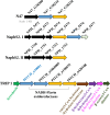
Fig 1
Fig 1
Comparison of the location of the 2-phenanthroyl-CoA reductase gene (PITCH_a10001) in the metagenome of culture TRIP_1 and the homologous genes in the metagenomes of the naphthalene-degrading cultures N47 and NaphS2. The genes in cultures N47 (N47_G38210, 5,6-dihydro-2-naphthoyl-CoA reductase) and NaphS2 (NPH_5476, 5,6-dihydro-2-naphthoyl-CoA reductase) that are similar to the 2-phenanthroyl-CoA reductase gene (PITCH_a10001 and PITCH_a1860005) in TRIP_1 are presented as blue arrows. Yellow arrows represent the homology between the 2-naphthoyl-CoA reductase gene in cultures N47 (N47_G38220), NaphS2 (NPH_5475 and NPH_1753), and culture TRIP_1 (PITCH_a190075 and PITCH_a420108). The image also indicates the localization of the oxidoreductase genes in the context of putative genes for β-oxidation (enoyl-CoA hydratase/hydrolase/isomerase, β-hydroxyacyl-CoA dehydrogenase, β-hydroxyacyl-CoA dehydratase, and β-oxoacyl-CoA thiolase).
Fig 2
Fig 2
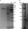
SDS-PAGE (a) and NATIVE-PAGE analysis (b) of 2-phenanthroyl-CoA reductase produced from the recombinant plasmid PASG-IBA103-PITCH_a10001. (a) M, protein MW standard; lane 1, E. coli cell-free extract; lane 2, 2-phenanthroyl-CoA reductase after purification with a Strep-Tactin resin. (b) M, protein MW standard; lane 2, purified 2-phenanthroyl-CoA reductase in blue native gel.
Fig 3
Fig 3
UV/vis spectra of the purified 2-phenanthroyl-CoA reductase as isolated and after reduction of the enzyme in steps of 0.05 mM sodium dithionite each. Complete reduction was achieved with 0.15 mM sodium dithionite, and the enzyme was re-oxidized by exposure to oxygen.
Fig 4
Fig 4
Protein-ligand interaction profile (PLIP) of FMN in (A) 2-phenanthroyl-CoA reductase and (B) 2-naphthoyl-CoA reductase. Blue solid lines show hydrogen bonds; gray dotted lines denote hydrophobic interactions; and yellow/orange dotted lines show salt bridges. PLIP of FAD in (C) 2-phenanthroyl-CoA reductase and (D) 2-naphthoyl-CoA reductase. Blue solid lines show hydrogen bonds; gray dotted lines show hydrophobic interactions; green dotted lines show π-stacking interactions; and white solid lines show water bridges. The labels of the interacting amino acid side chains are colored in the color corresponding to the interaction type (34, 35).
Fig 5
Fig 5
LC/MS chromatograms (Shimadzu single quadrupole mass-spectrometer) showing the time-dependent conversion of 2-phenanthroyl-CoA to the reduced product dihydro-2-phenanthroyl-CoA by purified 2-phenanthroyl-CoA reductase. The red line indicates the substrate 2-phenanthroyl-CoA (m/z = 972, positive ion mode) and the black line indicates the dihydro-2-phenanthroyl-CoA produced after 90 min (m/z = 974, positive ion mode). The position of the saturated bond in ring 3 of dihydro-2-phenanthroyl-CoA is not known and only shown exemplarily.
Fig 6
Fig 6
High-resolution mass spectrometry spectrum of the product dihydro-2-phenanthroyl-CoA (m/z = 974) in positive ion mode showing the fragmentation of the compound to coenzyme A (m/z = 768, the right side of the cleavage mark) and dihydro-2-phenanthroic acid (m/z = 224, the left side of the cleavage mark). The position of the saturated bond in ring 3 of dihydro-2-phenanthroyl-CoA is not known and only shown exemplarily.
Fig 7
Fig 7
Calculated energies (in kilojoules per mole) of the three possible dihydro-2-phenanthroyl-CoA isomers relative to the starting materials were calculated using B3LYP(CPCM)/6–311 + G(d,p). Isomer 3, 9,10-dihydro-2-phenanthroyl-CoA; isomer 4, 5,6-dihydro-2-phenanthroyl-CoA (IUPAC: 3,4-dihydro-7-phenanthroyl-CoA); and isomer 5, 7,8-dihydro-2-phenanthroyl-CoA (IUPAC: 1,2-dihydro-7-phenanthroyl-CoA).
Fig 8
Fig 8
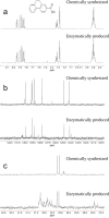
NMR spectra of the carboxylic acid of the isomer obtained by enzymatic reactions (bottom) and chemically synthesized 9,10-dihydrophenanthrene-2-carboxylic acid (top) highlighting their structural identity. (a) 1H NMR spectra of the protons on the aromatic rings (7.7–8.0 ppm) and of the methylene protons (2.9 ppm). (b) 13C NMR spectra of the protonated carbons in the aromatic rings. Due to the low sample quantity, the signals of the quaternary carbons could not be observed for the enzymatically produced sample. (c) 13C NMR spectra of the two methylene carbons (29.9 and 30.0 ppm). Full spectra are shown in the supporting information including a complete assignment of all resonances.
Fig 9
Fig 9
Proposed upper part of the anaerobic phenanthrene degradation pathway showing the elucidated anaerobic degradation reactions of phenanthrene to dihydro-2-phenanthroyl-CoA. Five possible isomers are shown as possible reaction products of 2-phenanthroyl-CoA reductase. Isomer 1, 3,4-dihydro-2-phenanthroyl-CoA; isomer 2, 4a,10a-dihydro-2-phenanthroyl-CoA; isomer 3, 9,10-dihydro-2-phenanthroyl-CoA; isomer 4, 5,6-dihydro-2-phenanthroyl-CoA (IUPAC: 3,4-dihydro-7-phenanthroyl-CoA); or isomer 5, 7,8-dihydro-2-phenanthroyl-CoA (IUPAC: 1,2-dihydro-7-phenanthroyl-CoA), are the most likely reduction products. Known enzyme reactions are depicted in black. The enzyme reaction studied here is shown in blue.
Fig 10
Fig 10
Proposed reduction mechanism of 2-phenanthroyl-CoA by 2-phenanthroyl-CoA reductase with possible resonance structure to the formation of 7,8-dihydro-2-phenanthroyl-CoA (IUPAC: 1,2-dihydro-7-phenanthroyl-CoA) (isomer 5 in Fig. 9). Compound b is a Meisenheimer complex-analogous transition state followed by stabilization by the CoA ester (compound c).

Similar articles

References

    1. Gao P, da Silva E, Hou L, Denslow ND, Xiang P, Ma LQ. 2018. Human exposure to polycyclic aromatic hydrocarbons: metabolomics perspective. Environ Int 119:466–477. doi:10.1016/j.envint.2018.07.017 - DOI - PubMed
    1. Adeniji AO, Okoh OO, Okoh AI. 2019. Levels of polycyclic aromatic hydrocarbons in the water and sediment of buffalo river estuary, South Africa and their health risk assessment. Arch Environ Contam Toxicol 76:657–669. doi:10.1007/s00244-019-00617-w - DOI - PMC - PubMed
    1. Meckenstock RU, Boll M, Mouttaki H, Koelschbach JS, Cunha Tarouco P, Weyrauch P, Dong X, Himmelberg AM. 2016. Anaerobic degradation of benzene and polycyclic aromatic hydrocarbons. J Mol Microbiol Biotechnol 26:92–118. doi:10.1159/000441358 - DOI - PubMed
    1. Friedman CL, Zhang Y, Selin NE. 2014. Climate change and emissions impacts on atmospheric PAH transport to the Arctic. Environ Sci Technol 48:429–437. doi:10.1021/es403098w - DOI - PubMed
    1. Lee Y, Lee Y, Jeon CO. 2019. Biodegradation of naphthalene, BTEX, and aliphatic hydrocarbons by Paraburkholderia aromaticivorans BN5 isolated from petroleum-contaminated soil. Sci Rep 9:860. doi:10.1038/s41598-018-36165-x - DOI - PMC - PubMed

LinkOut - more resources