. 2025 Mar-Apr;32(2):e70039.
doi: 10.1111/xen.70039.

Sex Differences in Maturation and Function of Neonatal Porcine Islets Upon Transplantation in Mice

Affiliations

Sex Differences in Maturation and Function of Neonatal Porcine Islets Upon Transplantation in Mice

Nerea Cuesta-Gomez et al. Xenotransplantation. 2025 Mar-Apr.

Abstract

Background: Neonatal porcine islets (NPIs) can mature into a mixed population of endocrine cells that can restore glucose control in mice, pigs, and non-human primates, representing a potential alternative islet source for clinical beta cell replacement therapy. However, it remains unclear how conditions in the recipient influence the maturation and function of these cells. Here, we investigated the impact of host sex on NPIs implanted under the kidney capsule of male and female B6.129S7-Rag1tm1Mom (B6/Rag-/-) mice.

Methods: Diabetic mice were transplanted with 3000 NPIs under the kidney capsule. All mice were monitored for reversal of hyperglycemia and glucose clearance at 8- and 20-weeks post-transplant. Grafts were assessed for cell composition and insulin content.

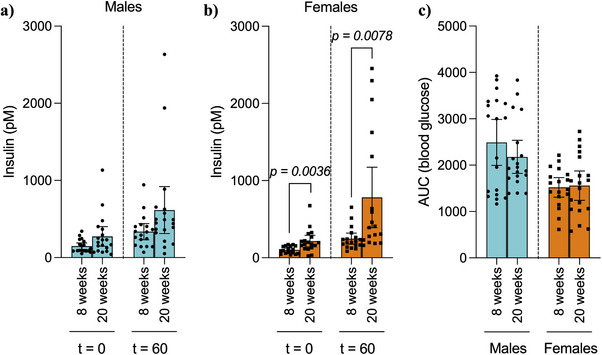
Results: Female mice demonstrated improved glucose clearance at 8- and 20-weeks post-transplant compared to their male counterparts. Improved glucose clearance correlated with accelerated diabetes reversal in females (8 weeks vs. 12 weeks in males) and increased rates of euglycemic achievement (17/18 in females vs. 14/19 in males). However, grafts collected from male mice exhibited an increased percentage of insulin-positive cells as well as increased insulin content.

Conclusion: The sex of the host influences the outcomes of NPI transplantation, showcasing the relevance of understanding the role of sex as a biological variable in islet transplantation.

Keywords: biological variables; diabetes; islet transplantation; neonatal porcine islets; sex differences; xenotransplantation.


Figures

FIGURE 1
FIGURE 1
Metabolic follow‐up of diabetic male and female B6.129S7‐Rag1tm1Mom (B6/Rag−/−) mice transplanted with neonatal porcine islets under the kidney capsule. (a) Weekly blood glucose measurements throughout the follow‐up period. Black arrow indicates graft retrieved, at which point mice reverted back to a hyperglycemic state. (b) Significance of weekly blood glucose values determined by area under the curve analysis. (c) Percentage of euglycemic animals throughout the follow‐up period. (d) Representation of time (weeks) to achieve euglycemia. Statistical difference amongst groups was conducted using the unpaired t‐test with Welch's correction. Kaplan‐Meier survival curves were compared via log‐rank statistical testing (Mantel‐Cox). Values are presented as means with standard deviation.
FIGURE 2
FIGURE 2
Intraperitoneal glucose challenge and stimulated porcine insulin secretion at 8 weeks post‐transplant. (a) Blood glucose levels, (b) porcine serum insulin levels, and (c) calculated stimulation index after an overnight fast and 60 min post‐intraperitoneal glucose administration (3 mg/g of weight). (d) Blood glucose levels during an IPGTT and (e) area under the curve for respective IPGTT. Statistical difference amongst groups was conducted using the unpaired t‐test with Welch's correction. Values are presented as means with standard deviation.
FIGURE 3
FIGURE 3
Intraperitoneal glucose challenge and stimulated porcine insulin secretion at 20 weeks post‐transplant. (a) Blood glucose levels, (b) porcine serum insulin levels, and (c) calculated stimulation index after an overnight fast and 60 min post‐intraperitoneal glucose administration (3 mg/g of weight). (d) Blood glucose levels during an IPGTT and (e) area under the curve for respective IPGTT. Statistical difference amongst groups was conducted using the unpaired t‐test with Welch's correction. Values are presented as means with standard deviation.
FIGURE 4
FIGURE 4
Comparison of basal and stimulated serum porcine insulin levels between 8‐ and 20‐weeks post‐transplant diabetic male and female B6.129S7‐Rag1tm1Mom (B6/Rag−/−) mice transplanted with neonatal porcine islets under the kidney capsule. (a) Male and (b) female porcine insulin levels after an overnight fast and 60 min post‐glucose administration (3 mg/g of weight) via intraperitoneal injection at 8‐ and 20‐weeks post‐transplantation. (c) Determination of the significance was measured as the area under the curve of blood glucose levels after an overnight fast and 15, 30, 60, 90, and 120 min after glucose (3 mg/g of weight) administration at 8‐ and 20‐weeks post‐transplantation. Statistical difference amongst groups was conducted using the unpaired t‐test with Welch's correction.
FIGURE 5
FIGURE 5
Morphological assessment of neonatal porcine islet grafts in diabetic male and female B6.129S7‐Rag1tm1Mom (B6/Rag−/−) mice. (a) Representative images of hematoxylin/Eosin, insulin (INS), and glucagon (GCG) staining of the NPI grafts in male and female mice. (b) Grafts in male and female mice were quantified for total graft area (mm2), (c) % total insulin, and (d) glucagon‐positive cells. (e) Grafts in the male and female mice were also quantified for total cellular porcine insulin content (µg) Statistical difference amongst groups was conducted using the unpaired t‐test with Welch's correction. Values are presented as means with standard deviation.

Similar articles

References

    1. Shapiro A. M., Lakey J. R., Ryan E. A., et al., “Islet Transplantation in Seven Patients With Type 1 Diabetes Mellitus Using a Glucocorticoid‐Free Immunosuppressive Regimen,” New England Journal of Medicine 343, no. 4 (2000): 230–238, 10.1056/NEJM200007273430401. - DOI - PubMed
    1. Marfil‐Garza B. A., Imes S., Verhoeff K., et al., “Pancreatic Islet Transplantation in Type 1 Diabetes: 20‐Year Experience From a Single‐Centre Cohort in Canada,” Lancet Diabetes & Endocrinology 10, no. 7 (2022): 519–532, 10.1016/S2213-8587(22)00114-0. - DOI - PubMed
    1. Ramzy A., Thompson D. M., Ward‐Hartstonge K. A., et al., “Implanted Pluripotent Stem‐Cell‐Derived Pancreatic Endoderm Cells Secrete Glucose‐Responsive C‐Peptide in Patients With Type 1 Diabetes,” Cell Stem Cell 28, no. 12 (2021): 2047–2061, 10.1016/j.stem.2021.10.003. e5. - DOI - PubMed
    1. Shapiro A. M. J., Thompson D., Donner T. W., et al., “Insulin Expression and C‐Peptide in Type 1 Diabetes Subjects Implanted With Stem Cell‐Derived Pancreatic Endoderm Cells in an Encapsulation Device,” Cell Reports Medicine 2, no. 12 (2021): 100466, 10.1016/j.xcrm.2021.100466. - DOI - PMC - PubMed
    1. Rezania A., Bruin J. E., Riedel M. J., et al., “Maturation of Human Embryonic Stem Cell‐Derived Pancreatic Progenitors Into Functional Islets Capable of Treating Pre‐Existing Diabetes in Mice,” Diabetes 61, no. 8 (2012): 2016–2029, 10.2337/db11-1711. - DOI - PMC - PubMed

Publication types