. 2025 May 14;16(5):e0315824.
doi: 10.1128/mbio.03158-24. Epub 2025 Apr 17.

Aeromonas hydrophila RTX adhesin has three ligand-binding domains that give the bacterium the potential to adhere to and aggregate a wide variety of cell types

Affiliations

Aeromonas hydrophila RTX adhesin has three ligand-binding domains that give the bacterium the potential to adhere to and aggregate a wide variety of cell types

Qilu Ye et al. mBio. .

Abstract

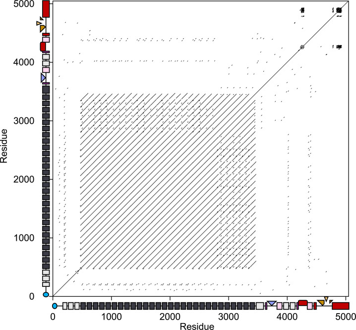
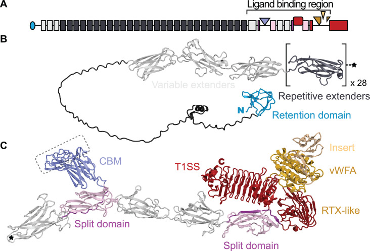
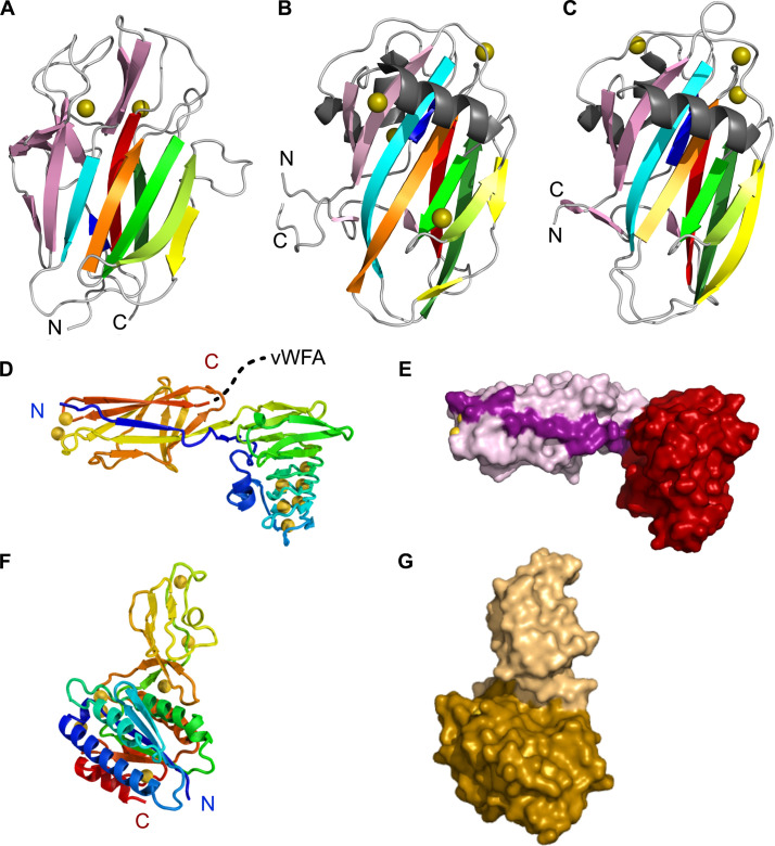
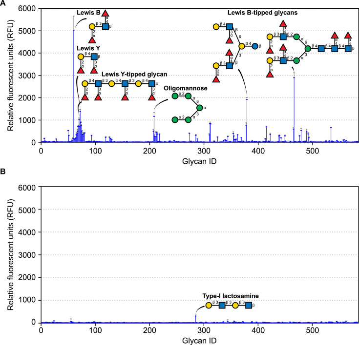
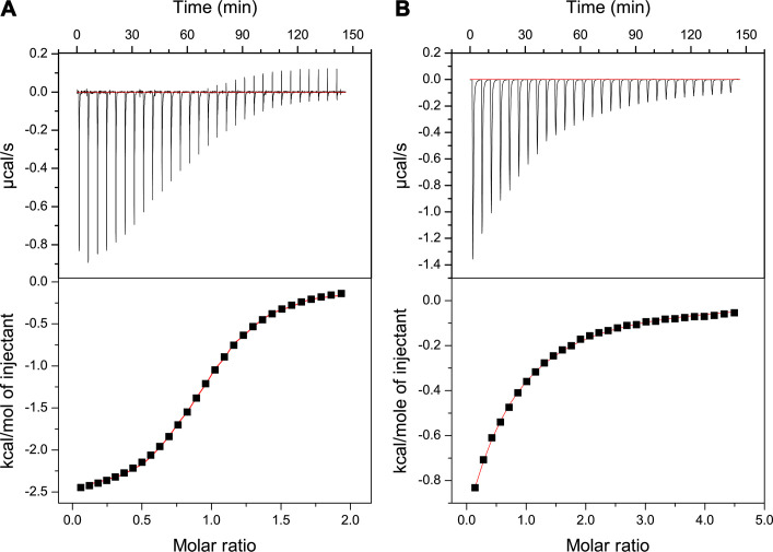
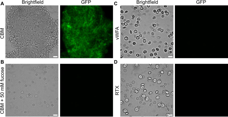
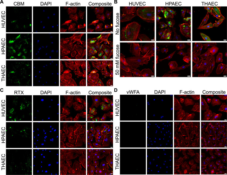
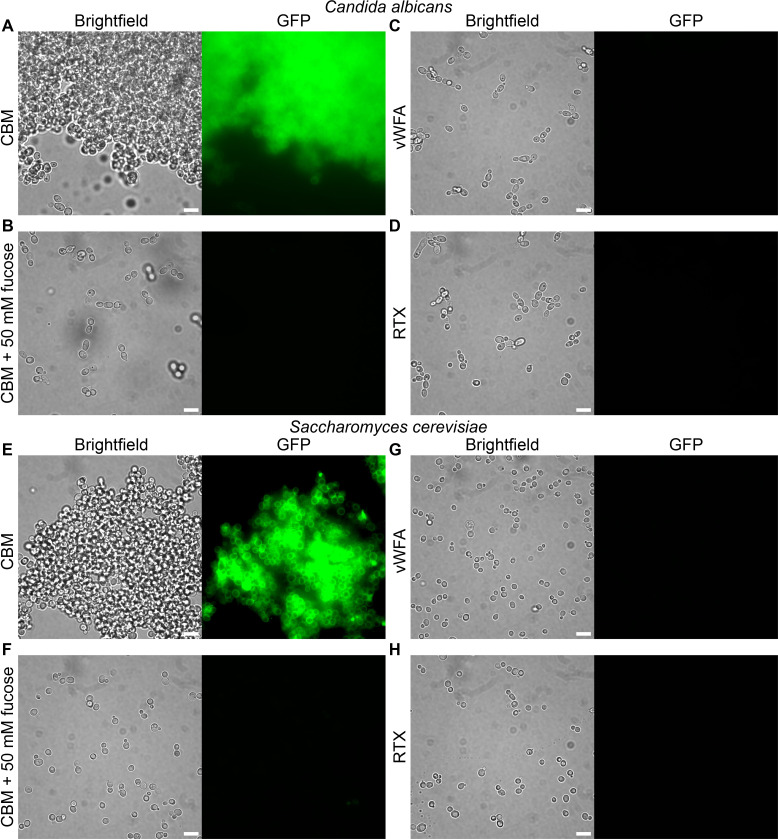
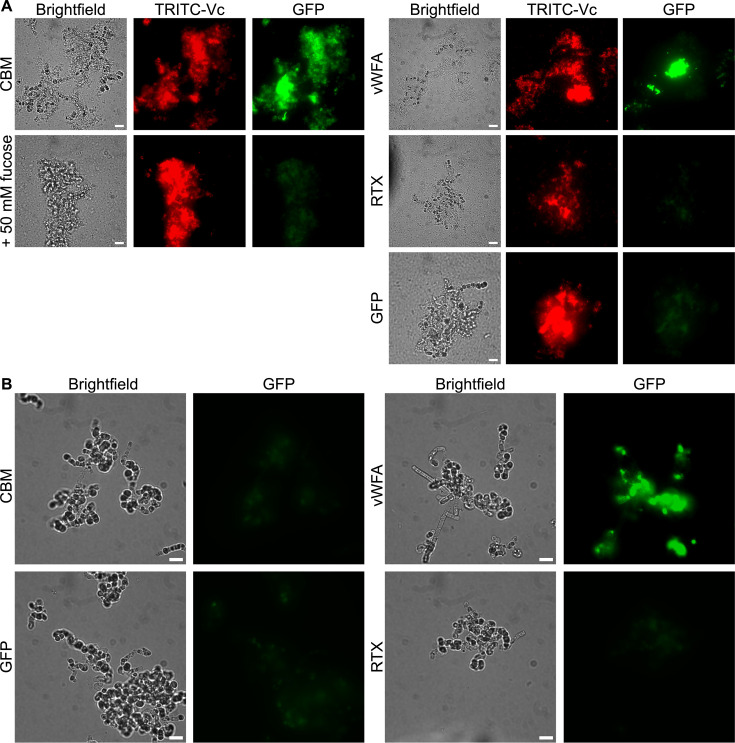
Bacteria often make initial contact with their hosts through the ligand-binding domains of large adhesin proteins. Recent analyses of repeats-in-toxin (RTX) adhesins in Gram-negative bacteria suggest that ligand-binding domains can be identified by the way they emerge from "split" domains within the adhesin. Here, using this criterion and an AlphaFold3 model of a 5047-residue RTX adhesin from Aeromonas hydrophila, we identified three different ligand-binding domains in this fibrillar protein. The crystal structures of the two novel domains were solved to 1.4 and 1.95 Å resolution, respectively, and demonstrate excellent agreement with their modeled structures. The other domain was recognized as a carbohydrate-binding module based on its beta-strand topology and confirmed by its micromolar affinity for fucosylated glycans, including the Lewis B and Y antigens. This lectin-like module, which was recombinantly produced with its companion split domain and nearby extender domain, bound to a wide variety of cells including yeasts, diatoms, erythrocytes, and human endothelial cells. In each case, 50 mM free fucose prevented this binding and may offer some protection from infection. The carbohydrate-binding module with its neighboring domains also caused aggregation of yeast and erythrocytes, which was again blocked by the addition of free fucose. The second putative ligand-binding domain has a beta-roll structure supported by a parallel alpha-helix, and the third is a homolog of a von Willebrand Factor A domain. These two domains bind to a more limited range of cell types, and their ligands have yet to be identified.IMPORTANCECharacterizing the ligand-binding domains of fibrillar adhesins is important for understanding how bacteria can colonize host surfaces and how this colonization might be blocked. Here, we show that the opportunistic pathogen, Aeromonas hydrophila, uses a carbohydrate-binding module (CBM) to attach to several different cell types. The CBM is one of three ligand-binding domains at the distal tip of the adhesin. Identifying the glycans bound by the CBM as Lewis B and Y antigens has helped explain the range of cell types that the bacterium will bind and colonize, and it has suggested sugars that might interfere with these processes. Indeed, fucose, which is a constituent of the Lewis B and Y antigens, is effective at 50 mM concentrations in blocking the attachment of the CBM to host cells. This will lead to the design of more effective inhibitors against bacterial infections.

Keywords: Aeromonas; adhesins; antimicrobial agents; binding proteins; biofilms; protein structure–function; receptor–ligand interaction.


Conflict of interest statement

The authors declare no conflict of interest.

Figures

Fig 1
Fig 1
Dot matrix analysis of AhLap against itself to detect repetitive regions. The x- and y-axes display the length of AhLap in amino acid residues alongside domain maps of the adhesin. The N-terminal retention domain is in blue, followed by grey and black rectangles representing the extender domains. In the ligand-binding region, Ig-like split domains are in pink, and the projected CBM, RTX domain, and vWFA domain are colored slate blue, dark red, and gold, respectively. The vWFA domain emerges from the C-terminal RTX domain (red). Dashes in the two-dimensional plot record matches with a threshold score of at least 30 using the BLOSUM62 matrix with a window size of 10 residues (37).
Fig 2
Fig 2
AlphaFold3 structure of AhLap in ribbon format in relation to the adhesin domain map. (A) Domain map of AhLap as presented in Fig. 1. (B) Structure of the retention domain (blue) and extender (light and dark grey) region of the adhesin. The square brackets around the dark grey extender domain indicate that there are 28 highly similar versions of this fold. The star denotes the end of the repetitive extender region. (C) Structure of the remainder of the AhLap adhesin from the star to the C-terminal end. The domains here in the ligand-binding region are colored as described in the Fig. 1 legend. The putative ligand-binding site on the CBM is indicated by the dashed line boundary. Note that the insert in the vWFA domain is colored yellow to distinguish it from the main section (gold). The N and C termini of these two adhesin halves are marked N in blue and C in red, respectively.
Fig 3
Fig 3
Structures of the three AhLap ligand-binding domains. (A) The AlphaFold3 model of AhLapCBM is shown as a nine-stranded beta-sandwich fold made up of a four-stranded antiparallel beta-sheet on one side and a five-stranded antiparallel beta-sheet on the other side. (B) Crystal structure of the CBM from MpIBP (PDB: 5J6Y) showing the similarity of its beta-strand topology (30). (C) Crystal structure of the CBM from MnLap (PDB: 6M8M), again showing the similarity of its beta-strand topology (38). (D) Crystal structure of the RTX-like two-domain construct (from residue 4168 to 4498) in rainbow ribbon format that traces the path of the polypeptide chain through the two domains. Numerous Ca2+ ions (gold spheres) are present, especially in the RTX solenoid domain. The dotted line from the C terminus leads into the vWFA domain. (E) Space-filling presentation of the RTX-like two-domain construct. The split domain strand that leads into the RTX domain is colored a darker pink than the rest of the split domain. (F) Crystal structure of one vWFA molecule from the crystal asymmetric unit. The rainbow ribbon tracing shows how the insert sequence buds out of the Rossman fold before the latter is complete. (G) Space-filling mode of the vWFA structure, with the main section corresponding to the Rossman fold colored gold and the insert sequence that is missing from some other vWFA domains colored beige.
Fig 4
Fig 4
Glycan array analysis. (A) Analysis using GFP-tagged AhLapCBM3 to probe 580+ mammalian glycans. Binding is shown in blue as relative fluorescence units with the standard deviation from four replicates indicated by error bars above the peaks. Six of the main glycans bound are shown in schematic representation where the sugar moieties are fucose (red triangles), galactose (yellow circles), glucose (blue squares), and mannose (green circles). (B) Analysis using GFP-tagged AhLapRTX domain showing the glycan structure of an interactor that is marginally above background levels.
Fig 5
Fig 5
Isothermal titration calorimetry. (A) Analysis of AhLapCBM3 binding Lewis B antigen. Top panel shows the thermogram for the interaction between Lewis B antigen and AhLapCBM3 plotted as µcal/s per event. The lower panel shows the fitted curve of these data from which the stoichiometry and KD were calculated. (B) Analysis of AhLapCBM3 binding free fucose. Top panel shows the thermogram for the interaction between l-fucose and AhLapCBM3 plotted as µcal/s per event. The lower panel shows the fitted curve of these data from which the KD was calculated.
Fig 6
Fig 6
Binding of AhLapCBM3 to human erythrocytes, with and without fucose present. (A) Erythrocytes incubated with GFP-tagged AhLapCBM3 construct that includes a split domain and one extender domain (Fig. 3A through C). The left-hand panel is viewed in brightfield and the right-hand panel is viewed to see the green fluorescence of GFP. (B) Erythrocytes incubated with the same GFP-tagged AhLapCBM3 construct in the presence of 50 mM fucose. (C) Erythrocytes incubated with the GFP-tagged AhLapvWFA construct. (D) Erythrocytes incubated with the GFP-tagged AhLapRTX construct. The white scale bar indicates 10 µm.
Fig 7
Fig 7
AhLapCBM3 binding to endothelial cells in the absence and presence of fucose. (A) The three images in the first column show fluorescence from the incubation of GFP-tagged AhLapCBM3 with human umbilical vein endothelial cells (HUVECs), human pulmonary artery endothelial cells (HPAECs), and telomerase-immortalized human aorta endothelial cells (THAECs). Images in the second column show the same cells stained with DAPI to visualize the nuclei. Images in the third column show the same cells stained with phalloidin to visualize the actin cytoskeleton. Images in the fourth column show composite images of the three staining patterns superimposed. (B) Composite staining images of the three endothelial cell types were repeated in the absence (top row) and presence (bottom row) of 50 mM fucose. The white scale bar indicates 10 µm. (C) The three images in the first column show fluorescence from the incubation of GFP-tagged AhLapRTX with HUVECs, HPAECs, and THAECs. Images in the second column show the same cells stained with DAPI to visualize the nuclei. Images in the third column show the same cells stained with phalloidin to visualize the actin cytoskeleton. Images in the fourth column show composite images of the three staining patterns superimposed. (D) The 12 panels shown here are equivalent to those in (A) and (C), except that the cells in the first column were probed with GFP-tagged AhLapvWFA.
Fig 8
Fig 8
AhLapCBM3 binding to yeasts in the presence and absence of fucose. (A) The two panels show Candida albicans incubated with GFP-labeled AhLapCBM3 and visualized in brightfield (left) and fluorescence (right). (B) The two panels repeat the conditions in (A) but with 50 mM fucose present. Panels (E) and (G) are repeats of (A) and (B) with Saccharomyces cerevisiae instead of C. albicans. (C) The two panels show C. albicans incubated with GFP-labeled AhLapvWFA and visualized in brightfield (left) and fluorescence (right). (D) The two panels show C. albicans incubated with GFP-labeled AhLapRTX and visualized in brightfield (left) and fluorescence (right). Panels (G) and (H) are repeats of (C) and (D) with S. cerevisiae instead of C. albicans. The white scale bar indicates 10 µm.
Fig 9
Fig 9
AhLap ligand-binding domain studies with diatoms, with and without V. cholerae present to generate biofilms. (A) The six panels on the left show binding of GFP-labeled AhLapCBM3 to a co-culture biofilm formed by TRITC-labeled V. cholerae on E. spinifer. The left-hand column of panels shows the brightfield view of the co-culture. The middle column shows the red TRITC fluorescence of V. cholerae colonizing E. spinifer. The right-hand column shows the green GFP fluorescence of bound AhLapCBM3. The top row of cells were incubated in the absence of fucose, and the bottom row of cells in the presence of 50 mM fucose. The nine panels on the right have the same horizontal sequence of brightfield, followed by TRITC staining to visualize V. cholerae, and GFP analysis. In the top sequence, the cells were incubated with GFP-labeled AhLapvWFA. In the middle sequence, the cells were labeled with AhLapRTX, and in the bottom sequence, the cells were mixed with free GFP to check for nonspecific labeling. (B) Here, diatoms in the absence of V. cholerae were incubated with the three GFP-tagged ligand-binding domains as indicated and with the GFP control. Brightfield and fluorescence images are compared side by side. The white scale bar indicates 10 µm.

Similar articles

Cited by

References

    1. Fernández-Bravo A, Figueras MJ. 2020. An update on the genus Aeromonas: taxonomy, epidemiology, and pathogenicity. Microorganisms 8:129. doi:10.3390/microorganisms8010129 - DOI - PMC - PubMed
    1. Janda JM, Abbott SL. 2010. The genus Aeromonas: taxonomy, pathogenicity, and infection. Clin Microbiol Rev 23:35–73. doi:10.1128/CMR.00039-09 - DOI - PMC - PubMed
    1. Cai W, Willmon E, Burgos FA, Ray CL, Hanson T, Arias CR. 2019. Biofilm and sediment are major reservoirs of virulent Aeromonas hydrophila (vAh) in catfish production ponds. J Aquat Anim Health 31:112–120. doi:10.1002/aah.10056 - DOI - PubMed
    1. Elhariry HM. 2011. Biofilm formation by Aeromonas hydrophila on green-leafy vegetables: cabbage and lettuce. Foodborne Pathog Dis 8:125–131. doi:10.1089/fpd.2010.0642 - DOI - PubMed
    1. Zepeda-Velazquez AP, Gómez-De-Anda F-R, Aguilar-Mendoza LF, Castrejón-Jiménez NS, Hernández-González JC, Varela-Guerrero JA, de-la-Rosa-Arana J-L, Vega-Sánchez V, Reyes-Rodríguez NE. 2023. Bullfrogs (Lithobates catesbeianus) as a potential source of foodborne disease. J Food Prot 86:100067. doi:10.1016/j.jfp.2023.100067 - DOI - PubMed

LinkOut - more resources