Functional Characterization of miR-216a-5p and miR-125a-5p on Pancreatic Cancer Stem Cells
- PMID: 40243417
- PMCID: PMC11988779
- DOI: 10.3390/ijms26072830
Functional Characterization of miR-216a-5p and miR-125a-5p on Pancreatic Cancer Stem Cells
Abstract
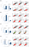
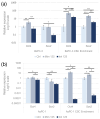
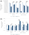
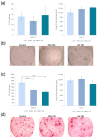
Pancreatic ductal adenocarcinoma (PDAC) is the third leading cause of cancer-related death. Its poor prognosis is closely related to late-stage diagnosis, which results from both nonspecific symptoms and the absence of biomarkers for early diagnosis. MicroRNAs (miRNAs) exert a regulatory role in numerous biological processes and their aberrant expression has been found in a broad spectrum of diseases, including cancer. Cancer stem cells (CSCs) represent a driving force for PDAC initiation, progression, and metastatic spread. Our previous research highlighted the interesting behavior of miR-216a-5p and miR-125a-5p related to PDAC progression and the CSC phenotype. The present study aimed to evaluate the effect of miR-216a-5p and miR-125a-5p on the acquisition or suppression of pancreatic CSC traits. BxPC-3, AsPC-1 cell lines, and their CSC-like models were transfected with miR-216a-5p and miR-125a-5p mimics and inhibitors. Following transfection, we evaluated their impact on the expression of CSC surface markers (CD44/CD24/CxCR4), ALDH1 activity, pluripotency- and EMT-related gene expression, and clonogenic potential. Our results show that miR-216a-5p enhances the expression of CD44/CD24/CxCR4 while negatively affecting the activity of ALDH1 and the expression of EMT genes. MiR-216a-5p positively influenced the clonogenic property. MiR-125a-5p promoted the expression of CD44/CD24/CxCR4 while inhibiting ALDH1 activity. It enhanced the expression of Snail, Oct-4, and Sox-2, while the clonogenic potential appeared to be affected. Comprehensively, our results provide further knowledge on the role of miRNAs in pancreatic CSCs. Moreover, they corroborate our previous findings about miR-216a-5p's potential dual role and miR-125a-5p's promotive function in PDAC.
Keywords: CSC; PDAC; functional study; miR-125a-5p; miR-216a-5p; miRNA; miRNA regulation.
Conflict of interest statement
The authors declare no conflicts of interest.
Figures


Similar articles
-
Unveiling the microRNA landscape in pancreatic ductal adenocarcinoma patients and cancer cell models.BMC Cancer. 2024 Oct 24;24(1):1308. doi: 10.1186/s12885-024-13007-w. BMC Cancer. 2024. PMID: 39448959 Free PMC article.
-
microRNA-21 Regulates Stemness in Pancreatic Ductal Adenocarcinoma Cells.Int J Mol Sci. 2022 Jan 24;23(3):1275. doi: 10.3390/ijms23031275. Int J Mol Sci. 2022. PMID: 35163198 Free PMC article.
-
miR-125a-3p is responsible for chemosensitivity in PDAC by inhibiting epithelial-mesenchymal transition via Fyn.Biomed Pharmacother. 2018 Oct;106:523-531. doi: 10.1016/j.biopha.2018.06.114. Epub 2018 Jul 11. Biomed Pharmacother. 2018. PMID: 29990840
-
The Multifaceted Role of miR-21 in Pancreatic Cancers.Cells. 2024 May 30;13(11):948. doi: 10.3390/cells13110948. Cells. 2024. PMID: 38891080 Free PMC article. Review.
-
Pancreatic cancer stem cell markers and exosomes - the incentive push.World J Gastroenterol. 2016 Jul 14;22(26):5971-6007. doi: 10.3748/wjg.v22.i26.5971. World J Gastroenterol. 2016. PMID: 27468191 Free PMC article. Review.
References
-
- Siegel R.L., Giaquinto A.N., Jemal A. Cancer Statistics. 2024. CA Cancer J. Clin. 2024;74:12–49. - PubMed
MeSH terms
Substances
LinkOut - more resources
Full Text Sources
Medical
Research Materials
Miscellaneous

