. 2025 Mar 21;26(7):2831.
doi: 10.3390/ijms26072831.

Reparixin as a Potential Antiepileptogenic Agent: Modulation of the CXCL1-CXCR1/2 Axis and Seizure Activity in a Kindling Rat Model of Temporal Lobe Epilepsy

Affiliations

Reparixin as a Potential Antiepileptogenic Agent: Modulation of the CXCL1-CXCR1/2 Axis and Seizure Activity in a Kindling Rat Model of Temporal Lobe Epilepsy

Nihan Çarçak et al. Int J Mol Sci. .

Abstract

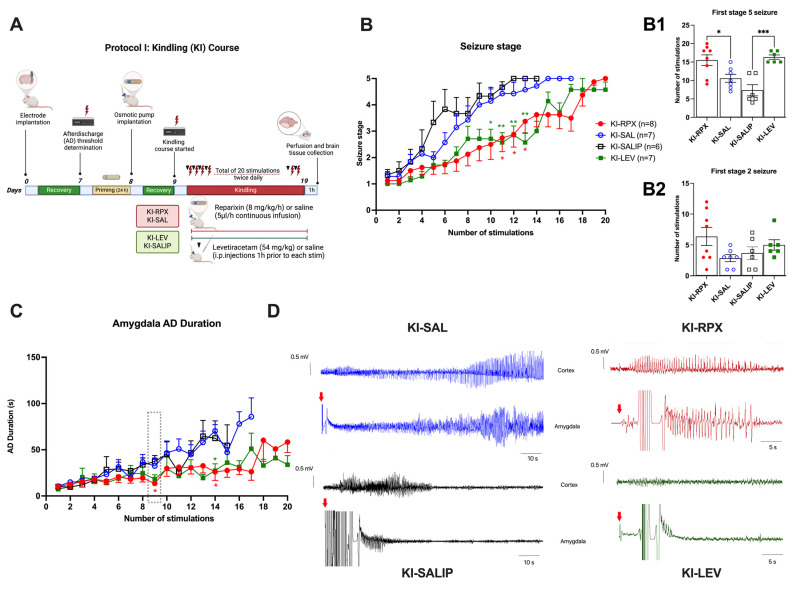
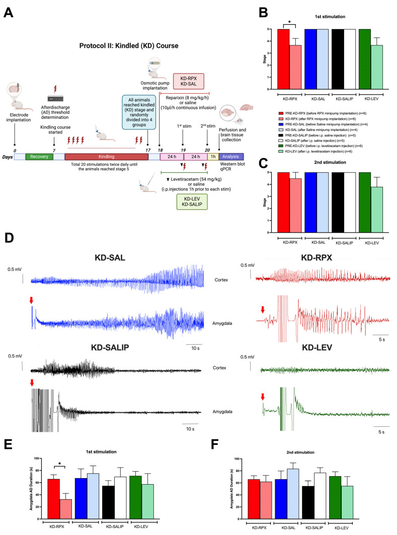
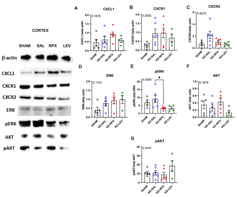
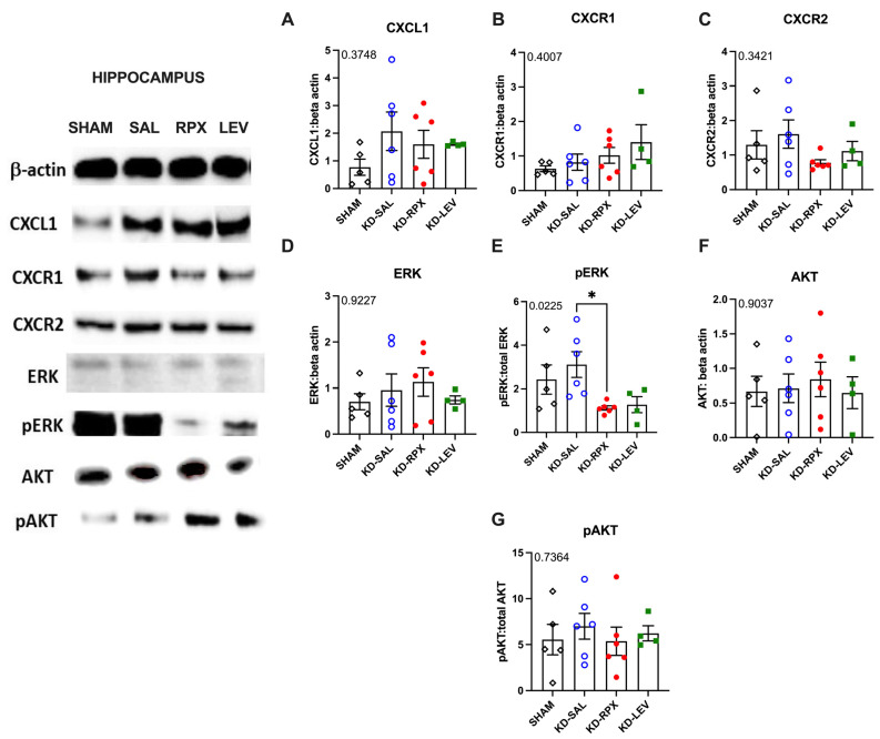
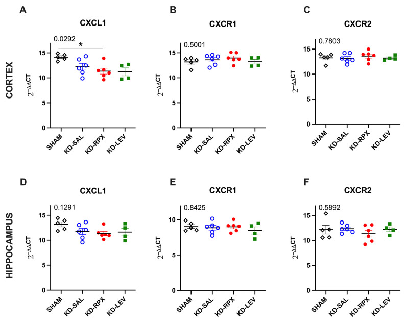
Chemokine (CXC motif) ligand 8 (CXCL8) is a pro-inflammatory chemokine binding to CXC motif receptors 1/2 (CXCR1/2). Patients with temporal lobe epilepsy (TLE) exhibit increased serum CXCL8 levels. CXC motif ligand 1 (CXCL1), a murine ortholog of CXCL8, has been implicated in seizure generation and neuronal loss. This study evaluated the antiepileptogenic and antiseizure effects of reparixin in amygdaloid kindling rat model of TLE. Reparixin was administered during the kindling period for 14 days, and seizures were induced twice daily via electrical stimulation. To assess the antiseizure effects, reparixin was administered to fully kindled animals, and stimulations were performed 24 and 48 h later. Levetiracetam, a broad-spectrum antiseizure drug, was administered intraperitoneally (i.p.) as positive control 1 h before each stimulation. Reparixin delayed secondary seizure generalization during kindling. Reparixin reduced seizure severity and after-discharge duration in fully kindled animals at 24 h from treatment initiation. CXCR1/2 and protein kinase B pathway proteins exhibited no significant changes; reparixin reduced the phospho-extracellular signal-regulated kinase (pERK)/ERK ratio in the cortex and hippocampus. CXCL1 expression was significantly decreased in the cortex. Reparixin exhibited antiepileptogenic and partial antiseizure effects by modulating the CXCL1-CXCR1/2 axis and reducing ERK signaling. Already in clinical trials on respiratory diseases, reparixin could be repurposed for epilepsy therapy.

Keywords: CXCL1; CXCR1/2 antagonist; amygdaloid kindling; antiepileptogenic; antiseizure therapy; neuroinflammation; reparixin.


Conflict of interest statement

L. Brandolini, A. Aramini, M. Allegretti and L. De Filippis are employed by Dompe’ Farmaceutici S.p.a. The other authors declare no conflicts of interest.

Figures

Figure 1
Figure 1
Antiepileptogenic effect of reparixin on kindling. Experimental plan (A) and seizure stages (B), the number of stimulations to reach first stage 5 seizure (B1) and stage 2 seizure (B2), and after-discharge (AD) durations (C) in the amygdala. The dotted line box indicates the 9th stimulation, during which EEG traces from the ipsilateral cortex and amygdala are represented for each group. In kindling (KI) groups treated with reparixin (KI-RPX, red traces) and levetiracetam (KI-LEV, green traces), after-discharges (ADs) induced by electrical stimulation of the amygdala are significantly shorter than in the control groups that received saline via an osmotic pump (KI-SAL, blue traces) or intraperitoneally (KI-SALIP, black traces). Red arrows indicate the timing of the electrical stimulation (D). Because of noise interference in EEG traces recorded from the amygdala, AD durations were evaluated using synchronized recordings from the ipsilateral cortex. Data are expressed as means ± standard errors of the mean. * p < 0.05, ** p < 0.01, *** p < 0.001. Experimental plan (A) was created in BioRender. https://BioRender.com where lightning bolts indicate electrical stimulations, and the black triangle represents the administration of levetiracetam one hour before each stimulation.
Figure 2
Figure 2
Antiseizure effect of reparixin in kindled (KD) rats. Experimental plan (A). The mean seizure stages after the first stimulation delivered 24 h after the osmotic pump implantation (B) and after the second stimulation delivered 48 h after the osmotic pump implantation (C). On the left, representative 110 s EEG traces from KD groups treated with saline via osmotic pump (KD-SAL, blue traces) or intraperitoneally (KD-SALIP, black traces) are indicated. On the right, representative 55 s traces from KD groups treated with reparixin (KD-RPX, red traces) or levetiracetam (54 mg/kg) (KD-LEV, green traces) are displayed. EEG traces, from both the amygdala and ipsilateral cortex showing ADs after the first stimulation (red arrows) delivered 24 h after the osmotic pump implantation (D). The mean AD durations in the ipsilateral amygdala after the first stimulation (E) and second stimulation (F). Data are expressed as means ± standard errors of the mean * p < 0.05. Experimental plan (A) was created in BioRender. https://BioRender.com, where lightning bolts indicate electrical stimulations, and the black triangle represents the administration of levetiracetam one hour before each stimulation.
Figure 3
Figure 3
The effects of reparixin and levetiracetam on the protein expression levels of CXCL1 (A), CXCR1 (B), CXCR2 (C), ERK (D), p-ERK/ERK (E), and AKT (F), and the ratios of p-AKT/AKT (G) in the parietal cortex of the KD groups treated with saline (KD-SAL), reparixin (KD-RPX), or levetiracetam (KD-LEV) and sham-operated nonepileptic animals (SHAM). Representative immunoblotting bands of the parietal cortex showing increased pERK protein levels under epileptic conditions and reduced by reparixin treatment. * p < 0.05 by Tukey’s post-hoc test. The number on the upper left corner of each panel is the p-value obtained by ANOVA. Data are expressed as means ± standard errors of the mean.
Figure 4
Figure 4
The effects of reparixin and levetiracetam on the protein expression levels of CXCL1 (A), CXCR1 (B), CXCR2 (C), ERK (D), p-ERK/ERK (E), and AKT (F) and the ratios of p-AKT/AKT (G) in the hippocampus of the KD groups treated with saline (KD-SAL), reparixin (KD-RPX), or levetiracetam (KD-LEV) and sham-operated nonepileptic animals (SHAM). Representative immunoblotting bands of the entire hippocampus showing increased pERK protein levels under epileptic conditions and reduced by reparixin treatment. * p < 0.05 by Tukey’s post-hoc test. The number on the upper left corner of each panel is the p-value obtained by analysis of variance (ANOVA). Data are expressed as means ± standard errors of the mean.
Figure 5
Figure 5
The effects of reparixin and levetiracetam on the mRNA expression levels of CXCL1, CXCR1, and CXCR2 in the cortex ((A), (B) and (C), respectively) and hippocampus ((D), (E) and (F), respectively) of the kindled (KD) groups treated with saline (KD-SAL), reparixin (KD-RPX), or levetiracetam (KD-LEV) and sham-operated nonepileptic animals (SHAM). * p < 0.05 by Tukey’s post-hoc test. The number on the upper left corner of each panel is the p-value obtained by analysis of variance (ANOVA). Horizontal lines indicate mean values. Data are expressed as means ± standard errors of the mean.

Similar articles

References

    1. Stafstrom C.E., Carmant L. Seizures and epilepsy: An overview for neuroscientists. Cold Spring Harb. Perspect. Med. 2015;5:a022426. doi: 10.1101/cshperspect.a022426. - DOI - PMC - PubMed
    1. Motovilov K., Maguire C., Briggs D., Melamed E. Altered cytokine profile in clinically suspected seronegative autoimmune associated epilepsy. medRxiv. 2024 doi: 10.1101/2024.09.13.24310337. - DOI
    1. Jauhari P., Kaur P., Gulati S., Meena A.K., Pandey T., Upadhyay A. Diagnostic and prognostic significance of serum interleukins in epileptic encephalopathy with spike wave activation in sleep (EE-SWAS) syndrome. Eur. J. Paediatr. Neurol. 2024;53:33–38. doi: 10.1016/j.ejpn.2024.09.006. - DOI - PubMed
    1. Kaczorowska M., Czekuć-Kryśkiewicz E., Dądalski M., Kotulska K. Immunological markers of drug resistant epilepsy and its response to immunomodulatory therapy with ACTH in children. Folia Neuropathol. 2023;61:360–370. doi: 10.5114/fn.2023.131662. - DOI - PubMed
    1. Milano C., Montali M., Barachini S., Burzi I.S., Pratesi F., Petrozzi L., Chico L., Morganti R., Gambino G., Rossi L., et al. Increased production of inflammatory cytokines by circulating monocytes in mesial temporal lobe epilepsy: A possible role in drug resistance. J. Neuroimmunol. 2024;386:578272. doi: 10.1016/j.jneuroim.2023.578272. - DOI - PubMed

LinkOut - more resources