. 2025 Mar 22;26(7):2883.
doi: 10.3390/ijms26072883.

The Role of FpfetC from Fusarium proliferatum in Iron Acquisition, Fumonisin B1 Production, and Virulence

Affiliations

The Role of FpfetC from Fusarium proliferatum in Iron Acquisition, Fumonisin B1 Production, and Virulence

Ling Wang et al. Int J Mol Sci. .

Abstract

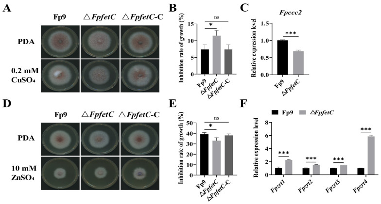
Iron is an essential micronutrient required for the fungal growth and propagation. Fusarium proliferatum is the causal agent of rice spikelet rot disease. In this study, we characterized the role of F. proliferatum multicopper ferroxidase (FpfetC), which mediated the oxidization of ferrous to ferric iron in the reductive system of iron assimilation. Deletion of FpfetC led to impaired growth under iron-deprived conditions, and the growth defect could be restored by exogenous iron. Compared to wild-type Fp9 strain, ΔFpfetC showed increased conidiation, resistance to copper stress, and sensitivity to zinc stress. FpfetC deficiency rendered a transcription remodeling of genes involved in high-affinity iron assimilation, iron homeostasis and iron storage. Moreover, production of fumonisin B1 (FB1) and transcript levels of fumonisin biosynthesis (Fpfums) genes were elevated in ΔFpfetC. ΔFpfetC exhibited hypervirulence to rice, accompanied with aggravation of invasive hyphae and activation of siderophore synthesis at the sites of inoculation. Additionally, disruption of FpfetC attenuated penetration ability to cellophane membrane under iron starvation. Taken together, these results demonstrated that FpfetC played important roles in iron uptake, conidiation, response to metal stress, fumonisin biosynthesis, and virulence in F. proliferatum.

Keywords: Fusarium proliferatum; fumonisins; iron acquisition; multicopper ferroxidase; virulence.


Conflict of interest statement

The authors declare no conflicts of interest.

Figures

Figure 1
Figure 1
Identification and expression of multicopper ferroxidase (FpfetC) in F. proliferatum. (A) Phylogenetic relationship of FpfetC protein and its homologs from different filamentous fungi. A phylogenetic tree was constructed by neighbor-joining method with 1000 bootstrap replicates using MEGA11 software (version 11.0). Bootstrap values were denoted at the supported node. The species names and GenBank accession numbers of the organisms were shown within the clade. (B) Expression of FpfetC gene under different concentrations of iron. The Fp9 strain was inoculated in MM media omitting iron at 28 °C for 3 days, then shifted into MM media supplemented with different concentrations of iron for 2 h. Expression value of FpfetC gene in Fp9 strain grown in MM media with BPS was artificially set as 1. Data are presented as mean ± standard deviation. Error bars denote standard deviation from three biological replicates. Different lowercase letters indicate significant differences as determined using ANOVA followed by Student’s t-tests at p < 0.05. There were three replicates for each sample. The experiment was repeated three times.
Figure 2
Figure 2
Effect of FpfetC on colony growth in F. proliferatum. (A) Colony morphology of ΔFpfetC cultured on PDA and CM media at 28 °C for 5 days. (B) Colony diameter of ΔFpfetC on PDA and CM media at 28 °C for 5 days. (C) Mycelial biomass of ΔFpfetC inocubated in PDB and CM liquid media at 28 °C for 4 days. (D) Hyphal tips of ΔFpfetC grown on PDA media at 28 °C for 36 h. Scale bars, 500 μm. (E) Colony morphology of ΔFpfetC cultured on MM media supplemented with different concentrations of iron at 28 °C for 5 days. (F) Colony diameter of ΔFpfetC cultured on MM media supplemented with different concentrations of iron at 28 °C for 5 days. Data are presented as mean ± standard deviation. Error bars denote standard deviation from three biological replicates. Asterisks indicate statistical significance as determined using ANOVA followed by Student’s t-tests (ns—not significant; ***—p < 0.001). Each experiment was carried out with three replicates and performed three times.
Figure 3
Figure 3
Impact of FpfetC on conidiation in F. proliferatum. (A) The amount of conidia of ΔFpfetC cultured in YEPD media at 28 °C. The sporulation was recorded at intervals of 12 h. (B) Relative expression levels of conidiation-related genes FpabaA, FpbrlA, and FpwetA. Expression value of each gene in Fp9 strain grown in YEPD media was artificially set as 1. (C) The amount of conidia of ΔFpfetC under different concentrations of iron. After culturing in MM media omitting iron at 28 °C for 3 days, ΔFpfetC was transferred into MM media supplemented with different concentrations of iron for 4 days. Data are presented as mean ± standard deviation. Error bars denote standard deviation from three biological replicates. Asterisks indicate statistical significance as determined using ANOVA followed by Student’s t-tests (ns—not significant; ***—p < 0.001). Different lowercase letters indicate significant differences at p < 0.05. Each experiment was carried out with three replicates and performed three times.
Figure 4
Figure 4
Involvement of FpfetC in expression of genes associated with iron metabolism. After preculturing in MM media omitting iron for 3 days at 28 °C, ΔFpfetC was transferred into MM media supplemented with 0.3 mM BPS (iron starvation, –Fe) or 0.03 mM FeSO4 (iron sufficiency, +Fe) for 2 h. Investigated genes were as follows: (A) FpfreB gene encoding ferric reductase and (B) FpftrA gene encoding iron permease, which were involved in reductive iron assimilation; (C) FpsidA gene encoding ornithine-N5-oxygenase, (D) FpsidC and (E) FpsidD genes encoding non-ribosomal peptide synthetases (NRPS), and (F) FpsidF gene encoding N5-transacylase, which were involved in siderophore biosynthesis; (G) Fpsit1A, (H) Fpsit1B, (I) Fpsit1C and (J) Fpsit2 genes encoding siderochrome-iron transporters, which were involved in ferrichrome-type siderophore transport; (K) FpmirA gene encoding enterobactin transporter, (L) FpmirB gene encoding TAFC importer and (M) FpmirD gene encoding fusarinine C transporter, which were involved in fusarinine-type siderophore transport; (N) FpcccA gene encoding vacuolar iron importer, which was involved in iron storage; (O) FphapX gene encoding bZIP type transcription factor and (P) FpsreA gene encoding GATA type transcription factor, which were iron regulators; (Q) FplysF gene encoding homoaconitase, (R) FphemA gene encoding 5-aminolevulinate synthase, (S) FpcycA gene encoding cytochrome C, and (T) FpacoA gene encoding aconitate hydratase, which were involved in iron consuming. Expression value of each gene in Fp9 strain in MM media with BPS was artificially set as 1. Data are presented as mean ± standard deviation. Error bars denote standard deviation from three biological replicates. Different lowercase letters indicate significant differences as determined using ANOVA followed by Student’s t-tests at p < 0.05. There were three replicates for each sample. The experiment was repeated three times.
Figure 5
Figure 5
Role of FpfetC on sensitivity to the excesses of copper or zinc in F. proliferatum. (A) Colony morphology of ΔFpfetC grown on PDA media with or without 0.2 mM CuSO4 at 28 °C for 5 days. (B) Inhibition rate of mycelial growth of ΔFpfetC on PDA media with 0.2 mM CuSO4. (C) Relative expression level of Fpccc2 gene encoding copper transport ATPase. After preculturing in PDB media for 48 h, ΔFpfetC was transferred to PDB media with 0.2 mM CuSO4 for 24 h. Expression value of Fpccc2 gene in Fp9 strain was artificially set as 1. (D) Colony morphology of ΔFpfetC grown on PDA media with or without 10 mM ZnSO4 at 28 °C for 5 days. (E) Inhibition rate of mycelial growth of ΔFpfetC on PDA media with 10 mM ZnSO4. (F) Relative expression levels of Fpzrts genes encoding zinc-regulated transporter. After preculturing in PDB media for 48 h, ΔFpfetC was transferred to PDB media with 10 mM ZnSO4 for 24 h. Expression values of Fpzrts genes in Fp9 strain were artificially set as 1. Data are presented as mean ± standard deviation. Error bars denote the standard deviation from three biological replicates. Asterisks indicate statistical significance as determined using ANOVA followed by Student’s t-tests (ns—not significant; *—p < 0.05; ***—p < 0.001). Each experiment was carried out with three replicates.
Figure 6
Figure 6
Effect of FpfetC on FB1 biosynthesis in F. proliferatum. (A) FB1 content produced by ΔFpfetC cultured in PDB media for 9 days. (B) FB1 content produced by ΔFpfetC cultured on cracked rice kernels for 14 days. (C) FB1 content produced by ΔFpfetC under different concentrations of iron. After being grown in MM media omitting iron at 28 °C for 4 days, ΔFpfetC was transferred into MM media supplemented with different concentrations of iron at 28 °C for 5 days. (D) Culture of ΔFpfetC grown in MM media with different concentrations of iron at 28 °C for 5 days. (E) Relative expression levels of Fpfum genes responsible for fumonisin biosynthesis. Expression value of each gene in Fp9 strain was artificially set as 1. Data are presented as mean ± standard deviation. Error bars denote standard deviation from three biological replicates. Asterisks indicate statistical significance as determined using ANOVA followed by Student’s t-tests (ns—not significant; **—p < 0.01; ***—p < 0.001). Three replicates were used for each sample, and the experiment was performed three times.
Figure 7
Figure 7
Influence of FpfetC on virulence in F. proliferatum. (A) Disease symptom of rice spikelets inoculated with ΔFpfetC at 21 days post-infection (dpi). (B) Disease index of rice spikelets inoculated with ΔFpfetC at 21 dpi. (C) FB1 accumulation on rice spikelets inoculated with ΔFpfetC at 21 dpi. (D) Relative expression levels of genes (FpsidA, FpsidC, and FpsidF) associated with siderophore biosynthesis at sites of inoculation challenged by ΔFpfetC at 48 h post-infection (hpi). Expression value of each gene in Fp9 strain was artificially set as 1. (E) Invasive hyphae on endepidermis of rice glumes infected with ΔFpfetC at 24, 48, and 72 hpi under scanning electron micrograph (SEM). Arrows indicate representative hyphae. Scale bars, 100 μm. (F) Ultrastructure of rice glumes challenged by ΔFpfetC at 72 hpi under transmission electron micrograph (TEM). Sg indicates starch grain. Chl indicates chloroplast. Scale bars, 500 nm. (G) Penetration of ΔFpfetC on cellophane membranes. ΔFpfetC was grown on MM media overlaid with cellophane membranes containing different concentrations of iron at 28 °C for 3 days (Before). After removing the cellophane, the plates were cultured for additional 3 days (After). Data are presented as mean ± standard deviation. Error bars denote standard deviation from three biological replicates. Asterisks indicate statistical significance as determined by using ANOVA followed by Student’s t-tests (ns—not significant; ***—p < 0.001). Each experiment was performed in triplicate.
Figure 8
Figure 8
Schematic model for acquisition and transport of iron affected by FpfetC in F. proliferatum. Reductive iron assimilation is depicted in orange, composed of ferric reductase (FpfreB), multicopper ferroxidase (FpfetC), and iron permease (FpftrA). Siderophore biosynthesis enzymes are shown in purple, composed of ornithine-N5-oxygenase (FpsidA), non-ribosomal peptide synthetases (FpsidC and FpsidD), N5-transacylase (FpsidF), and transacetylases (FpsidG and FpsidL). Ferrichrome-type siderophore transporters are demonstrated in blue, composed of Fpsit1A, Fpsit1B, Fpsit1C, and Fpsit2. Fusarinine-type siderophore transporters are indicated in green, composed of enterobactin transporter (FpmirA), TAFC importer (FpmirB), and fusarinine C transporter (FpmirD). Vacuolar iron storage protein FpcccA is represented in gray. Environmental (chelated) iron is shown in brown dots.

Similar articles

References

    1. Bogdan A.R., Miyazawa M., Hashimoto K., Tsuji Y. Regulators of iron homeostasis: New players in metabolism, cell death, and disease. Trends Biochem. Sci. 2016;41:274–286. doi: 10.1016/j.tibs.2015.11.012. - DOI - PMC - PubMed
    1. Galaris D., Barbouti A., Pantopoulos K. Iron homeostasis and oxidative stress: An intimate relationship. Biochim. Biophys. Acta – Mol. Cell Res. 2019;1866:118535. doi: 10.1016/j.bbamcr.2019.118535. - DOI - PubMed
    1. Haas H. Fungal siderophore metabolism with a focus on Aspergillus fumigatus. Nat. Prod. Rep. 2014;31:1266–1276. doi: 10.1039/C4NP00071D. - DOI - PMC - PubMed
    1. Misslinger M., Hortschansky P., Brakhage A.A., Haas H. Fungal iron homeostasis with a focus on Aspergillus fumigatus. Biochim. Biophys. Acta – Mol. Cell Res. 2021;1868:118885. doi: 10.1016/j.bbamcr.2020.118885. - DOI - PubMed
    1. Hu G., Chen S.H., Qiu J., Bennett J.E., Myers T.G., Williamson P.R. Microevolution during serial mouse passage demonstrates FRE3 as a virulence adaptation gene in Cryptococcus neoformans. mBio. 2014;5:e00941-14. doi: 10.1128/mBio.00941-14. - DOI - PMC - PubMed

LinkOut - more resources