. 2025 Mar 24;26(7):2937.
doi: 10.3390/ijms26072937.

Circ_RUSC2 Sequesters miR-661 and Elevates TUSC2 Expression to Suppress Colorectal Cancer Progression

Affiliations

Circ_RUSC2 Sequesters miR-661 and Elevates TUSC2 Expression to Suppress Colorectal Cancer Progression

Yixin Shi et al. Int J Mol Sci. .

Abstract

Despite advancements in diagnostic efficiency, colorectal cancer (CRC) remains a leading cause of cancer-related mortality, with increasing incidence rates. Circular RNA (circRNA) is a closed-loop, generally stable noncoding RNA that functions as a sponge for microRNAs in CRC. The purpose of this study was to investigate the function and underlying mechanism of circ_RUSC2, a new circRNA, in CRC. The expression levels of circ_RUSC2, miR-661, and TUSC2 were assessed using qRT-PCR, Western blot, and immunohistochemistry. Functional assays, including CCK-8, Transwell, and scratch wound healing, were performed to evaluate cell proliferation, migration, and invasion. RNA pull-down and actinomycin D assays were used to study RNA interactions and stability. In both CRC cells and tissues, miR-661 was markedly elevated, while circ_RUSC2 expression was considerably reduced. Poor differentiation, distant metastases, lymph node metastases, and an advanced stage were all strongly correlated with either miR-661 overexpression or circ_RUSC2 downregulation. circ_RUSC2 was more stable compared to its linear RUSC2 mRNA. CRC cell invasion, migration, and proliferation were suppressed by circ_RUSC2 ectopic expression; this inhibitory effect was restored by a miR-661 mimic. Circ_RUSC2 served as miR-661's sponge. TUSC2 counteracted the effects of miR-661, which stimulated CRC cell proliferation, migration, and invasion. At the post-transcriptional level, miR-661 controlled the expression of TUSC2 in CRC cells. In comparison to the negative control, circ_RUSC2 expression was markedly reduced, and its half-life was shortened by methyltransferase-like 3 (METTL3) knockdown. Circ_RUSC2 is a stable cytoplasmic circRNA. Circ_RUSC2 inhibits CRC cell malignant phenotypes via the miR-661/TUSC2 axis. The onset and progression of CRC are linked to the downregulation of Circ_RUSC2. circ_RUSC2 might become more stable through N6-methyladenosine (m6A) methylation regulated by METTL3. According to our research, circ_RUSC2 might be a new biomarker and treatment target for CRC.

Keywords: METTL3; TUSC2; circ_RUSC2; colorectal cancer; microRNA-661.


Conflict of interest statement

The authors declare no conflicts of interest.

Figures

Figure 1
Figure 1
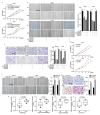
Circ_RUSC2 expression is decreased in CRC cells and tissues and is associated with poor clinicopathologic features. (A,B) Comparison of circ_RUSC2 expression in DLD-1, LoVo, HCT116, and RKO cells and CRC tissues, and CCD841 CoN cells, and normal adjacent colorectal tissues by qRT-PCR. (C) Relative expression level of circ_RUSC2 in CRC tissues and normal adjacent colorectal tissues of each case. (D) Analysis of clinicopathologic significance between relative expression of circ_RUSC2 and TNM stage, lymph node metastasis, distant metastasis, and tumor size. 18S rRNA was the internal control of circ_RUSC2. Data were indicated as mean ± SD from 3 independent experiments. **** p < 0.0001.
Figure 2
Figure 2
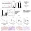
Circ_RUSC2 is a stable cytoplasmic circRNA in CRC cells. (A) Schematic diagram of circ_RUSC2 formation from exon 2 of the RUSC2 gene at chromosome 9p13. (B) Relative expression of circ_RUSC2 and linear RUSC2 mRNA by qRT-PCR after RNase R treatment on DLD-1 and LoVo cells. The expression level of circ_RUSC2 or linear RUSC2 mRNA without RNase R treatment was set as 1. (C) Relative expression of circ_RUSC2 and linear RUSC2 mRNA by qRT-PCR after actinomycin D treatment on DLD-1 and LoVo cells every 4 h for 24 h. The expression level of circ_RUSC2 or linear RUSC2 mRNA at 0 h was set as 1. GAPDH was the internal control of linear RUSC2 mRNA. (D) Percentage of U6, 18S rRNA, and circ_RUSC2 in the cytoplasmic and nuclear fraction. U6 was a nuclear RNA marker, and 18S rRNA was a cytoplasmic RNA marker. Data were shown as mean ± SD from 3 independent experiments. *** p < 0.001, **** p < 0.0001.
Figure 3
Figure 3
Circ_RUSC2 binds with miR-661 as a ceRNA in CRC. (A) Predicted circ_RUSC2-bound miRNA by databases and the binding sites between circ_RUSC2 and miR-661. (B) Enrichment of circ_RUSC2 by biotin-labeled miR-661 mimic. The biotin-miR-NC group was the negative control. (C) Comparison of miR-661 expression in DLD-1, LoVo, HCT116, and RKO cells and CCD841 CoN cells. (D) Comparison of miR-661 expression in the CRC tissues and paired normal colorectal tissues. (E) Relative expression level of miR-661 in CRC tissues and normal adjacent colorectal tissues of each case. (F) Spearman correlation analysis of circ_RUSC2 and miR-661 expression in the CRC cohort. Data are shown as mean ± SD from 3 independent experiments. ** p < 0.01, *** p < 0.001, **** p < 0.0001.
Figure 4
Figure 4
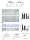
Circ_RUSC2 suppresses the capacities of CRC cell proliferation, migration, and invasion via sponging an oncomiR, miR-661. (AC) DLD-1 and LoVo cells were transfected with circ_NC or circ_RUSC2, and mimic-NC or miR-661 mimic. Cell viability (A), migratory (B), and invasive (C) abilities were determined by CCK-8, and scratch wound healing and Transwell chamber assay with Matrigel. (DF) DLD-1 and LoVo cells were transfected with mimic-NC or miR-661 mimic. Cell viability (D), migratory (E), and invasive (F) abilities were determined by CCK-8, scratch wound healing, and Transwell chamber assay with Matrigel. (G) Correlation of miR-661 expression with TNM stage, lymph node metastasis, distant metastasis, and tumor size. Data are shown as mean ± SD from 3 independent experiments. * p < 0.05, ** p < 0.01, *** p < 0.001, **** p < 0.0001.
Figure 5
Figure 5
miR-661 negatively regulates TUSC2 in CRC cells. (A) Predicted binding site between miR-661 and 3’UTR of TUSC2 mRNA. (B) Enrichment of TUSC2 mRNA by biotin-labeled miR-661 probe pull-down. GAPDH was the internal control. (C) Relative expressions of TUSC2 mRNA and protein in DLD-1 and LoVo cells transfected with miR-661 or mimic-NC, and miR-661 inhibitor or inhibitor-NC. (D) Relative expression of TUSC2 mRNA in DLD-1, LoVo, HCT116, and RKO cells and CRC tissues, and CCD841 CoN cells, and normal adjacent colorectal tissues by qRT-PCR. (E) Comparison of TUSC2 mRNA between tumor tissues and paired adjacent normal intestinal tissues in each case. (F) TUSC2 protein expression in the CRC cell lines and CCD841 CoN cells by Western blot. β-actin was the internal control. (G) Association of miR-661 and TUSC2 mRNA expressions by Spearman correlation analysis. (H) Clinicopathologic significance analysis of TUSC2 mRNA expression in CRC patients. Data are shown as mean ± SD from 3 independent experiments. (I) Immunohistochemistry showed that the expression level of TUSC2 was decreased in tumor tissues compared with normal tissue *** p < 0.001, **** p < 0.0001.
Figure 6
Figure 6
Circ_RUSC2 increases TUSC2 protein expression through sponging miR-661 in CRC cells. (AD) DLD-1 and LoVo cells were transfected with inhibitor-NC or miR-661 inhibitor, and si-NC or si-TUSC2. Cell viability (A), migratory (B), and invasive (C) abilities were determined by CCK-8, scratch wound healing, and Transwell chamber assay with Matrigel, and TUSC2 protein was detected by Western blot (D). (E) DLD-1 and LoVo cells were transfected with circ_NC or circ_RUSC2, and mimic-NC or miR-661 mimic. TUSC2 protein was examined by Western blot. (F) Spearman correlation analysis of circ_RUSC2 and TUSC2 mRNA expressions in the CRC cohort. Data are shown as mean ± SD from 3 independent experiments. * p < 0.05, ** p < 0.01, *** p < 0.001, **** p < 0.0001.
Figure 7
Figure 7
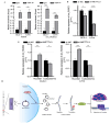
METTL3-mediated m6A modification increases the stability of circ_RUSC2 expression. (A) The MeRIP experiment proved the existence of m6A methylation site of circ_RUSC2. (B) Relative expression of circ_RUSC2 after knocking down METTL3. (C) The expression difference of circ_RUSC2 in the nucleus and cytoplasm after knocking down METTL3. (D) Mechanism of circ_RUSC2. Data are shown as mean ± SD from 3 independent experiments. * p < 0.05, ** p < 0.01, *** p < 0.001, **** p < 0.0001.

Similar articles

Cited by

References

    1. Ou Q., Chang Y., Liu J., Yan H., Chen L., Guo D., Zhang S.F. Mapping the intellectual structure and landscape of colorectal cancer immunotherapy: A bibliometric analysis. Hum. Vaccin. Immunother. 2024;20:2323861. doi: 10.1080/21645515.2024.2323861. - DOI - PMC - PubMed
    1. Cao W., Chen H., Yu Y., Li N., Chen W. Changing profiles of cancer burden worldwide and in China: A secondary analysis of the global cancer statistics 2020. Chin. Med. J. 2021;134:783–791. doi: 10.1097/CM9.0000000000001474. - DOI - PMC - PubMed
    1. He M., Pan Y., You C., Gao H. CircRNAs in cancer therapy tolerance. Clin. Chim. Acta. 2024;558:119684. doi: 10.1016/j.cca.2024.119684. - DOI - PubMed
    1. Mao J., Lu Y. Roles of circRNAs in the progression of colorectal cancer: Novel strategies for detection and therapy. Cancer Gene Ther. 2024;31:831–841. doi: 10.1038/s41417-024-00739-x. - DOI - PubMed
    1. Kosik K. Molecular biology: Circles reshape the RNA world. Nature. 2013;495:322–324. doi: 10.1038/nature11956. - DOI - PubMed

MeSH terms

LinkOut - more resources