. 2025 Mar 25;26(7):2976.
doi: 10.3390/ijms26072976.

Potential Role of Malassezia restricta in Pterygium Development

Affiliations

Potential Role of Malassezia restricta in Pterygium Development

Martina Paradzik Simunovic et al. Int J Mol Sci. .

Abstract

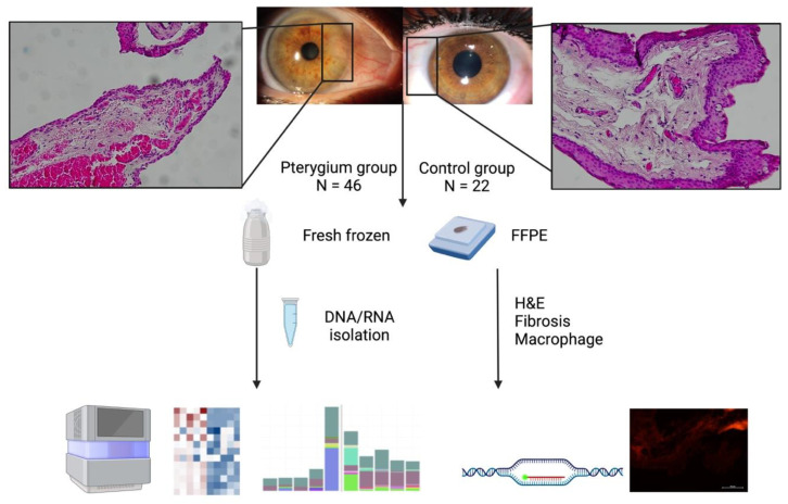
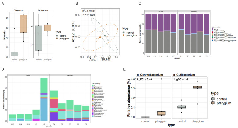
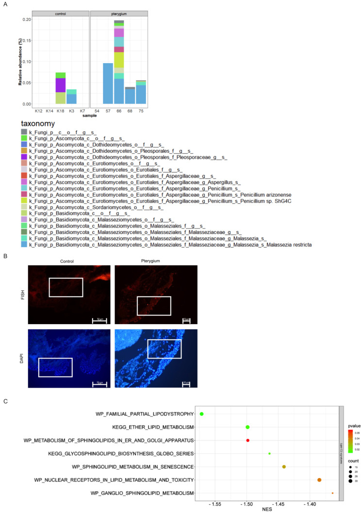
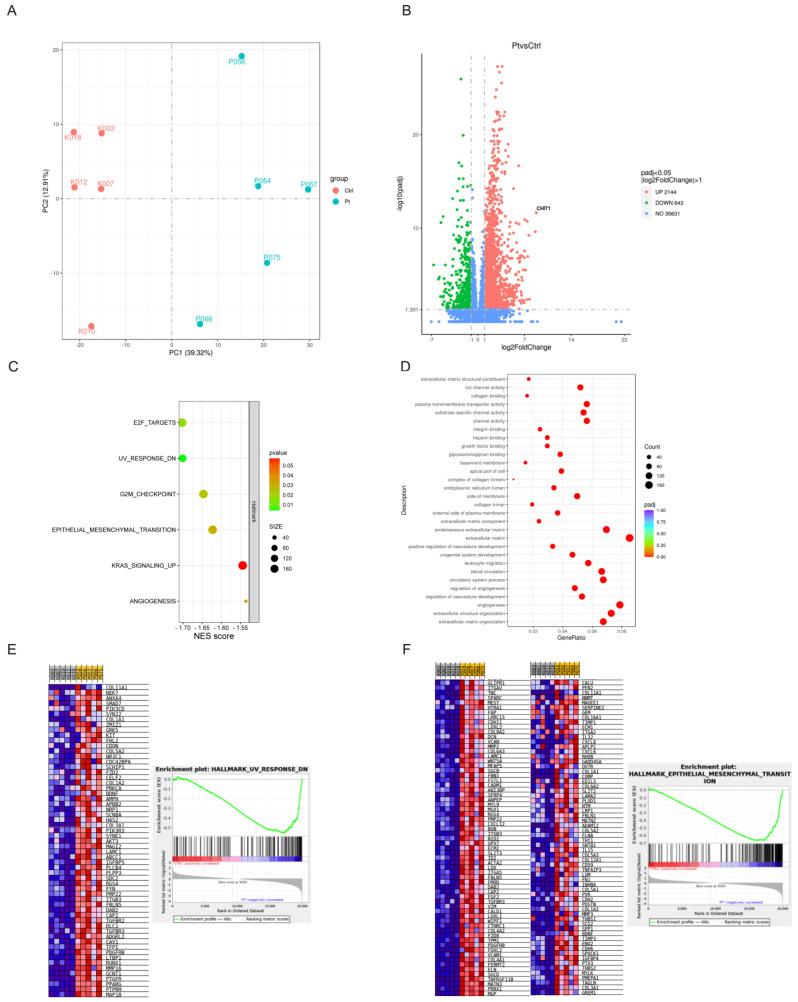
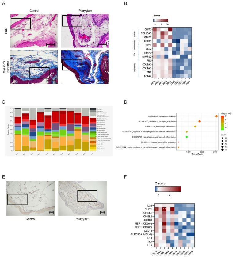
Pterygium is a condition affecting the ocular surface, marked by a triangular-shaped growth of fibrotic tissue extending from the nasal conjunctiva toward the corneal center, potentially causing visual impairment. While ultraviolet (UV )light exposure is the primary risk factor for pterygium, its underlying cause remains unclear. In order to better understand the true genesis of pterygium development, we investigated pterygium tissue and compared it with healthy conjunctiva controls. Given the eye's direct environmental exposure, we analyzed the microbiota composition using metagenomic sequencing of pterygium tissue to identify microbes potentially associated with this condition. Metagenomic sequencing revealed a higher prevalence of the fungus Malassezia restricta in five pterygium samples, confirmed by in situ hybridization. The CHIT1 gene, which plays a role in antifungal defenses, displayed the highest expression in five pterygium tissue samples compared to healthy conjunctiva controls, suggesting the potential involvement of Malassezia restricta in pterygium development. Gene expression profiling of pterygium highlighted an IL-33 and IL-4 gene expression signature, along with an increased presence of M2 macrophages, emphasizing their role in promoting fibrosis-a hallmark feature of pterygium. The detection of Malassezia restricta in the pterygium samples and associated molecular changes provides novel insights into the ocular microbiome and raises the possibility of Malassezia's involvement in pterygium pathology.

Keywords: CHIT1 gene; Malassezia restricta; ocular microbiome; pterygium.


Conflict of interest statement

The authors declare no conflicts of interest.

Figures

Figure 1
Figure 1
The work flow of the experiment. Created in BioRender. Paradzik Simunovic, M. (2025) https://BioRender.com/f95m049, accessed on 11 March 2025.
Figure 2
Figure 2
Metagenomic analysis of pterygium. Taxonomic analysis of pterygium compared with healthy conjunctiva. (A) Even though not statistically significant, pterygium samples have, on average, higher alpha diversity (observed species and Shannon index) compared to controls. (B) No significant differences in beta diversity (Bray–Curtis distance) were found between groups. (C) Kingdom-level relative abundance (%). (D,E) Corynebacterium and Cutibacterium genera were statistically differentially abundant between pterygium and healthy conjunctiva.
Figure 3
Figure 3
Malassezia restricta presence in pterygium. (A) Relative abundance of fungal species across pterygium and healthy control samples. (B) Conformation of Malassezia restricta with fluorescein in situ hybridization. An indicative part of the section is squared. (C) Lipid gene signatures.
Figure 4
Figure 4
Gene expression profile of pterygium. (A) PCA plot. (B) The differentially expressed genes between pterygium and controls visualized in volcano plot. (C) Gene ontology. (D) HALLMARK gene signature GSEA. (E) UV response in HALLMARK gene signature. (F) Epithelial–mesenchymal transition in HALLMARK gene signature. Hallmark gene sets enriched in Pterygium phenotype. FDR < 25% total of 6.
Figure 5
Figure 5
Alternately activated M2 macrophages in pterygium. (A) Fibrosis. (B) Inflammation and fibrosis. (C) Macrophage CD staining. (D) IL33 signature. (E) Immunohistochemical staining for a marker of M2 macrophage CD163 was present in pterygium and almost absent from control tissues. (F) Genes involved in alternative activation of macrophages. An indicative part of the section is squared.

Similar articles

Cited by

References

    1. Rezvan F., Khabazkhoob M., Hooshmand E., Yekta A., Saatchi M., Hashemi H. Prevalence and Risk Factors of Pterygium: A Systematic Review and Meta-Analysis. Surv. Ophthalmol. 2018;63:719–735. doi: 10.1016/j.survophthal.2018.03.001. - DOI - PubMed
    1. Liu L., Wu J., Geng J., Yuan Z., Huang D. Geographical Prevalence and Risk Factors for Pterygium: A Systematic Review and Meta-Analysis. BMJ Open. 2013;3:e003787. doi: 10.1136/bmjopen-2013-003787. - DOI - PMC - PubMed
    1. Chui J., Coroneo M.T., Tat L.T., Crouch R., Wakefield D., Di Girolamo N. Ophthalmic Pterygium: A Stem Cell Disorder with Premalignant Features. Am. J. Pathol. 2011;178:817–827. doi: 10.1016/j.ajpath.2010.10.037. - DOI - PMC - PubMed
    1. Suarez M.F., Echenique J., López J.M., Medina E., Irós M., Serra H.M., Fini M.E. Transcriptome Analysis of Pterygium and Pinguecula Reveals Evidence of Genomic Instability Associated with Chronic Inflammation. Int. J. Mol. Sci. 2021;22:12090. doi: 10.3390/ijms222112090. - DOI - PMC - PubMed
    1. Wanzeler A.C.V., Barbosa I.A.F., Duarte B., Borges D., Barbosa E.B., Kamiji D., Huarachi D.R.G., de Melo M.B., Alves M. Mechanisms and Biomarker Candidates in Pterygium Development. Arq. Bras. Oftalmol. 2019;82:528–536. doi: 10.5935/0004-2749.20190103. - DOI - PubMed

LinkOut - more resources