. 2025 Mar 25;26(7):3002.
doi: 10.3390/ijms26073002.

Administration of Noggin Suppresses Fibrinogen Leakage into the Brain in the Acute Phase After Traumatic Brain Injury in Mice

Affiliations

Administration of Noggin Suppresses Fibrinogen Leakage into the Brain in the Acute Phase After Traumatic Brain Injury in Mice

Miho Yasunaga et al. Int J Mol Sci. .

Abstract

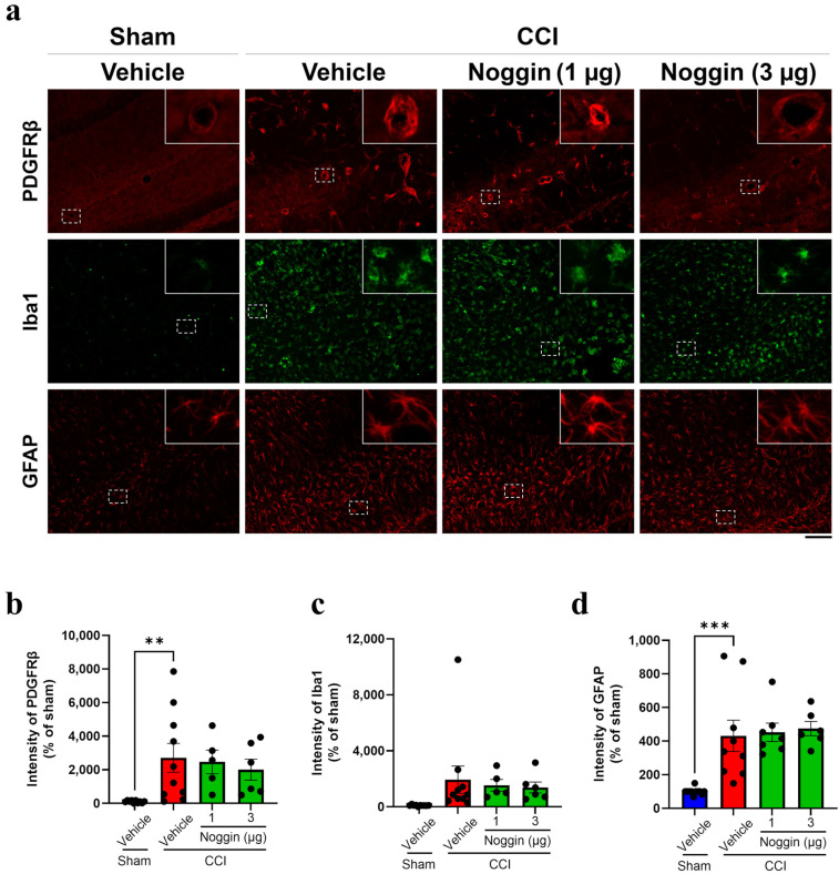
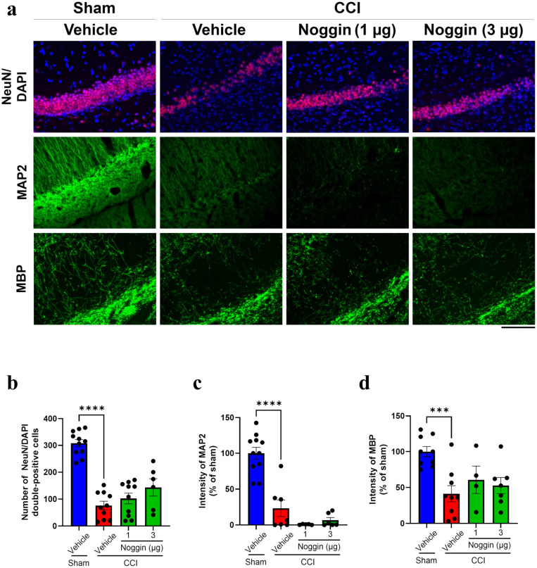
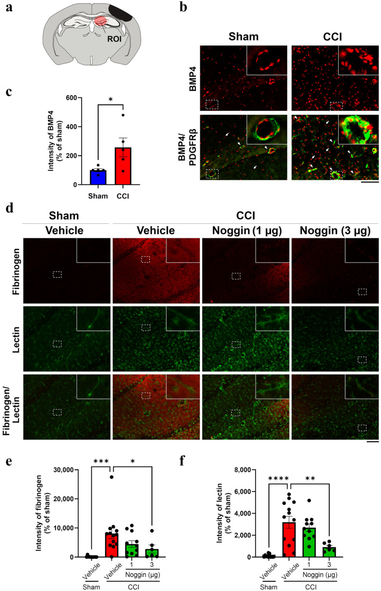
Traumatic brain injury (TBI) causes neurovascular unit (NVU) dysfunction, including hyperpermeability of the blood-brain barrier to fibrinogen, glial activation, and neuronal damage, possibly leading to secondary brain damage. However, no known substance can inhibit its pathogenesis. In this study, we investigated noggin, a bone morphogenetic protein (BMP) 4 inhibitor, as a TBI pathogenesis-inhibiting substance. We induced acute TBI in C57BL/6J mice through a controlled cortical impact (CCI) and evaluated the effects of noggin on fibrinogen leakage into the brain and NVU-constituting cells, including pericytes, microglia, astrocytes, and neurons. CCI mice showed increased BMP4 levels and extravascular fibrinogen in the hippocampus. Noggin treatment significantly suppressed fibrinogen leakage four days post-CCI in a dose-dependent manner. Immunofluorescence staining revealed that noggin administration did not inhibit the activation of NVU cells such as pericytes, microglia, and astrocytes, which were characterized by increased PDGFRβ, Iba1, and GFAP expression levels, respectively. On postoperative day 4, CCI mice showed neuronal cell and myelinated neuronal fiber loss, which were not significantly affected by noggin administration. In conclusion, noggin administration suppresses fibrinogen leakage into the brain in the acute phase after TBI. However, the suppression of fibrinogen leakage through noggin administration did not alleviate neuronal damage and activation of NVU cells during the acute phase of TBI.

Keywords: bone morphogenetic protein 4; fibrinogen leakage; neuronal damage; neurovascular unit; noggin; traumatic brain injury.


Conflict of interest statement

The authors declare no conflicts of interest. The funders had no role in the design of the study, in the collection, analyses, or interpretation of data, in the writing of the manuscript, or in the decision to publish the results.

Figures

Figure 2
Figure 2
Effect of noggin on the PDGFRβ, Iba1, and GFAP expression levels in the ipsilateral hippocampus of controlled cortical impact (CCI) mice on postoperative day 4. (a) The top panels show PDGFRβ immunoreactivity (red). The middle panels show Iba1 immunoreactivity (green). The bottom panels show GFAP immunoreactivity (red). Scale bar: 100 μm. (b) Quantification of PDGFRβ immunoreactivity, vehicle + sham: n = 10, vehicle + CCI: n = 10, 1 μg noggin + CCI: n = 5, 3 μg noggin + CCI: n = 6. (c) Quantification of Iba1 immunoreactivity, vehicle + sham: n = 10, vehicle + CCI: n = 10, 1 μg noggin + CCI: n = 5, 3 μg noggin + CCI: n = 6. (d) Quantification of GFAP immunoreactivity. Vehicle + sham: n = 11, vehicle + CCI: n = 9, 1 μg noggin + CCI: n = 7, 3 μg noggin + CCI: n = 6. CCI mice were treated with noggin at 1 or 3 μg/mouse for 4 days from the operative day to postoperative day 3. The blue, red and green bars represent the vehicle + sham, vehicle + CCI and noggin + CCI groups, respectively. The point represents an individual mouse. Data are expressed as the mean ± SEM. ** p < 0.01, *** p < 0.001.
Figure 3
Figure 3
Effect of noggin on NeuN, MAP2, and MBP expression levels in the ipsilateral hippocampus of controlled cortical impact (CCI) mice on postoperative day 4. (a) The top panels show images of NeuN (red) and DAPI (blue) double-labeled staining. The middle panels show MAP2 immunoreactivity (green). The bottom panels show MBP immunoreactivity (green). Scale bar: 100 μm. (b) The number of NeuN/DAPI double-positive cells. Vehicle + sham: n = 11, vehicle + CCI: n = 10, 1 μg noggin + CCI: n = 10, 3 μg noggin + CCI: n = 6. (c) Quantification of MAP2 immunoreactivity. Vehicle + sham: n = 11, vehicle + CCI: n = 7, 1 μg noggin + CCI: n = 5, 3 μg noggin + CCI: n = 6. (d) Quantification of MBP immunoreactivity. Vehicle + sham: n = 9, vehicle + CCI: n = 9, 1 μg noggin + CCI: n = 4, 3 μg noggin + CCI: n = 7. CCI mice were treated with 1 or 3 μg/mouse for 4 days from the operative day to postoperative day 3. The blue, red and green bars represent the vehicle + sham, vehicle + CCI and noggin + CCI groups, respectively. Each data point represents an individual mouse. Data are expressed as the mean ± SEM. *** p < 0.001, **** p < 0.0001.
Figure 1
Figure 1
BMP4 immunoreactivity in the ipsilateral hippocampus of controlled cortical impact (CCI) mice and effect of noggin on extravascular fibrinogen leakage on postoperative day 4. (a) The brain diagram shows the lesion and region of interest (ROI), including the hippocampus CA1 and dentate gyrus. (b) The top panels show bone morphogenetic protein (BMP) 4 immunoreactivity (red) in sham and CCI mice on postoperative day 4. The bottom panels show merged BMP4 (red) and PDGFRβ (green) immunoreactivity images. The arrowheads and arrows in the bottom panels indicate BMP4 immunoreactivity in the brain vessels and parenchyma, respectively. Scale bar: 100 μm. (c) Quantification of BMP4 immunoreactivity. Sham: n = 5, CCI: n = 5. The blue and red bars represent the sham and CCI group, respectively. Each point represents an individual mouse. Data are expressed as the mean ± SEM. (d) CCI mice were treated with noggin at 1 or 3 μg/mouse for 4 days from the operative day to postoperative day 3. The top panels show fibrinogen immunoreactivity (red). The middle panels show brain vessels revealed through lectin staining (green). Each bottom panel shows the merged image of the top and middle panels. Scale bar: 100 μm. (e) Quantification of fibrinogen immunoreactivity. Vehicle + sham: n = 12, vehicle + CCI: n = 13, 1 μg noggin + CCI: n = 11, 3 μg noggin + CCI: n = 6. (f) Quantification of lectin intensity. Vehicle + sham: n = 12, vehicle + CCI: n = 13, 1 μg noggin + CCI: n = 11, 3 μg noggin + CCI: n = 6. The blue, red and green bars represent the vehicle + sham, vehicle + CCI and noggin + CCI groups, respectively. Each point represents an individual mouse. Data are expressed as the mean ± SEM. * p < 0.05, ** p < 0.01, *** p < 0.001, **** p < 0.0001.

Similar articles

References

    1. VanItallie T.B. Traumatic brain injury (TBI) in collision sports: Possible mechanisms of transformation into chronic traumatic encephalopathy (CTE) Metab. Clin. Exp. 2019;100s:153943. doi: 10.1016/j.metabol.2019.07.007. - DOI - PubMed
    1. Ling H., Hardy J., Zetterberg H. Neurological consequences of traumatic brain injuries in sports. Mol. Cell. Neurosci. 2015;66:114–122. doi: 10.1016/j.mcn.2015.03.012. - DOI - PubMed
    1. Pearn M.L., Niesman I.R., Egawa J., Sawada A., Almenar-Queralt A., Shah S.B., Duckworth J.L., Head B.P. Pathophysiology Associated with Traumatic Brain Injury: Current Treatments and Potential Novel Therapeutics. Cell. Mol. Neurobiol. 2017;37:571–585. doi: 10.1007/s10571-016-0400-1. - DOI - PMC - PubMed
    1. Jochems D., van Rein E., Niemeijer M., van Heijl M., van Es M.A., Nijboer T., Leenen L.P.H., Houwert R.M., van Wessem K.J.P. Incidence, causes and consequences of moderate and severe traumatic brain injury as determined by Abbreviated Injury Score in the Netherlands. Sci. Rep. 2021;11:19985. doi: 10.1038/s41598-021-99484-6. - DOI - PMC - PubMed
    1. Brett B.L., Gardner R.C., Godbout J., Dams-O’Connor K., Keene C.D. Traumatic Brain Injury and Risk of Neurodegenerative Disorder. Biol. Psychiatry. 2022;91:498–507. doi: 10.1016/j.biopsych.2021.05.025. - DOI - PMC - PubMed

MeSH terms

LinkOut - more resources