. 2025 Mar 28;26(7):3146.
doi: 10.3390/ijms26073146.

Pathological Changes in Extracellular Matrix Composition Orchestrate the Fibrotic Feedback Loop Through Macrophage Activation in Dupuytren's Contracture

Affiliations

Pathological Changes in Extracellular Matrix Composition Orchestrate the Fibrotic Feedback Loop Through Macrophage Activation in Dupuytren's Contracture

Elizabeth Heinmäe et al. Int J Mol Sci. .

Abstract

Dupuytren's contracture belongs to a group of fibrotic diseases that have similar mechanisms but lack effective treatment and prevention options. The excessive accumulation of connective tissue in Dupuytren's disease leads to palmar fibrosis that results in contracture deformities. The present study aimed to investigate how the tissue microenvironment in Dupuytren's contracture affects the phenotypic differentiation of macrophages, which leads to an inflammatory response and the development of chronicity in fibrotic disease. We utilized a decellularization-based method combined with proteomic analysis to identify shifts in extracellular matrix composition and the surrounding tissue microenvironment. We found that the expression of several matricellular proteins, such as MFAP4, EFEMP1 (fibulin-3), and ANGPTL2, was elevated in Dupuytren's tissue. We show that, in response to the changes in the extracellular matrix of Dupuytren's contracture, macrophages regulate the fibrotic process by cytokine production, promote myofibroblast differentiation, and increase the fibroblast migration rate. Moreover, we found that the extracellular matrix of Dupuytren's contracture directly supports the macrophage-to-myofibroblast transition, which could be another contributor to Dupuytren's disease pathogenesis. Our results suggest that interactions between macrophages and the extracellular matrix should be considered as targets for novel fibrotic disease treatment and prevention strategies in the future.

Keywords: Dupuytren’s contracture; extracellular matrix; fibrosis; macrophages; myofibroblasts.


Conflict of interest statement

The authors declare no conflicts of interest.

Figures

Figure 1
Figure 1
Proteomic profile of the changes in the ECM of Dupuytren’s contracture. (A) Experimental layout of the proteomic analysis. For more details, see the Section 4. (B) Heatmap of differentially enriched proteins in Dupuytren’s contracture and control palmar fascia tissue samples. (C) Volcano plot showing significantly up- and downregulated proteins in Dupuytren’s contracture ECM.
Figure 2
Figure 2
Analysis of signalling pathways potentially involved in DD. KEGG pathway analysis was performed to identify differentially expressed genes in Dupuytren’s contracture. Colour gradient shows statistical significance for each analysed pathway, and the numbers refer to genes involved in each signalling pathway.
Figure 3
Figure 3
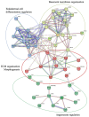
STRING protein–protein interactions between the proteins upregulated in DD. STRING network analysis of proteins with FC > 1.2 and p-value < 0.05 is shown. Four clusters were identified using a k-means approach, visualised in four different colours. The interactions between different clusters are illustrated with dotted lines, and intracluster interactions are shown with solid lines. The interactions shown are sourced from databases (light blue), experimental data (pink), textmining (green), co-expression (black), gene co-occurrence (dark blue), and protein homology (purple).
Figure 4
Figure 4
Immunofluorescence analysis of DD-associated markers in patient tissue sections. The representative samples (A) and relative quantification of the fluorescence signals of ANGPTL2, MFAP4, and EFEMP1 expression by mean integrated density (B) are shown. The scale bar is 200 μm. Results are presented as mean + SD; n = 7; *** p < 0.001.
Figure 5
Figure 5
Effects of DD-tissue-derived ECM on macrophage cytokine production. Primary human macrophages were stimulated with decellularized homogenized DD or control palmar fascia ECM, and cytokine production was analysed. Relative RNA induction of IL6, TNF, IL1B, IL10, and TGFB was quantified using RT-qPCR (A), the graphs depict fold changes compared to unstimulated macrophages, n = 3–5. IL-6, TNF, and IL-10 protein concentrations in the cell culture medium were quantified using ELISA (B). n = 7; * p < 0.05. Results are presented as mean + SD.
Figure 6
Figure 6
Co-expression of the myofibroblast marker α-SMA and the macrophage marker CD68 in DD palmar tissue sections. (A) Representative images of 3 DD and control patient tissue sections are shown. White arrows indicate areas of α-SMA and CD68 proximity in DD tissue samples. The scale bar is 200 μm. (B) The boxed region of the fibrotic sample from DD patient no. 2 is shown at higher magnification with channels separated and merged. Scale bar is 50 μm.
Figure 7
Figure 7
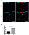
Exposure to DD tissue-derived ECM promotes MMT. Human monocyte-derived macrophages were stimulated for 72 h with decellularized homogenized DD or control palmar fascia ECM and stained for the myofibroblast marker α-SMA and the macrophage marker CD68. Representative images (A) and the quantification of 3 biological replicates (B) are shown. Results are presented as mean + SD and compared to control; * p < 0.05; the scale bar is 200 μm.
Figure 8
Figure 8
DD ECM-exposed macrophages support fibroblast proliferation and promote fibroblast differentiation into myofibroblasts. Human monocyte-derived macrophages were stimulated for 72 h with decellularized homogenized DD or control palmar fascia ECM, and subsequently, the cell culture medium was used to stimulate fibroblasts for 48 h (A). Cell proliferation, type I collagen, and α-SMA relative expression were quantified (B), n = 3. The scale bar is 200 μm. Results are presented as mean + SD and compared to control; * p < 0.05; ** p < 0.01; *** p < 0.001.
Figure 9
Figure 9
DD ECM-exposed macrophages signal to fibroblasts to promote their migration. Human monocyte-derived macrophages were stimulated for 72 h with control palmar fascia ECM (Ctrl), decellularized homogenized DD ECM, or M1/M2 cytokines as a control. Subsequently, the cell culture media were used to stimulate tissue-cultured fibroblasts for 48 h. Fibroblasts were allowed to migrate through a transwell chamber for 24 h. Representative images (A) and the quantitated results of the transwell assay (B). Results are presented as mean + SD and compared to Ctrl. Scale bar is 200 µm. n = 3; * p < 0.05; ** p < 0.01 compared to control palmar fascia ECM.

Similar articles

References

    1. Hynes R.O., Naba A. Overview of the matrisome—An inventory of extracellular matrix constituents and functions. Cold Spring Harb. Perspect. Biol. 2012;4:a004903. doi: 10.1101/cshperspect.a004903. - DOI - PMC - PubMed
    1. Mecham R.P. Overview of extracellular matrix. Curr. Protoc. Cell Biol. 2012;10:10.1.1–10.1.16. doi: 10.1002/0471143030.cb1001s57. - DOI - PubMed
    1. Lu P., Takai K., Weaver V.M., Werb Z. Extracellular matrix degradation and remodeling in development and disease. Cold Spring Harb. Perspect. Biol. 2011;3:a005058. doi: 10.1101/cshperspect.a005058. - DOI - PMC - PubMed
    1. Xue M., Jackson C.J. Extracellular Matrix Reorganization During Wound Healing and Its Impact on Abnormal Scarring. Adv. Wound Care. 2015;4:119–136. doi: 10.1089/wound.2013.0485. - DOI - PMC - PubMed
    1. Cardenas-Leon C.G., Maemets-Allas K., Klaas M., Lagus H., Kankuri E., Jaks V. Matricellular proteins in cutaneous wound healing. Front. Cell Dev. Biol. 2022;10:1073320. doi: 10.3389/fcell.2022.1073320. - DOI - PMC - PubMed

Substances

LinkOut - more resources