. 2025 Mar 29;26(7):3159.
doi: 10.3390/ijms26073159.

The Role of Puccinia polysora Underw Effector PpEX in Suppressing Plant Defenses and Facilitating Pathogenicity

Affiliations

The Role of Puccinia polysora Underw Effector PpEX in Suppressing Plant Defenses and Facilitating Pathogenicity

Qiang Su et al. Int J Mol Sci. .

Abstract

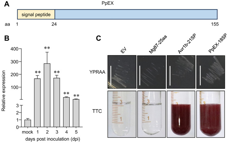
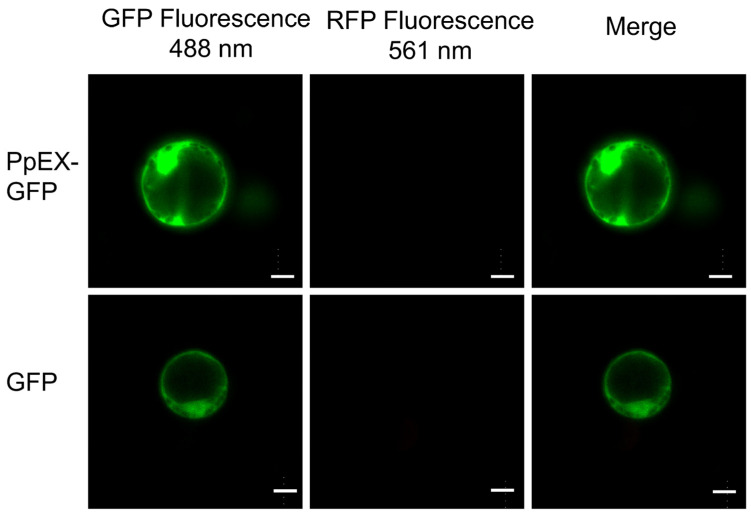
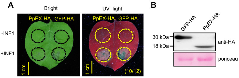
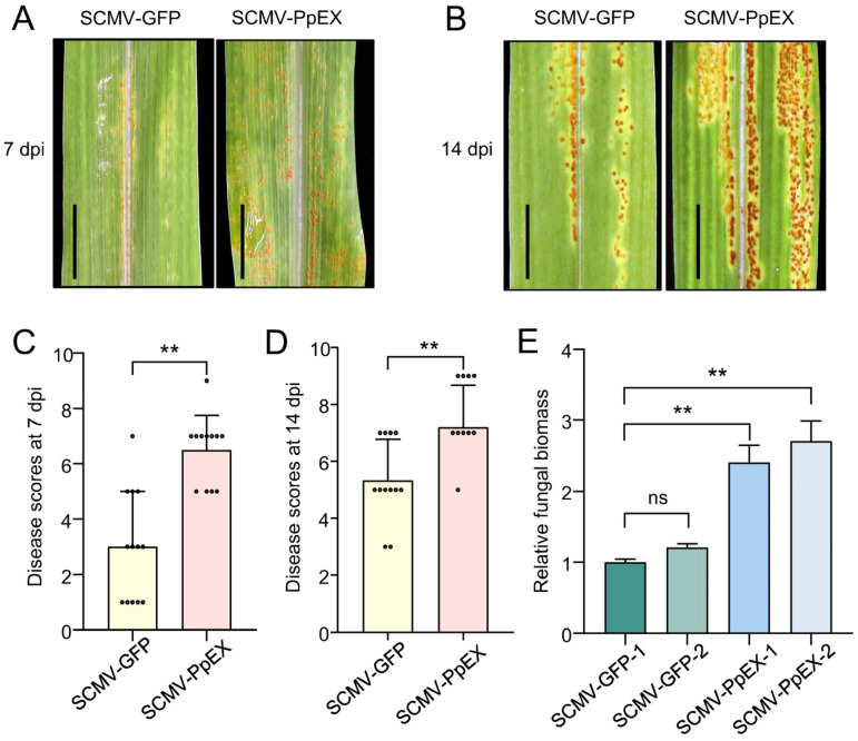
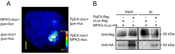
Puccinia polysora Underw, the pathogen that causes southern corn rust (SCR), delivers effectors to manipulate host immune responses. However, the mechanisms by which these effectors modulate host defenses are not well characterized. In this study, we found that the P. polysora effector PpEX is highly upregulated during infection. PpEX suppresses plant immune responses that are initiated by chitin, including the activation of mitogen-activated protein kinases (MAPKs) and the expression of pathogenesis-related (PR) genes. Maize plants transiently expressing PpEX exhibited higher pathogen infection rates, larger colony areas, and greater fungal biomass on their leaves compared to the control group. By employing TurboID proximity labeling technology coupled with mass spectrometry analysis, we discovered potential target proteins of PpEX in maize. The split-luciferase system enabled us to identify ZmMPK3, a component of the MAPK signaling pathway, as an interacting partner of PpEX among the candidate proteins. This interaction was subsequently confirmed by co-immunoprecipitation (Co-IP) experiments. Additionally, we verified that ZmMPK3 plays a positive role in regulating maize resistance to SCR. Thus, PpEX may function as a virulence effector that dampens plant PTI immunity by interacting with ZmMPK3 and impeding the MAPK signaling pathway.

Keywords: PpEX; Puccinia polysora Underw; ZmMPK3; effector; southern corn rust.


Conflict of interest statement

The authors declare no conflicts of interest.

Figures

Figure 1
Figure 1
The secreted protein PpEX is highly expressed during P. polysora infection. (A) Primary structure features of PpEX are shown. (B) Quantitative analysis of PpEX transcripts was performed using RNA extracted from germinated spores as a control and from leaves of the B73 maize cultivar infected with GD1913 urediniospores at various time points post-inoculation (1, 2, 3, 4, 5 dpi). P. polysora tubulin served as an endogenous control. The expression ratio in germinated spores was normalized to a value of 1. The presented data represents the average with the standard error of the mean from three independent biological replicates. Significance was assessed using an unpaired two-tailed Student’s t-test, where ** indicates p < 0.01. (C) Confirmation of the signal peptide activity of PpEX was carried out by inserting the 21 aa signal peptides from Avr1b, 18 aa putative signal peptides PpEX, and the first 25 amino acids of the non-secretory Mg87 into the pSUC2 vector, which was then introduced into the yeast strain YTK12. Yeast colonies were grown on YPRAA medium with raffinose. Invertase secretion was detected by monitoring the reduction of 2,3,5-triphenyltetrazolium chloride (TTC); a red color change indicates active invertase. Bar = 1 cm.
Figure 2
Figure 2
Subcellular localization of the PpEX-GFP fusion protein was performed using protoplasts from maize etiolated seedlings, which exhibited no spontaneous fluorescence under RFP conditions. Bar = 10 μm.
Figure 3
Figure 3
PpEX suppresses INF1-triggered cell death. (A) PpEX suppresses INF1-triggered cell death in N. benthamiana leaves. Agrobacterium suspensions (OD600 of 0.2) carrying GFP-HA and PpEX-HA were introduced into the leaf tissues 24 h prior to INF1 infiltration (left panel). Images were captured 3 days after the INF1 treatment. The corresponding leaves were also visualized under ultraviolet illumination (right panel). The circle represents the injection area. (B) Western blot analysis confirmed the expression of PpEX-HA and GFP-HA.
Figure 4
Figure 4
The inhibitory effect of PpEX on the maize immune response. Maize plants infected with SCMV-PpEX were inoculated with chitin, and the inhibitory effect of the effector protein PpEX on MAPK activation (A), ROS burst (B), PR1 (C), and PR5 (D) gene expression was then detected. The ** (p < 0.01) indicates significant differences between treatments and WT, as determined by Student’s t-test. Error bars represent the standard deviation. The red arrow represents the target protein.
Figure 5
Figure 5
PpEX promotes infection of P. Polysora on maize. Maize plants infected by SCMV-GFP and SCMV-PpEX were inoculated with P. polysora GD1913. After 7 days of inoculation, the disease grades (A,C) and fungal biomass (E) were assessed. After 14 days of inoculation, the disease grades (B,D) were assessed. The ** (p < 0.01) indicates significant differences between treatments and the wild type (WT), as determined by Student’s t-test. The small circles in the bar chart represent the number of maize samples in the experiment. Error bars represent the standard deviation. Bar = 1 cm.
Figure 6
Figure 6
PpEX interacts with ZmMPK3 in vivo. (A) Split-luciferase analysis of the interaction between PpEX and ZmMPK3 in N. benthamiana leaves. The β-glucuronidase-cLuc (gus-cLuc) and β-glucuronidase-nLuc (gus-nLuc) were used as negative controls. The pseudocolor bar shows the range of luminescence intensity. Bar = 1 cm. (B) The interaction of PpEX and ZmMPK3 was detected by co-immunoprecipitation. Total proteins were extracted from leaves co-expressing PpEX-flag and ZmMPK3-nLuc-HA. The input proteins (Input) and proteins extracted with Flag magnetic beads were analyzed by immunoblotting with anti-Flag and anti-HA antibodies. The cLuc-flag protein served as a negative control, with component indicators at the top and protein combinations and antibody indicators on the left. The blue pentagram indicates cLuc-Flag, while the green pentagram indicates PpEX-flag.
Figure 7
Figure 7
Knocking down ZmMPK3 with CMV-VIGS decreased resistance to P. polysora in B73 maize plants. (A) qRT-PCR analysis of ZmMPK3 expression in CMV-infected B73 plants at 15 dpi. (B) Disease scores of CMV-infected B73 plants at 7 dpi with P. polysora. The small circles in the bar chart represent the number of maize samples in the experiment. (C) Urediospore symptoms on leaves of CMV-infected B73 plants at 7 dpi with P. polysora. dpi indicates days post-inoculation. Significance was determined by an unpaired two-tailed Student’s t-test (** p < 0.01). Error bars represent the standard deviation. Bar = 1 cm.

Similar articles

References

    1. Aime M.C. Toward resolving family-level relationships in rust fungi (Uredinales) Mycoscience. 2006;47:112–122. doi: 10.1007/S10267-006-0281-0. - DOI
    1. Mei J., Zhou S., Liu W. Gene-for-gene-mediated resistance to southern corn rust in maize. Trends Plant Sci. 2023;28:255–258. doi: 10.1016/j.tplants.2022.12.002. - DOI - PubMed
    1. Sun Q., Li L., Guo F., Zhang K., Dong J., Luo Y., Ma Z. Southern corn rust caused by Puccinia polysora Underw: A review. Phytopathol. Res. 2021;3:25. doi: 10.1186/s42483-021-00102-0. - DOI
    1. Deng C., Leonard A., Cahill J., Lv M., Li Y., Thatcher S., Li X., Zhao X., Du W., Li Z., et al. The RppC-AvrRppC NLR-effector interaction mediates the resistance to southern corn rust in maize. Mol. Plant. 2022;15:904–912. doi: 10.1016/j.molp.2022.01.007. - DOI - PubMed
    1. Jones J.D.G., Dangl J.L. The plant immune system. Nature. 2006;444:323–329. doi: 10.1038/nature05286. - DOI - PubMed

MeSH terms

LinkOut - more resources