. 2025 Mar 29;26(7):3197.
doi: 10.3390/ijms26073197.

Ex Vivo Overactivation of Lymphocyte Subsets in Fibrotic Hypersensitivity Pneumonitis Is Blunted by a Sphingosine-1-Phosphate Receptor Ligand

Affiliations

Ex Vivo Overactivation of Lymphocyte Subsets in Fibrotic Hypersensitivity Pneumonitis Is Blunted by a Sphingosine-1-Phosphate Receptor Ligand

Olivier Courtemanche et al. Int J Mol Sci. .

Abstract

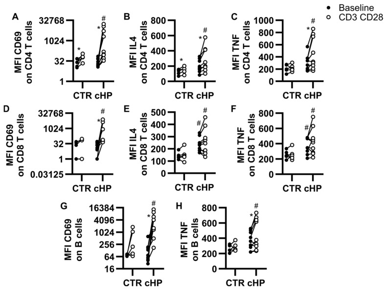
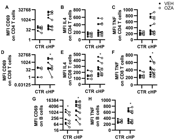
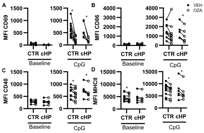
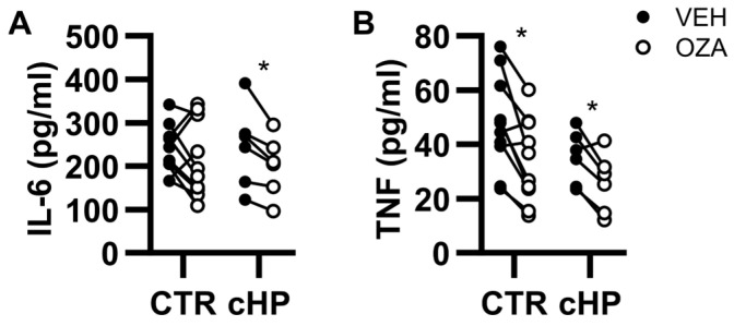
Lymphocytes are central to the pathogenesis of hypersensitivity pneumonitis and a strong body of evidence supports that lymphocytes are modulated by sphingosine-1-phosphate receptor-modifying drugs. This exploratory study aimed to determine if a pharmacological sphingosine-1-phosphate receptor ligand interfered with the activation of lymphocytes obtained from fibrotic hypersensitivity pneumonitis patients. Peripheral blood mononuclear cells of 12 patients and 10 control subjects were submitted to CD3/CD28 stimulation, isolated B cells were incubated with a TLR9 ligand; and we tested how these stimulations were impacted by ozanimod, a sphingosine-1-phosphate receptor ligand. T cell and B cell subsets from patients overexpressed CD69 and cytokines such as TNF and IL-4 in response to CD3/CD28 stimulation, compared to controls. In patients with fibrotic hypersensitivity pneumonitis, ozanimod alleviated CD3/CD28 induction of CD69, IL-4, and TNF in CD8, but not CD4 T cells. In isolated B cells stimulated with a TLR9 ligand, ozanimod reduced cell surface expression of CD69, CD86, and CD40, as well as TNF and IL-6 accumulation in supernatant. We conclude that lymphocyte subsets are functionally impacted in patients with fibrotic hypersensitivity pneumonitis and that ozanimod can interfere ex vivo with the overactivation of B cells and CD8 T cells in response to specific stimuli.

Keywords: BAFF; CD69; S1P; S1P1; S1P5; TLR9; TNF; cytokine; extrinsic allergic alveolitis; ozanimod.


Conflict of interest statement

D.M. received research funding (no honorariums) from Bristol Myers Squibb to perform a clinical trial on the impact of ozanimod in COVID-19 patients from 2020 to 2023.

Figures

Figure 1
Figure 1
Activation profiles of lymphocyte subsets in PBMC cultures. Peripheral blood mononuclear cells (PBMC) from control (CTR) subjects or patients with fibrotic hypersensitivity pneumonitis (fHP) were cultured in the absence (baseline, black dots) or in the presence of anti-CD3/CD28-coupled microbeads (CD3 CD28, empty dots). Median fluorescence intensity (MFI) for CD69 (A,D,G), IL-4 (B,E), and TNF (C,F,H) was assessed on CD4 T cells (AC), CD8 T cells (DF) and B cells (G,H). Two-way ANOVA was used for control vs. fHP comparisons and # denotes a significant difference against homologous baseline or CD3/CD28 condition. Within groups, the effect of CD3/CD28 stimulation vs. baseline was assessed using paired bidirectional t-tests, and * denotes a significant difference. Dots represent individual values, which are connected with a black line for the same individual. * p < 0.05, n = 5 to 9 per group.
Figure 2
Figure 2
Ozanimod inhibits lymphocyte subset activation in PBMC cultures stimulated with anti-CD3/CD28-coupled microbeads. Peripheral blood mononuclear cells (PBMC) from the control group (CTR) or patients with fibrotic hypersensitivity pneumonitis (fHP) incubated with anti-CD3/CD28-coupled microbeads in the presence of a vehicle (VEH, black dots) or ozanimod (1 µM, empty dots). Median fluorescence intensity (MFI) for CD69 (A,D,G), IL-4 (B,E), and TNF (C,F,H) were assessed on CD4 T cells (AC), CD8 T cells (DF), and B cells (G,H). The effect of ozanimod vs. vehicle was assessed using paired bidirectional t-tests and * denotes a significant difference. Dots represent individual values, which are connected with a black line for the same individual. * p < 0.05, n = 5 to 9 per group.
Figure 3
Figure 3
Ozanimod inhibits CpG-induced B cell activation in isolated B cell cultures. Isolated B cells from individual control subjects (CTR) or fibrotic hypersensitivity pneumonitis (fHP) patients were cultured for 24 h with medium only (Baseline) or CpG ODN2006 3 μg/mL (CpG) in the absence (VEH, black dots) or presence of ozanimod (1 µM; empty dots), Median fluorescence intensity (MFI) of CD69 (A), CD86 (B), CD40 (C), and MHCII (D) was assessed. n = 5 to 10 per group. Paired bidirectional t-tests were performed for VEH vs. ozanimod comparisons. * p < 0.05.
Figure 4
Figure 4
Ozanimod reduces CpG-induced TNF and IL-6 supernatant accumulation in isolated B cell cultures. Isolated B cells from individual control (CTR) subjects or fibrotic hypersensitivity pneumonitis (fHP) patients were incubated with CpG ODN2006 (3 μg/mL) in the absence (black dots) or presence of ozanimod (1 µM; empty dots) for 24 h. The concentration of IL-6 (A) and TNF (B) in the cell-free supernatant was quantified using ELISA. In total, 6 to 10 individuals were included for each experimental condition based on the availability of B cells. Paired bidirectional t-tests were performed for VEH vs. ozanimod comparisons. * p < 0.05.

Similar articles

References

    1. Vasakova M., Morell F., Walsh S., Leslie K., Raghu G. Hypersensitivity Pneumonitis: Perspectives in Diagnosis and Management. Am. J. Respir. Crit. Care Med. 2017;196:680–689. doi: 10.1164/rccm.201611-2201PP. - DOI - PubMed
    1. Thorne P.S., Adamcakova-Dodd A., Kelly K.M., O’Neill M.E., Duchaine C. Metalworking fluid with mycobacteria and endotoxin induces hypersensitivity pneumonitis in mice. Am. J. Respir. Crit. Care Med. 2006;173:759–768. doi: 10.1164/rccm.200405-627OC. - DOI - PMC - PubMed
    1. Huppe C.A., Blais-Lecours P., Bernatchez E., Lauzon-Joset J.F., Duchaine C., Rosen H., Dion G., McNagny K.M., Blanchet M.R., Morissette M.C., et al. S1P1 Contributes to Endotoxin-enhanced B Cell Functions Involved in Hypersensitivity Pneumonitis. Am. J. Respir. Cell Mol. Biol. 2020;63:209–218. doi: 10.1165/rcmb.2019-0339OC. - DOI - PMC - PubMed
    1. Bueno M., Zank D., Buendia-Roldan I., Fiedler K., Mays B.G., Alvarez D., Sembrat J., Kimball B., Bullock J.K., Martin J.L., et al. PINK1 attenuates mtDNA release in alveolar epithelial cells and TLR9 mediated profibrotic responses. PLoS ONE. 2019;14:e0218003. doi: 10.1371/journal.pone.0218003. - DOI - PMC - PubMed
    1. Fernandez Perez E.R., Kong A.M., Raimundo K., Koelsch T.L., Kulkarni R., Cole A.L. Epidemiology of Hypersensitivity Pneumonitis among an Insured Population in the United States: A Claims-based Cohort Analysis. Ann. Am. Thorac. Soc. 2018;15:460–469. doi: 10.1513/AnnalsATS.201704-288OC. - DOI - PubMed

MeSH terms

LinkOut - more resources