. 2025 Mar 30;26(7):3214.
doi: 10.3390/ijms26073214.

Hydroxyurea Mitigates Heme-Induced Inflammation and Kidney Injury in Humanized Sickle Cell Mice

Affiliations

Hydroxyurea Mitigates Heme-Induced Inflammation and Kidney Injury in Humanized Sickle Cell Mice

William Kwaku Agbozo et al. Int J Mol Sci. .

Abstract

Kidney disorders significantly contribute to morbidity and mortality in sickle cell disease (SCD). Acute kidney injury (AKI), a major risk factor for chronic kidney disease (CKD), often arises from intravascular hemolysis, where plasma cell-free heme drives AKI through inflammatory and oxidative stress mechanisms. Hydroxyurea (HU), a well-established SCD-modifying therapy, improves clinical outcomes, but its effects on systemic heme and inflammatory mediators of kidney injury remain underexplored. This study evaluated HU's impact on plasma heme, pro-inflammatory mediators, kidney injury, and renal histopathology in a sickle cell mouse model. Townes humanized sickle cell mice (HbSS) and non-sickle (HbAA) controls were treated with HU or vehicle for two weeks. HU significantly reduced total plasma heme, lactate dehydrogenase, and pro-inflammatory cytokines (CXCL10, VEGF-A, IFN-γ) in HbSS mice. HU reduced renal injury biomarkers (cystatin C, NGAL) and improved renal histopathology, evidenced by reduced vascular congestion, glomerulosclerosis, and tubular damage. Interestingly, HU did not alter the levels of kidney repair biomarkers (clusterin and EGF). These findings suggest that HU mitigates kidney injury by reducing the deleterious effects of circulating heme and inflammation, supporting its potential to slow or prevent progressive kidney injury in SCD.

Keywords: acute kidney injury; heme; hydroxyurea; inflammation; sickle cell disease.


Conflict of interest statement

The authors declare no conflict of interest.

Figures
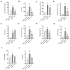
Figure 1
Figure 1
Hydroxyurea modulates markers of systemic hemolysis and inflammation relevant to kidney injury in Townes humanized sickle cell mice. Plasma levels of hemolysis markers: (A) total plasma heme and (B) lactate dehydrogenase; (C) white blood cell count; (D) neutrophil count, and serum pro-inflammatory markers: (E) interleukin-6 (IL-6); (F) C-X-C motif chemokine ligand 10 (CXCL10); (G) interferon-gamma (IFN-γ); (H) vascular endothelial growth factor-A (VEGF-A); (I) murine monocyte chemoattractant protein-5 (MCP-5); and anti-inflammatory marker: (J) interleukin-10 (IL-10) were assessed in 12-week-old Townes mice after two weeks of treatment with either the vehicle or hydroxyurea (50 mg/kg/day) (n = 7–8 per group). Data are presented as the mean ± SEM. HbAA indicates the genetic control mice; HbSS, SCD mice. * p < 0.05 vs. HbAA vehicle; # p < 0.05 vs. HbSS vehicle.
Figure 2
Figure 2
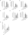
Hydroxyurea mitigates kidney injury in Townes humanized sickle cell mice. Urinary levels of kidney injury biomarkers: (A) cystatin C and (B) neutrophil gelatinase-associated lipocalin (NGAL) and kidney repair biomarkers (C) clusterin and (D) epidermal growth factor (EGF) were measured in 12-week-old sickle cell (HbSS) mice and genetic control (HbAA) mice (n = 7–8 per group) and after 2 weeks of treatment with hydroxyurea (50 mg/kg/day) or the vehicle. Data are presented as the mean ± SEM. Changes in biomarker levels pre- and post-treatment were analyzed using paired ratio t-tests (two-tailed). ns: not significant; p < 0.05 (*); p < 0.01 (**); p < 0.001 (***).
Figure 3
Figure 3
Hydroxyurea attenuates kidney damage in Townes humanized sickle cell mice. Histological analysis of renal tissues from sickle cell (HbSS) and non-sickle control (HbAA) mice following vehicle or hydroxyurea (50 mg/kg/day) treatment for 2 weeks at 12 weeks of age (n = 7–8 per group). Quantification of (A) vasa recta congestion, (B) glomerular congestion, (C) Bowman’s capsule thickening, (D) glomerulosclerosis, (E) tubular brush border loss, (F) intensity of tubular iron deposition, and (G) kidneyl heme oxygenase-1 expression. Data represent the mean ± SEM. * p < 0.05 vs. HbAA vehicle; # p < 0.05 vs. HbSS vehicle. Panel (H) shows halved kidneys with mild medulla congestion in a vehicle-treated HbAA mouse (green arrowheads), severe medulla congestion in a vehicle-treated HbSS mouse, and mild congestion in an HU-treated HbSS mouse (red arrowheads). Panel (I) shows representative Masson trichrome–stained sections, highlighting normal, uncongested glomeruli in a vehicle-treated HbAA mouse (green arrowheads), severe congestion in a vehicle-treated HbSS mouse, and moderate congestion in an HU-treated HbSS mouse (black arrowheads). Panel (J) depicts representative PAS-stained sections, showing a normal Bowman’s capsule membrane in a vehicle-treated HbAA mouse (green arrowheads), moderate thickening in a vehicle-treated HbSS mouse, and mild thickening in an HU-treated HbSS mouse (black arrowheads). Panel (K) shows Masson trichrome–stained sections, indicating normal glomeruli with no sclerosis in a vehicle-treated HbAA mouse (green arrowheads), moderate glomerulosclerosis in a vehicle-treated HbSS mouse, and mild glomerulosclerosis in an HU-treated HbSS mouse (black arrowheads). Panel (L) shows PAS-stained sections of cortical tubules, with a stained epithelial cell brush border in a vehicle-treated HbAA mouse (green arrowheads), severe tubular brush border loss in a vehicle-treated HbSS mouse, and mild tubular brush border loss in an HU-treated HbSS mouse (black arrowheads). Panel (M) shows Perl’s Prussian blue–stained renal cortex sections, demonstrating no tubular iron deposits in a vehicle-treated HbAA mouse and iron deposits in a vehicle-treated and HU-treated HbSS mouse (black arrowheads). Panel (N) shows immunohistochemistry staining of anti-heme oxygenase-1 in cortex sections with nearly absent HO-1 expression in a vehicle-treated HbAA mouse and mild expression in both an HU- and vehicle-treated sickle cell mouse (black arrowheads). Original magnification, ×40.
Figure 3
Figure 3
Hydroxyurea attenuates kidney damage in Townes humanized sickle cell mice. Histological analysis of renal tissues from sickle cell (HbSS) and non-sickle control (HbAA) mice following vehicle or hydroxyurea (50 mg/kg/day) treatment for 2 weeks at 12 weeks of age (n = 7–8 per group). Quantification of (A) vasa recta congestion, (B) glomerular congestion, (C) Bowman’s capsule thickening, (D) glomerulosclerosis, (E) tubular brush border loss, (F) intensity of tubular iron deposition, and (G) kidneyl heme oxygenase-1 expression. Data represent the mean ± SEM. * p < 0.05 vs. HbAA vehicle; # p < 0.05 vs. HbSS vehicle. Panel (H) shows halved kidneys with mild medulla congestion in a vehicle-treated HbAA mouse (green arrowheads), severe medulla congestion in a vehicle-treated HbSS mouse, and mild congestion in an HU-treated HbSS mouse (red arrowheads). Panel (I) shows representative Masson trichrome–stained sections, highlighting normal, uncongested glomeruli in a vehicle-treated HbAA mouse (green arrowheads), severe congestion in a vehicle-treated HbSS mouse, and moderate congestion in an HU-treated HbSS mouse (black arrowheads). Panel (J) depicts representative PAS-stained sections, showing a normal Bowman’s capsule membrane in a vehicle-treated HbAA mouse (green arrowheads), moderate thickening in a vehicle-treated HbSS mouse, and mild thickening in an HU-treated HbSS mouse (black arrowheads). Panel (K) shows Masson trichrome–stained sections, indicating normal glomeruli with no sclerosis in a vehicle-treated HbAA mouse (green arrowheads), moderate glomerulosclerosis in a vehicle-treated HbSS mouse, and mild glomerulosclerosis in an HU-treated HbSS mouse (black arrowheads). Panel (L) shows PAS-stained sections of cortical tubules, with a stained epithelial cell brush border in a vehicle-treated HbAA mouse (green arrowheads), severe tubular brush border loss in a vehicle-treated HbSS mouse, and mild tubular brush border loss in an HU-treated HbSS mouse (black arrowheads). Panel (M) shows Perl’s Prussian blue–stained renal cortex sections, demonstrating no tubular iron deposits in a vehicle-treated HbAA mouse and iron deposits in a vehicle-treated and HU-treated HbSS mouse (black arrowheads). Panel (N) shows immunohistochemistry staining of anti-heme oxygenase-1 in cortex sections with nearly absent HO-1 expression in a vehicle-treated HbAA mouse and mild expression in both an HU- and vehicle-treated sickle cell mouse (black arrowheads). Original magnification, ×40.

Similar articles

References

    1. Ofori-Acquah S.F. Sickle Cell Disease as a Vascular Disorder. Expert. Rev. Hematol. 2020;13:645–653. doi: 10.1080/17474086.2020.1758555. - DOI - PubMed
    1. Thomson A.M., McHugh T.A., Oron A.P., Teply C., Lonberg N., Vilchis Tella V., Wilner L.B., Fuller K., Hagins H., Aboagye R.G., et al. Global, Regional, and National Prevalence and Mortality Burden of Sickle Cell Disease, 2000–2021: A Systematic Analysis from the Global Burden of Disease Study 2021. Lancet Haematol. 2023;10:e585–e599. doi: 10.1016/S2352-3026(23)00118-7. - DOI - PMC - PubMed
    1. McCormick M., Richardson T., Warady B.A., Novelli E.M., Kalpatthi R. Acute Kidney Injury in Paediatric Patients with Sickle Cell Disease Is Associated with Increased Morbidity and Resource Utilization. Br. J. Haematol. 2020;189:559–565. doi: 10.1111/bjh.16384. - DOI - PubMed
    1. Batte A., Menon S., Ssenkusu J., Kiguli S., Kalyesubula R., Lubega J., Mutebi E.I., Opoka R.O., John C.C., Starr M.C., et al. Acute Kidney Injury in Hospitalized Children with Sickle Cell Anemia. BMC Nephrol. 2022;23:110. doi: 10.1186/s12882-022-02731-9. - DOI - PMC - PubMed
    1. Roy N.B., Carpenter A., Dale-Harris I., Dorée C., Estcourt L.J. Interventions for Chronic Kidney Disease in People with Sickle Cell Disease. Cochrane Database Syst. Rev. 2023;8:CD012380. doi: 10.1002/14651858.CD012380.pub3. - DOI - PMC - PubMed

MeSH terms

LinkOut - more resources