. 2025 Apr 1;26(7):3264.
doi: 10.3390/ijms26073264.

Oncogenic KRASG12D Transfer from Platelet-like Particles Enhances Proliferation and Survival in Non-Small Cell Lung Cancer Cells

Affiliations

Oncogenic KRASG12D Transfer from Platelet-like Particles Enhances Proliferation and Survival in Non-Small Cell Lung Cancer Cells

Jorge Ceron-Hernandez et al. Int J Mol Sci. .

Abstract

In the tumor context, platelets play a significant role in primary tumor progression, dissemination and metastasis. Analysis of this interaction in various cancers, such as non-small cell lung cancer (NSCLC), demonstrate that platelets can both transfer and receive biomolecules (e.g. RNA and proteins) to and from the tumor at different stages, becoming tumor-educated platelets. To investigate how platelets are able to transfer oncogenic material, we developed in vitro platelet-like particles (PLPs), from a differentiated MEG-01 cell line, that stably carry RNA and protein of the KRASG12D oncogene in fusion with GFP. We co-cultured these PLPs with NSCLC H1975 tumor cells to assess their ability to transfer this material. We observed that the generated platelets were capable of stably expressing the oncogene and transferring both its RNA and protein forms to tumor cells using qPCR and imaging techniques. Additionally, we found that coculturing PLPs loaded with GFP-KRASG12D with tumor cells increased their proliferative capacity at specific PLP concentrations. In conclusion, our study successfully engineered an MEG-01 cell line to produce PLPs carrying oncogenic GFP-KRASG12D simulating the tumor microenvironment, demonstrating the efficient transfer of this oncogene to tumor cells and its significant impact on enhancing proliferation.

Keywords: KRAS; non-small cell lung cancer; oncogene transfer; platelet-like-particles.


Conflict of interest statement

The authors declare no conflicts of interest.

Figures
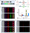
Figure 1
Figure 1
Generation and differentiation of MEG-01 cell line expressing the fusion protein GFP-KRASG12D. (A) Schematic representation of the lentiviral vectors designs: Empty vector and GFP-KRASG12D vector. LTR: long terminal repeats. EF1α and mPGK: promoters. (B) Schematic representation of MEG-01 cell line differentiation and PLP production on day 12. (C) Representative ImageStream images of undifferentiated MEG-01 EV (top) and GFP-KRASG12D (bottom) cell lines. Channels indicate the localization and expression levels for Hoechst (nucleic DNA, purple), CD61 (red) and GFP-KRASG12D (green); the last channel corresponds to the merged channels, except for the brightfield. (D) Representative ImageStream images of differentiated MEG-01 EV (top) and GFP-KRASG12D (bottom) cell lines. Channels indicate the same as in panel (C). (E) Mean fluorescence intensity (MFI) comparison of CD61 (left) and GFP-KRASG12D (right) in MEG-01 EV and GFP-KRASG12D non-differentiated and differentiated. Data represent the mean ± SD for three independent experiments. Statistics were assessed with ANOVA two-way plus Tukey multiple comparison test (statistically significant differences: ns = non-significant, * p < 0.05, *** p < 0.001). (F) Representative ImageStream images of PLP EV (top) and GFP-KRASG12D (bottom) produced from MEG-01 on day 12. Channels indicate the same as in panel (C).
Figure 2
Figure 2
Production of PLP charged with GFP-KRASG12D from differentiated MEG-01 cell line. (A) Immunocytofluorescence of differentiated MEG-01 EV (top) and GFP-KRASG12D (bottom). Channels indicate the localization and expression levels for CD61 (red), GFP-KRASG12D (green) and DAPI (nucleic DNA, blue); the last channel corresponds to the merged channels. The arrows indicate the proplatelet formation. Scale bar = 10 μm. (B) qRT-PCR analysis showing the expression of GP9, GP2B and KRAS genes in differentiated MEG-01 EV and GFP-KRASG12D and PLP produced at day 12. (C) qRT-PCR analysis showing the expression of the fusion gene in differentiated MEG-01 EV and GFP-KRASG12D and PLP produced at day 12. (D) Characterization of fusion gene cDNA coding sequence (mutation corresponds to c.G35A). (E) Western blot analysis detecting the GFP-KRASG12D fusion protein in both MEG-01 differentiated and PLP. GAPDH is used as a loading control. Molecular weights: GFP-KRASG12D (51 kDa) and GAPDH (37 kDa). Data in plots represent mean ± SD for three independent experiments. Statistics were assessed with two-tailed unpaired Student’s t-test (statistically significant differences: *** p < 0.001).
Figure 3
Figure 3
PLP fusion and GFP-KRASG12D transfer to H1975 tumor cells. (A) Immunocytofluorescence of H1975 cells co-cultured with PLP EV (top) and GFP-KRASG12D (bottom). Channels indicate the localization and expression levels for DiD (gray), CD61 (red), GFP-KRASG12D (green), DAPI (nucleic DNA, blue) and merge channel. The last image corresponds to ortho Z-stack slide. Scale bar = 10 μm. (B) Representative ImageStream images of co-culture of H1975 cells with PLP EV (top) and GFP-KRASG12D (bottom). Channels indicate the localization and expression levels for Hoechst (nucleic DNA, purple), CD61 (red) and GFP-KRASG12D (green); the last channel corresponds to the merged channels, except for the brightfield (BF). (C) qRT-PCR analysis showing the expression of KRAS gene in co-culture of H1975 cells with PLP EV and GFP-KRASG12D. (D) qRT-PCR analysis showing the expression of the fusion gene in co-culture of H1975 cells with PLP EV and GFP-KRASG12D. Data in plots represent the mean ± SD for three independent experiments. Statistics were assessed with two-tailed unpaired Student’s t-test (statistically significant differences: * p < 0.05).
Figure 4
Figure 4
Evaluation of the effect in proliferation of PLP GFP-KRASG12D in H1975 tumor cells. (A) Proliferation assay of H1975 cells co-culture with different concentrations of PLP EV and PLP GFP-KRASG12D. (B) Proliferation assay of H1975 cells co-culture with PLP EV and PLP GFP-KRASG12D and treated with MRTX1133 specific KRASG12D inhibitor. Data in plots represent the mean ± SEM for three independent experiments. Statistics were assessed with two-tailed unpaired Student’s t-test (statistically significant differences: * p < 0.05, ** p < 0.01).

References

    1. Clemetson K.J. Platelets and Primary Haemostasis. Thromb. Res. 2012;129:220–224. doi: 10.1016/j.thromres.2011.11.036. - DOI - PubMed
    1. Gremmel T., Iii A.L.F., Michelson A.D. Platelet Physiology. Semin. Thromb. Hemost. 2016;42:191–204. doi: 10.1055/s-0035-1564835. - DOI - PubMed
    1. Gay L.J., Felding-Habermann B. Contribution of Platelets to Tumour Metastasis. Nat. Rev. Cancer. 2011;11:123–134. doi: 10.1038/nrc3004. - DOI - PMC - PubMed
    1. Labelle M., Begum S., Hynes R.O. Direct Signaling between Platelets and Cancer Cells Induces an Epithelial-Mesenchymal-like Transition and Promotes Metastasis. Cancer Cell. 2011;20:576–590. doi: 10.1016/j.ccr.2011.09.009. - DOI - PMC - PubMed
    1. Best M.G., Sol N., Kooi I., Tannous J., Westerman B.A., Rustenburg F., Schellen P., Verschueren H., Post E., Koster J., et al. RNA-Seq of Tumor-Educated Platelets Enables Blood-Based Pan-Cancer, Multiclass, and Molecular Pathway Cancer Diagnostics. Cancer Cell. 2015;28:666–676. doi: 10.1016/j.ccell.2015.09.018. - DOI - PMC - PubMed

MeSH terms

Substances