Review
. 2025 Apr 3;26(7):3367.
doi: 10.3390/ijms26073367.

Parkinson's Disease: The Neurodegenerative Enigma Under the "Undercurrent" of Endoplasmic Reticulum Stress

Affiliations
Review

Parkinson's Disease: The Neurodegenerative Enigma Under the "Undercurrent" of Endoplasmic Reticulum Stress

Xiangrui Kong et al. Int J Mol Sci. .

Abstract

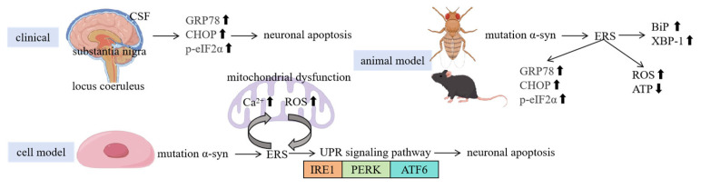
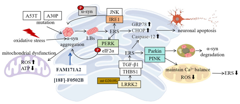
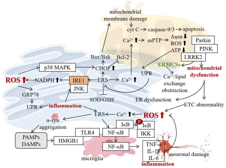
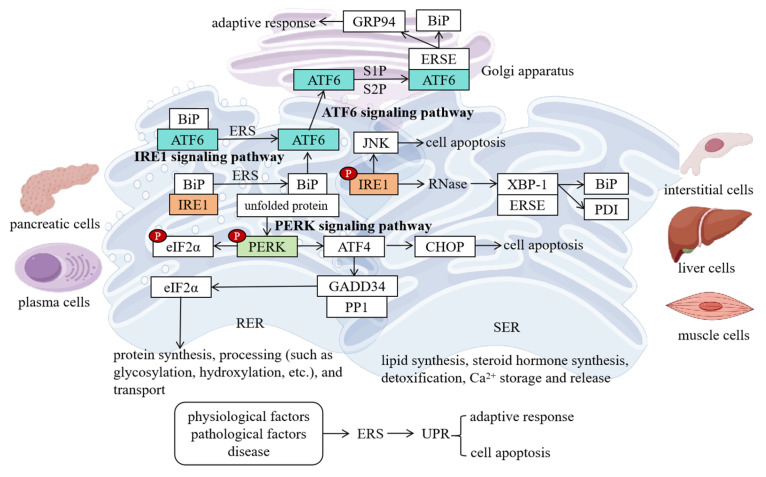
Parkinson's disease (PD), a prevalent neurodegenerative disorder, demonstrates the critical involvement of endoplasmic reticulum stress (ERS) in its pathogenesis. This review comprehensively examines the role and molecular mechanisms of ERS in PD. ERS represents a cellular stress response triggered by imbalances in endoplasmic reticulum (ER) homeostasis, induced by factors such as hypoxia and misfolded protein aggregation, which activate the unfolded protein response (UPR) through the inositol-requiring enzyme 1 (IRE1), protein kinase R-like endoplasmic reticulum kinase (PERK), and activating transcription factor 6 (ATF6) pathways. Clinical, animal model, and cellular studies have consistently demonstrated a strong association between PD and ERS. Abnormal expression of ERS-related molecules in PD patients' brains and cerebrospinal fluid (CSF) correlates with disease progression. In animal models (e.g., Drosophila and mice), ERS inhibition alleviates dopaminergic neuronal damage. Cellular experiments reveal that PD-mimicking pathological conditions induce ERS, while interactions between ERS and mitochondrial dysfunction promote neuronal apoptosis. Mechanistically, (1) pathological aggregation of α-synuclein (α-syn) and ERS mutually reinforce dopaminergic neuron damage; (2) leucine-rich repeat kinase 2 (LRRK2) gene mutations induce ERS through thrombospondin-1 (THBS1)/transforming growth factor beta 1 (TGF-β1) interactions; (3) molecules such as Parkin and PTEN-induced kinase 1 (PINK1) regulate ERS in PD. Furthermore, ERS interacts with mitochondrial dysfunction, oxidative stress, and neuroinflammation to exacerbate neuronal injury. Emerging therapeutic strategies show significant potential, including artificial intelligence (AI)-assisted drug design targeting ERS pathways and precision medicine approaches exploring non-pharmacological interventions such as personalized electroacupuncture. Future research should focus on elucidating ERS-related mechanisms and identifying novel therapeutic targets to develop more effective treatments for PD patients, ultimately improving their quality of life.

Keywords: Parkinson’s disease; endoplasmic reticulum stress; molecular mechanisms; therapeutic strategies; unfolded protein response; α-synuclein.


Conflict of interest statement

The authors declare no competing interests.

Figures

Figure 2
Figure 2
ERS is closely linked to the pathological mechanisms of PD, with clinical, animal, and cellular studies highlighting its critical role. Clinical evidence reveals significant upregulation of ERS markers (e.g., GRP78, CHOP, p-eIF2α) in PD patients’ brain regions such as the SN and locus coeruleus, as well as in CSF, correlating positively with disease severity and suggesting ERS-driven neuronal apoptosis. In animal models, Drosophila expressing mutant α-syn and MPTP-induced mouse PD models exhibit ERS activation (e.g., increased BiP expression and XBP-1 splicing), while ERS suppression alleviates dopaminergic neuron damage and improves motor function. Cellular experiments further confirm that mutant α-syn overexpression or MPP+ treatment triggers ER unfolded protein accumulation and mitochondrial dysfunction, activating the UPR pathways (IRE1, PERK, ATF6). However, persistent stress ultimately leads to apoptosis, with ERS and mitochondrial damage (Ca2+ overload, ROS surge) forming a vicious cycle that synergistically exacerbates neuronal degeneration. These findings collectively underscore ERS as a multifaceted contributor to PD pathogenesis and a potential therapeutic target. (↑ represents increase; ↓ represents decrease).
Figure 3
Figure 3
In PD, the molecular mechanism of ERS involves the interaction of multiple factors. The vicious cycle between alpha syn and ERS, where gene mutations or abnormal aggregation of alpha syn (such as A53T, A30P) lead to misfolding and the formation of LBs, directly triggering ERS (upregulation of GRP78 and CHOP expression). Meanwhile, ERS activates the UPR pathway, further promoting alpha syn aggregation and forming a positive feedback loop. Knocking out FAM171A2 can block the pathological spread of α-syn and improve symptoms. PET tracer [18F]-F0502B can visualize alpha syn aggregation. LRRK2 gene mutations drive ERS through the THBS1/TGF-β1 pathway. Common LRRK2 mutations (such as G2019S) enhance kinase activity, upregulate THBS1 expression, and bind to TGF-β1 to activate its signaling pathway, disrupting UPR homeostasis and leading to elevated ERS markers and mitochondrial damage. The synergistic protective effect of Parkin and PINK1, ERS-induced Parkin expression, clears misfolded proteins through the UPS and initiates mitochondrial autophagy, reducing the ERS burden; PINK1 regulates mitochondrial Ca2+ homeostasis and synergizes with Parkin to clear damaged mitochondria and reduce ROS production. Lack of Parkin or PINK1 can exacerbate ERS sensitivity and neuronal apoptosis. (“p” represents phosphorylation; ↑ represents increase; ↓ represents decrease).
Figure 4
Figure 4
In PD, ERS forms a complex vicious cycle with mitochondrial dysfunction, oxidative stress, and neuroinflammation, collectively driving disease progression. ERS activates the mPTP via calcium overload, leading to mitochondrial membrane potential collapse, reduced ATP synthesis, and a ROS burst, while simultaneously activating proapoptotic pathways (CHOP/Bax/Bak) to exacerbate mitochondrial damage. Mitochondrial dysfunction-induced energy metabolism collapse and ROS feedback further disrupt ER homeostasis, creating a bidirectional detrimental loop. Structural disorganization of ERMCS and mitophagy defects caused by PINK1/Parkin mutations amplify this cross-damage. Additionally, ERS activates NADPH oxidase through the IRE1-JNK pathway, while mitochondrial ROS leakage directly triggers oxidative stress. ROS oxidizes ER proteins and calcium channels, impairing their function and forming a mutually reinforcing cycle, compounded by weakened antioxidant systems (SOD/GSH). ERS also activates the TLR/NF-κB pathway via misfolded protein release (e.g., α-syn), inducing pro-inflammatory cytokines (TNF-α, IL-1β), which, in turn, suppress ER folding capacity and activate JNK/p38 pathways to aggravate ERS. Microglial activation synergizes with oxidative stress, forming an “ERS–inflammation–oxidative stress” triangular vicious cycle. Ultimately, apoptotic cascades, metabolic imbalance, and synaptic dysfunction lead to irreversible dopaminergic neuron loss. Targeting key nodes in these interconnected networks (e.g., inhibiting ERS, enhancing mitophagy, or blocking inflammatory signaling) may offer novel therapeutic strategies for PD. (“p” represents phosphorylation; ↑ represents increase; ↓ represents decrease).
Figure 1
Figure 1
The ER is a membranous network structure in eukaryotic cells, divided into the RER and the SER. The RER, studded with ribosomes, is responsible for the synthesis and modification of secretory proteins (e.g., glycosylation) and is highly developed in cells with active secretory functions. The SER lacks ribosomes and performs diverse roles, including steroid hormone synthesis (e.g., in adrenal cells), detoxification (e.g., in liver cells), calcium ion storage (e.g., in the sarcoplasmic reticulum regulating muscle contraction), and membrane lipid synthesis. The ER also participates in cellular signaling through calcium homeostasis regulation. When ER homeostasis is disrupted (e.g., due to hypoxia, misfolded protein accumulation, or calcium imbalance), ERS is triggered, prompting the cell to activate the UPR via three core pathways: (1) the IRE1 pathway, upon activation, cleaves XBP-1 mRNA to generate the transcription factor XBP-1, which upregulates chaperones (e.g., BiP) to enhance the protein-folding capacity; prolonged stress activates the JNK pathway to promote apoptosis; (2) the PERK pathway phosphorylates eIF2α to inhibit global protein synthesis while inducing ATF4 expression to activate stress adaptation genes (e.g., antioxidant genes); chronic activation upregulates the pro-apoptotic factor CHOP; (3) the ATF6 pathway is transported to the Golgi apparatus, where it is cleaved to release its active fragment, which enters the nucleus to activate ER function-related genes. These pathways synergistically reduce the unfolded protein burden and restore homeostasis. If stress persists and remains unresolved, the UPR shifts toward apoptosis. (“p” represents phosphorylation).

Similar articles

Cited by

  • Actual Data on Essential Trace Elements in Parkinson's Disease.
    Popescu C, Munteanu C, Spînu A, Andone I, Bistriceanu R, Postoiu R, Suciu A, Giuvara S, Vlădulescu-Trandafir AI, Aurelian SM, Pop NL, Ciobanu V, Onose G. Popescu C, et al. Nutrients. 2025 May 29;17(11):1852. doi: 10.3390/nu17111852. Nutrients. 2025. PMID: 40507121 Free PMC article. Review.

References

    1. Kalia L.V., Lang A.E. Parkinson’s disease. Lancet. 2015;386:896–912. doi: 10.1016/S0140-6736(14)61393-3. - DOI - PubMed
    1. Tolosa E., Garrido A., Scholz S.W., Poewe W. Challenges in the diagnosis of Parkinson’s disease. Lancet Neurol. 2021;20:385–397. doi: 10.1016/S1474-4422(21)00030-2. - DOI - PMC - PubMed
    1. Ghemrawi R., Khair M. Endoplasmic Reticulum Stress and Unfolded Protein Response in Neurodegenerative Diseases. Int. J. Mol. Sci. 2020;21:6127. doi: 10.3390/ijms21176127. - DOI - PMC - PubMed
    1. Zhang J., Guo J., Yang N., Huang Y., Hu T., Rao C. Endoplasmic reticulum stress-mediated cell death in liver injury. Cell Death Dis. 2022;13:1051. doi: 10.1038/s41419-022-05444-x. - DOI - PMC - PubMed
    1. Wang D., Qu S., Zhang Z., Tan L., Chen X., Zhong H.-J., Chong C.-M. Strategies targeting endoplasmic reticulum stress to improve Parkinson’s disease. Front. Pharmacol. 2023;14:1288894. doi: 10.3389/fphar.2023.1288894. - DOI - PMC - PubMed