Review
. 2025 Apr 4;26(7):3377.
doi: 10.3390/ijms26073377.

Basic Pathological Mechanisms in Peripheral Nerve Diseases

Affiliations
Review

Basic Pathological Mechanisms in Peripheral Nerve Diseases

Angelo Schenone et al. Int J Mol Sci. .

Abstract

Pathological changes and the cellular and molecular mechanisms underlying axonopathy and myelinopathy are key to understanding a wide range of inherited and acquired peripheral nerve disorders. While the clinical indications for nerve biopsy have diminished over time, its diagnostic value remains significant in select conditions, offering a unique window into the pathophysiological processes of peripheral neuropathies. Evidence highlights the symbiotic relationship between axons and myelinating Schwann cells, wherein disruptions in axo-glial interactions contribute to neuropathogenesis. This review synthesizes recent insights into the pathological and molecular underpinnings of axonopathy and myelinopathy. Axonopathy encompasses Wallerian degeneration, axonal atrophy, and dystrophy. Although extensively studied in traumatic nerve injury, the mechanisms of axonal degeneration and Schwann cell-mediated repair are increasingly recognized as pivotal in non-traumatic disorders, including dying-back neuropathies. We briefly outline key transcription factors, signaling pathways, and epigenetic changes driving axonal regeneration. For myelinopathy, we discuss primary segmental demyelination and dysmyelination, characterized by defective myelin development. We describe paranodal demyelination in light of recent findings in nodopathies, emphasizing that it is not an exclusive indicator of demyelinating disorders. This comprehensive review provides a framework to enhance our understanding of peripheral nerve pathology and its implications for developing targeted therapies.

Keywords: axon; myelin; nerve; neuropathies; peripheral nervous system.


Conflict of interest statement

The authors declare no conflicts of interest.

Figures

Figure 1
Figure 1
Graphical representation of axonal (Wallerian) degeneration. 1 = axon; 2 = site of axonal crush; 3 = Schwann cell; 4 = basal lamina; 5 = macrophages phagocytizing myelin and axonal debris; 6 = proliferating Schwann cells; 7 = regeneration and remyelination result in internodal distances distal to the injury that are uniformly shorter compared to those formed during developmental myelination.
Figure 2
Figure 2
Molecular pathways involved in axonal degeneration and regeneration. Following axotomy, the axon separated from its soma enters a lag phase, during which its morphology remains intact while several subcellular processes unfold, ultimately resulting in energy deficits, ROS production, and calpain activation, leading to the execution phase. NMNAT2 plays a critical role in axon survival and is continuously transported from the nucleus to the axon; thus, axotomy and impaired axonal transport result in a drop in axoplasmic NMNAT2. The MAPK pathway and the atypical ubiquitin ligase complex drive the degradation of vesicle-associated and soluble NMNAT2, respectively. NMNAT2 sustains constant NAD+ levels and low NMN concentration. A reduction in axoplasmic NMNAT2 leads to decreased NAD+ and elevated NMN concentrations, triggering SARM1 activation. SARM1 activation depletes NAD+ and NADP+, resulting in ATP depletion, mitochondrial dysfunction with ROS production, and Ca++ influx, culminating in calpain activation. Increased Axed levels have also been described in Drosophila models. During the execution phase, axon fragmentation occurs. Plgf released from the damaged axon facilitates the constriction of actin filaments in SCs, promoting axon fragmentation. SCs then undergo both biochemical and morphological changes, transitioning into repair SCs. The formation of repair SCs and subsequent fiber regeneration are driven by multiple transcription factors, signaling pathways, and epigenetic modifications, some of which are illustrated in the figure and described in Section 2.1.3. ATP = adenosine triphosphate; MAPK = mitogen-activated protein kinase; NAD+ = nicotinamide adenine dinucleotide; NADP+ = nicotinamide adenine dinucleotide phosphate; NMN = nicotinamide mononucleotide; NMNAT2 = nicotinamide mononucleotide adenylyltransferase 2; Plgf = placental growth factor; ROS = reactive oxygen species; SARM1 = sterile alpha/Armadillo/Toll-Interleukin receptor homology domain 1 protein; SC = Schwann cell.
Figure 3
Figure 3
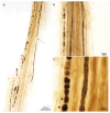
Axonopathy. Wallerian degeneration. Initially, there is retraction of the paranodal myelin ((a), arrow) and widening of the Schmidt–Lanterman clefts, where myelin degradation into short segments (“myelin ovoids”, asterisks) begins. Teased nerve fiber preparation, (a) 10×; (b) 40×; (c) 100× oil, * = myelin ovoids.
Figure 4
Figure 4
Axonopathy. Activity of macrophages and Schwann cells in the sural nerve of a patient with vasculitis. In panel (a), a macrophage outlined with a white dotted line is shown, and an enlarged view is provided in panel (b). (teased nerve fiber preparation; magnification: (a) 60× oil immersion; (b) 100× oil immersion).
Figure 5
Figure 5
Axonopathy. Macrophage and Schwann cell activity in sciatic nerves of a 120-day-old mSOD1-G93A mouse. (a) Several macrophages (red arrows) are shown in the endoneurium in a 0.8 µm thick toluidine blue-stained Epon section (magnification: 100× oil immersion). (b) Immunohistochemical analysis of the mSOD1-G93A mouse sciatic nerve (green: neurofilament marker NF200; red: macrophage marker CD68; blue: nuclear marker DAPI) (40× magnification). (c,d) Electron micrographs of an activated macrophage (c) and Schwann cell (d), respectively (magnification: (c) 750×; (d) 1500×).
Figure 6
Figure 6
Axonopathy. Regeneration of the nerve fiber. (a,b) Clusters of small regenerating axons enwrapped by the original Schwann cell basal lamina can be seen by light microscopy. (b) Magnified view of the region marked in a to emphasize one of these axon clusters (red arrow). (1 µm thick toluidine blue-stained Epon section, magnification: (a) 40×; (b) 100× oil).
Figure 7
Figure 7
Collagen pockets (red arrows) surrounded by Schwann cell processes (electron microscopy, 4.400×).
Figure 8
Figure 8
Axonopathy. Axonal atrophy. Several fibers exhibit a reduced axon diameter relative to the myelin sheath thickness (1 µm thick toluidine blue-stained Epon section, 100× oil immersion).
Figure 9
Figure 9
Axonopathy. Axonal dystrophy. (a,b) Giant axonal neuropathy. Many myelinated fibers appear distended and display attenuated myelin sheaths. (b) Magnified view of the region marked in a to emphasize one of these fibers. (1 μm thick toluidine blue-stained Epon sections; magnification: (a) 40×; (b) 100× oil immersion).
Figure 10
Figure 10
Myelinopathy. Segmental demyelination. Selective damage to the Schwann cell or the myelin itself results in myelin sheath degeneration and segmental demyelination, morphologically characterized by the loss of one or more internodes with relative preservation of axonal integrity. (teased nerve fiber preparation; magnification: (a) 20×; (b) 60× oil immersion).
Figure 11
Figure 11
Scheme of segmental demyelination. 1 = axon; 2 = Schwann cell; 3 = basal lamina; 4 = myelinopathic injury; 5 = macrophages phagocytizing myelin debris; 6 = regeneration with shorter internodes.
Figure 12
Figure 12
Myelinopathy. Onion bulbs. (a) Onion bulbs in a patient with CMT1A (1 μm thick toluidine blue-stained Epon section, 100× oil immersion). (b) Onion bulbs in a patient with anti-myelin-associated glycoprotein neuropathy (1 μm thick toluidine blue-stained Epon section, 100× oil immersion). (c) Onion bulbs in a patient with multifocal acquired demyelinating sensory and motor neuropathy (Lewis–Sumner syndrome) (1 μm thick toluidine blue-stained Epon section, 100× oil immersion).
Figure 13
Figure 13
Paranodal demyelination. (ac) Several instances of paranodal demyelination characterized by the typical widening of the nodes of Ranvier (indicated by dotted white parentheses). Teased nerve fiber preparation (magnification: (a) 60× oil immersion; (b,c) 100× oil immersion).
Figure 14
Figure 14
Myelinopathy. Tomacula. (a,b) Teased nerve fiber preparation shows focal thickenings of the myelin membrane (magnification: (a) 60× oil immersion; (b) 100× oil immersion). (c) Sural nerve biopsy displaying fibers with abnormally thick myelin sheath relative to axon diameter (magnification: 100× oil immersion).

References

    1. Watson J.C., Dyck P.J.B. Peripheral Neuropathy: A Practical Approach to Diagnosis and Symptom Management. Mayo Clin. Proc. 2015;90:940–951. doi: 10.1016/j.mayocp.2015.05.004. - DOI - PubMed
    1. Burns T.M., Mauermann M.L. The Evaluation of Polyneuropathies. Neurology. 2011;76:S6–S13. doi: 10.1212/WNL.0b013e31820c3622. - DOI - PubMed
    1. Prada V., Massucco S., Venturi C., Geroldi A., Bellone E., Mandich P., Minuto M., Varaldo E., Mancardi G., Grandis M., et al. Diagnostic Value of Sural Nerve Biopsy: Retrospective Analysis of Clinical Cases from 1981 to 2017. Front. Neurol. 2019;10:1218. doi: 10.3389/fneur.2019.01218. - DOI - PMC - PubMed
    1. Sommer C.L., Brandner S., Dyck P.J., Harati Y., Lacroix C., Lammens M., Magy L., Mellgren S.I., Morbin M., Navarro C., et al. Peripheral Nerve Society Guideline on Processing and Evaluation of Nerve Biopsies. J. Peripher. Nerv. Syst. 2010;15:164–175. doi: 10.1111/j.1529-8027.2010.00276.x. - DOI - PubMed
    1. Vallat J.M., Funalot B., Magy L. Nerve Biopsy: Requirements for Diagnosis and Clinical Value. Acta Neuropathol. 2011;121:313–326. doi: 10.1007/s00401-011-0804-4. - DOI - PubMed

MeSH terms

LinkOut - more resources